Development of the Face and Neck
Learning Objectives
New Key Terms
Branchial apparatus (brang-ke-al ap-pah-ra-tis), grooves
Branchial arch (brang-ke-al): first, second, third, fourth, fifth, sixth
Cartilage (kar-ti-lij): Meckel’s (mek-els), Reichert’s (rike-erts)
Cervical cysts (ser-vi-kal)
Cleft lip (kleft)
Hyoid arch (hi-oid)
Intermaxillary segment (in-ter-mak-si-lare-ee)
Mandibular arch (man-dib-you-lar), processes
Nasal pits (nay-zil)
Oronasal membrane (or-oh-nay-zil)
Pharyngeal pouches (fah-rin-je-il)
Placodes (plak– odz): lens, nasal (nay-zil), otic (o-tik)
Primitive pharynx (fare-inks)
Process(es): frontonasal (frun-to-nay-zil), lateral nasal, maxillary (mak-si-lare-ee), medial nasal
Stomodeum (sto-mo-de-um)
Development of the Face
Dental professionals must have a clear understanding about the development of the face to further relate the underlying structural relationships to any developmental disturbances that may be present.
Overview of Facial Development
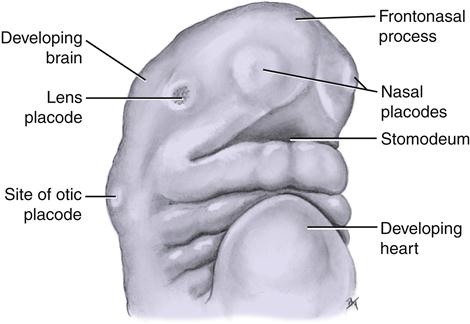
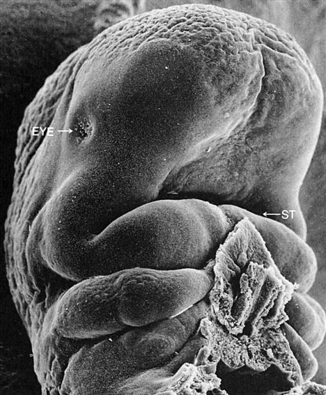
The face and its associated tissue begin to form during the fourth week of prenatal development within the embryonic period (Box 4-1). During this time, the rapidly growing brain of the embryo bulges over the oropharyngeal membrane and developing heart (Figures 4-1 and 4-2). The area of the future face is now squeezed between the developing brain and heart with the formation of the three embryonic layers and resultant embryonic folding (see Figure 3-15).
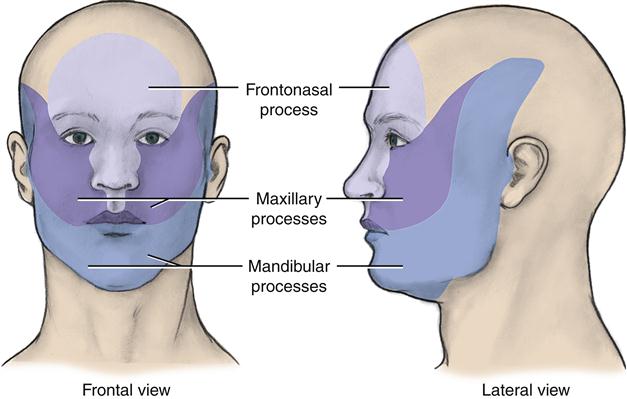
All three embryonic layers are involved in facial development: ectoderm, mesoderm, and endoderm (Table 4-1). Facial development includes the formation of the primitive mouth, mandibular arch, maxillary process, frontonasal process, and nose. Facial development depends on the five major facial processes (or prominences) that form during the fourth week and surround the embryo’s primitive mouth: single frontonasal process and paired maxillary and mandibular processes (Figure 4-3). Thus these facial processes become the centers of growth for the face. If the adult face is divided into thirds—upper, midface, and lower parts—these parts roughly correspond to the centers of facial growth. The upper part of the face is derived from the frontonasal process, the midface from the maxillary processes, and the lower from the mandibular processes.
TABLE 4-1
Embryonic Development of the Face
| EMBRYONIC STRUCTURES | ORIGIN | FUTURE TISSUES |
| Stomodeum | Ectodermal depression enlarged by disintegration of oropharyngeal membrane | Oral cavity proper |
| Mandibular arch (first branchial arch) | Fused mandibular processes and neural crest cells | Lower lip, lower face, mandible with associated tissues (other arch derivatives shown in Table 4-2) |
| Maxillary process(es) | Superior and anterior swelling(s) from mandibular arch and neural crest cells | Midface, upper lip sides, cheeks, secondary palate, posterior part of maxilla with associated tissues, zygomatic bones, part of temporal bones |
| Frontonasal process | Ectodermal tissue and neural crest cells | Medial and lateral nasal processes |
| Nasal pits | Nasal placodes | Nasal cavities |
| Medial nasal process(es) | Frontonasal process medial to nasal pits | Middle of nose, philtrum region, intermaxillary segment |
| Intermaxillary segment | Fused medial nasal processes | Anterior part of maxilla with associated tissues, primary palate, nasal septum |
| Lateral nasal process(es) | Frontonasal process lateral to nasal pits | Nasal alae |
The facial development that starts in the fourth week will be completed later in the twelfth week, within the fetal period. The face changes shape considerably as it grows. Thus, overall facial proportions develop during the fetal period. It important to note that the development of the associated oral structures is occurring at the same time and is discussed in Chapters 5 and 6.
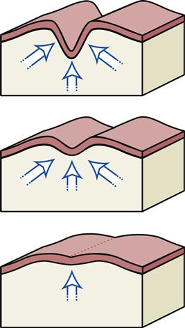
Most of the facial structures develop by fusion of swellings or tissue on the same surface of the embryo (Figure 4-4). A cleft, or furrow, is initially located between these adjacent swellings due to proliferation, differentiation, and morphogenesis (see Table 3-3). However, with facial fusion, these furrows are usually eliminated as the underlying mesenchyme migrates into the furrow, making the embryonic facial surface smooth. This migration takes place because adjacent mesenchyme grows and merges beneath the external ectoderm during the maturation of the structure. In some cases, a slight groove or line maybe left on the facial surface, showing where the fusion of the swellings took place. Differing from the fusion that takes place on the facial surface is the type of fusion that occurs during development of the palate (see Figure 5-1). In contrast to facial fusion, palatal fusion involves the fusion of swellings or tissue from different surfaces of the embryo, such as that which occurs with the fusion of the neural tube (see Figure 3-10, C).
The overall growth of the face is in both an inferior and anterior direction in relationship to the cranial base. The growth of the upper face is initially the most rapid, in keeping with its association with the developing brain. Subsequently, the forehead ceases to grow significantly after age 12. In contrast, the middle and lower parts of the face grow more slowly over a prolonged period of time and finally cease to grow late in puberty. The eruption of the permanent third molars at approximately 17 to 21 years of age marks the end of the major growth of the lower two thirds of the face. The underlying facial bones, also developing at this time, depend on centers of bone formation by intramembranous ossification (see Figure 8-12).
Stomodeum and Oral Cavity Formation
The primitive mouth is now the stomodeum (or stomatodeum), which initially appeared as a shallow depression in the embryonic surface ectoderm at the cephalic end before the fourth week (see Figures 4-1 and 4-2). At this time, the stomodeum is limited in depth by the oropharyngeal membrane. This temporary membrane, consisting of external ectoderm overlying endoderm, was formed during the third week of prenatal development. The membrane also separates the stomodeum from the primitive pharynx. The primitive pharynx is the cranial part of the foregut, the beginning of the future digestive tract.
Stay updated, free dental videos. Join our Telegram channel

VIDEdental - Online dental courses


