CHAPTER 4

DENTAL MORPHOLOGY
MARGARIDA HENRIQUE

Dental morphology is unique for each person, like a fingerprint. Thus, there is a huge variation of tooth shapes in nature. Although this huge variation of dental anatomy, there has been much research to discover common features in esthetically pleasant teeth arrangements and shapes…
Dental morphology
A restorative treatment, the assumption of which is to perform a reconstruction of the patient biology, function, structure, and consequently also esthetics, cannot be done without the knowledge of tooth morphology. A dentist who understands the morphology of hard tissues can rebuild the missing tissues in a way that meets the appropriate criteria.
NATURAL ESTHETIC RESULTS
A knowledge of dental morphology is fundamental in the practice of dentistry. We want to replicate nature and give biological, functional, structural and, consequently, esthetic restorations to our patients. Nature is so well designed that each morphological characteristic of a tooth has a certain function, so if you have developed knowledge about morphology, you will know how to restore a tooth fulfilling all these premises. That is why dental professionals should search for this knowledge in a cognitive, visual and, most importantly, practical way.1–4
Dental morphology is so complex and variable that there are no absolute truths. However, every tooth has some basic characteristic features, and when integrated into a smile according to some established esthetic parameters, it produces an esthetically pleasant result, a beautiful smile.
It is important to note that esthetics, although not well defined, is a universal language, and frequently people of the same society or culture agree about the beauty of a person or not. Indeed, this high concordance in judging beauty in the same culture can simply reflect the media, which is today highly competitive and a dictator of trends. However, research from diverse cultures suggests that the shared beauty ideals do not depend on a preformed image.5
In dentistry, esthetic treatments enhance a patient’s natural beauty, restore and reproduce the natural characteristics of teeth and smile.6 There is always the need to search for more beautiful restorations, and many esthetic criteria that should be present in a beautiful smile can be proposed.
Esthetic dentistry emphasizes the natural beauty of the patient and his smile …
TOOTH STRUCTURE
When we think about dental morphology is important to have knowledge about the tooth structure per se. In Fig 4-1, we have a polarized photograph of a tooth cut of approximately 1 mm, in which we can appreciate the various components of a tooth.

Fig 4-1
Tooth structure and its components.
We know that a tooth is composed of several tissues: enamel, cementum, dentin, and pulp. And that it is divided into two parts: the crown that is exposed in the oral cavity, and the root that is covered by the cementum and submerged and anchored to the bone tissue through the periodontal ligament. To better understand this structure, we will briefly describe the basic characteristics of each of its components (Fig 4-2).
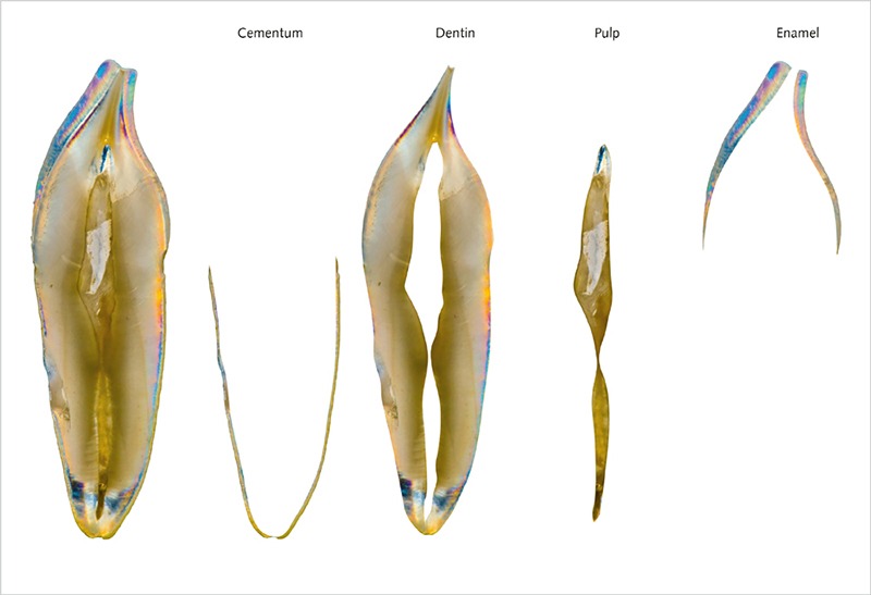
Fig 4-2
Tooth components.
 Cementum (Fig 4-3)
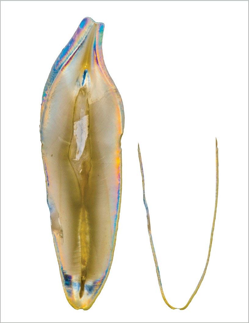
Fig 4-3
Schematic division of the cementum.
Very thin (50–100 µm) mineralized connective tissue covers all the root area dentin, from the cervical anatomical line to the apex. Its principal function is the connection of the fibers of the periodontal ligament.
We can compare it with bone since its hardness and chemical composition are similar. It is not vascularized, and it lacks proper innervation. It also has no ability to be remodeled and is generally more resistant to resorption than the bone.
It presents a pearly white color, is darker and more opaque than enamel but less yellow than dentin.
Composition:
- Cellular components: cementoblasts and cementocytes
- Calcified extracellular matrix (46–50% inorganic matter, 22% organic matter, and 32% water)7
 REMEMBER
The natural tooth is polychromatic, which means that it has a large variety of colors and nuances that can be observed and perceived by the human brain. To reproduce all the internal characteristics of a tooth is not an easy task, and the dentist should have the knowledge and artistic sense to identify all the details and define the different characteristics of each one.
 Pulp (
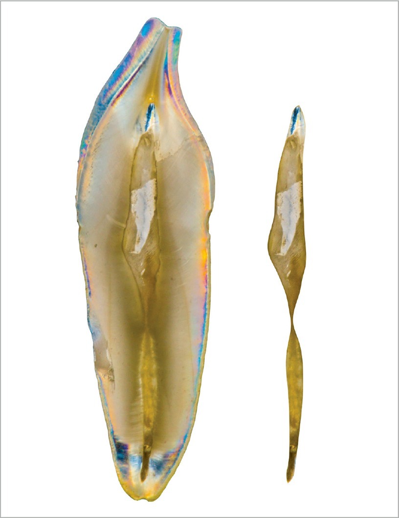
Fig 4-4
Schematic division of the pulp.
Conjunctive tissue organ is located in the center of each tooth, constituted by numerous structures like arteries, veins, lymphatic system, and nerves. Its principal function is to form the dentin.
In newly erupted teeth, the pulp is big, and it progressively reduces as the tooth
is completed.
Pulp maintains its tissue-forming activity (secondary dentin), especially when caries advances into the pulp. Sometimes the pulp chamber can be totally obliterated by the secondary dentin to protect the pulp.
It has a coronal portion (pulp chamber) and a root portion (pulp or root canal). The opening of the pulp in the apex is constricted, and it is called apical foramen. The pulp connects directly with the periodontal ligament in this area through the periapex. The blood vessels and nerve fibers enter and exit the pulp through it.7,8
Approximated composition:
- 75% water
- 25% organic matter (cells and extracellular matrix)
- Dentin–pulp complex (Fig 4-5)
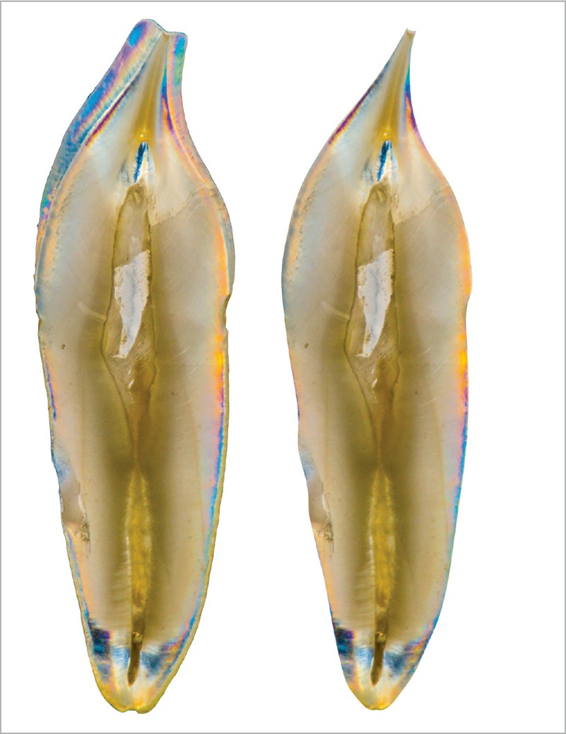
Fig 4-5
Schematic division of the dentin–pulp complex. - Structural unit – odontoblasts prolongations are included on the dentin
- Functional unit – the pulp maintains the vitality of the dentin, and the dentin protects the pulp
- Their embrionary origin is common
 Dentin (
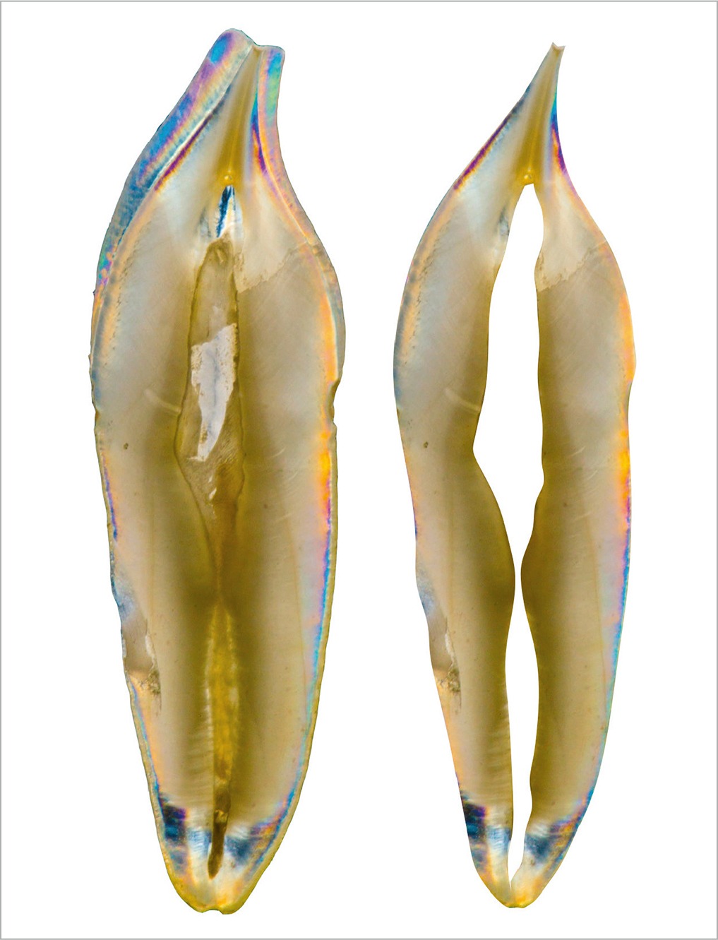
Fig 4-6
Schematic division of the dentin.
This is mineralized tissue that forms the largest volume of a tooth. We can distinguish two basic components: mineralized matrix and dentinal tubules that cross all its thickness and lodge the odontoblastic processes, which are prolongations of the odontoblasts, responsible for the production of the collagen matrix and calcification of the dentin.
The odontoblasts are located in the most peripheral region of the pulp, and their prolongations are included in the dentin; that is why dentin and pulp are considered a unit: a dentin–pulp complex (see Fig 4-5).
It presents with yellowish-white color, but it may vary from one individual to another, and also throughout life. It is less translucent than the enamel due to its lower degree of mineralization.
Approximate composition:
- 70% inorganic matter (hydroxyapatite crystals)
- 18% organic matter (collagen fibers)
- 12% water
This composition is variable between the different regions of the dentin:
- Microhardness = 0.57 to 1.13 GPa
- Young elasticity modulus = 17.6 to 22.9 GPa; Vickers hardness = 60–70 HV; Compressive strength = 200–350 MPa
The natural tooth is polychromatic, which means that it has a large variety of colors and nuances that can be observed and perceived by the human brain. To reproduce all the internal characteristics of a tooth is not an easy task, and the dentist should have the knowledge and artistic sense to identify all the details and define the different characteristics of each one. Dentin and enamel have a rich composition and detail of optical characteristics.
 TIP
TIP
Dentin anatomy is a way to understand internal teeth characteristics.
Dentin is responsible for the basic color of the tooth (hue), and enamel modulates the chroma and the value of the hue according to its thickness. Comprehension of the thickness, both of the enamel and dentin necessary to emulate the natural tooth, is fundamental to achieving a great esthetic result. To better understand it, I did the removal of the enamel layer of several teeth following the protocol suggested by Bazos and Magne, submerging the natural teeth in 10% hydrochloric acid (HCL) under ultrasonic vibration for 20 minutes, and I have obtained the structures shown in Fig 4-7. We can observe the dentoenamel junction (DEJ) surface, and understand that its morphology has a crucial role on the enamel surface morphology.9,10
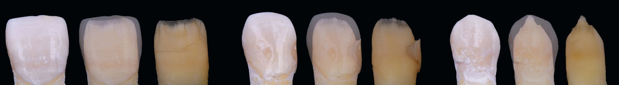
Fig 4-7
Natural teeth treated with hydrochloric acid (HCL) at 10% to remove all the enamel layer and have a better comprehension of the dentin morphology.
To better understand and train the morphology of the dentin surface, I have tried to reproduce it with a characterized wax-up of each of the anterior teeth (13-12-11-41-42-43) as shown in Figs 4-8 and 4-9.
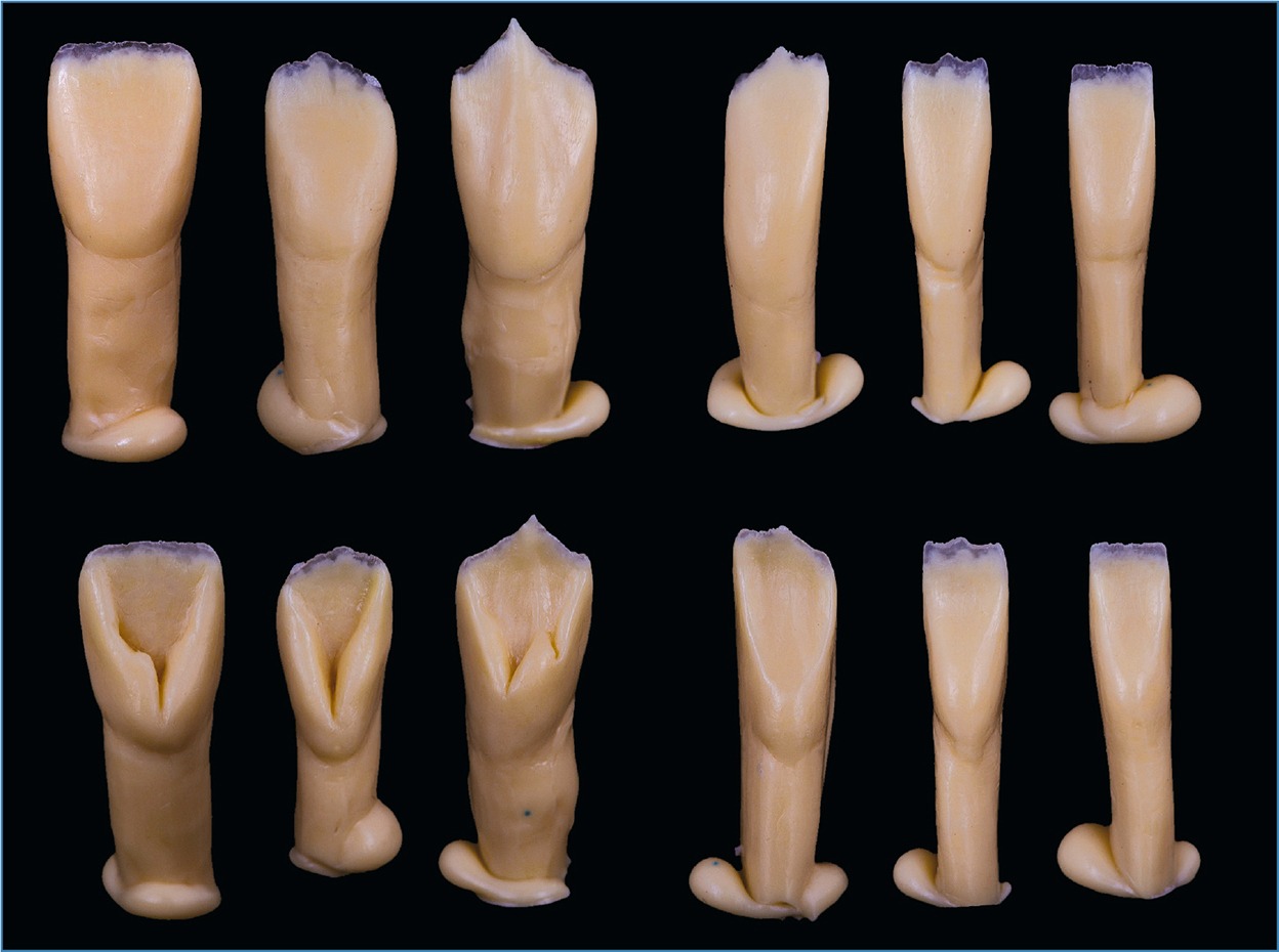
Fig 4-8
Total wax dentin cores to simulate dentin morphology, facial and lingual view (note the simulation of the mantle of dentin).
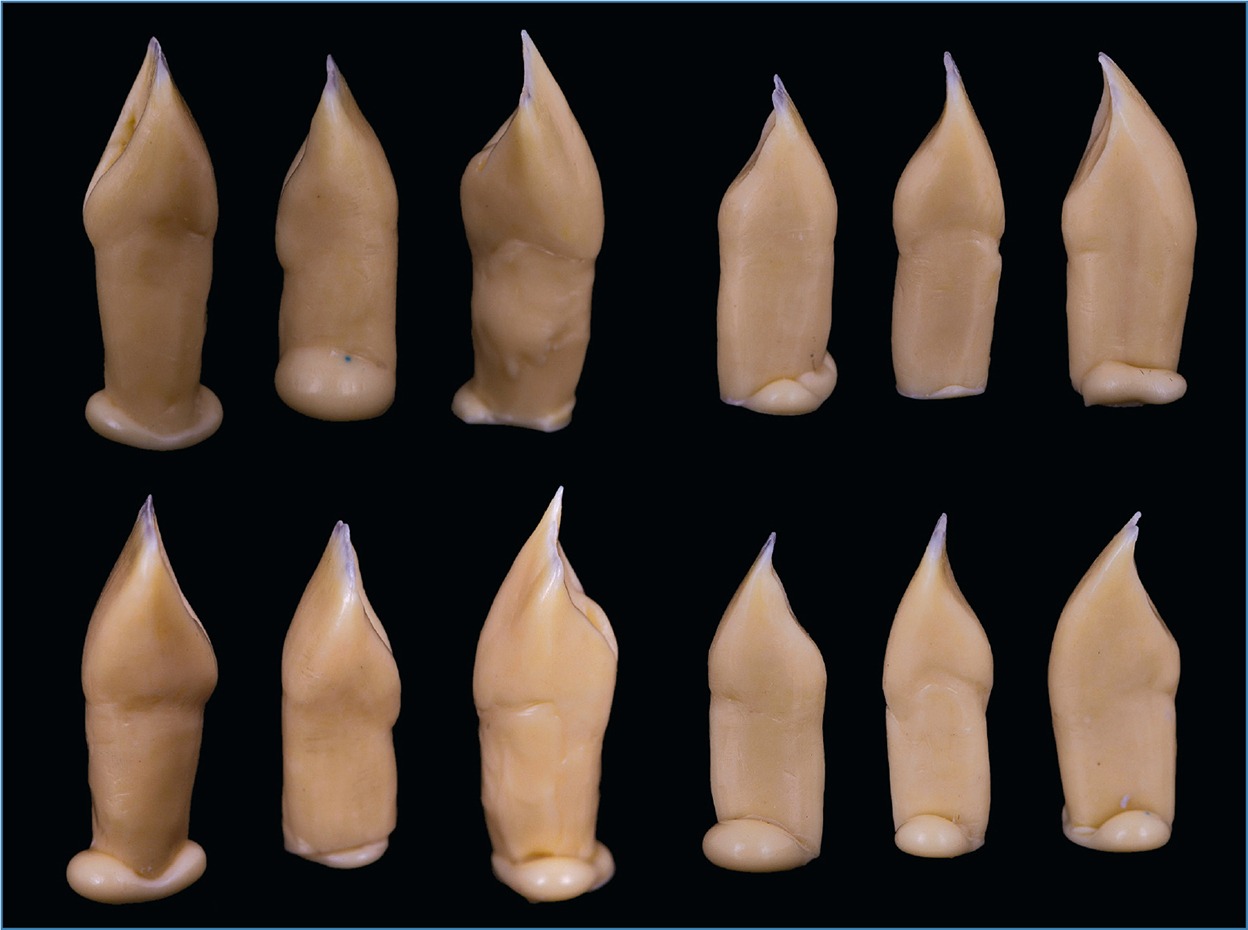
Fig 4-9
Total wax dentin cores to simulate dentin morphology, mesial and distal view (note the simulation of the mantle of dentin).
 DEJ – dentinoenamel junction (Fig 4-10)

Fig 4-10
Scheme indicating the dentinoenamel junction (DEJ) position.
Well delimitated line resultant of the union of the enamel with the dentin.7 A less simplistic view suggests that the DEJ complex should also include the inner aprismatic enamel and the mantle dentin.11 The DEJ and the outer enamel surface are essential features of the tooth’s three-dimensional structures and significantly affect its visual appearance. It is known that the shape of the DEJ is very similar to the shape of the outer surface enamel.9,12,13
 JAC – amelocementary junction (Fig 4-11)

Fig 4-11
Scheme indicating the amelocementary junction (JAC) position.
Also known as the cervical anatomic line. There are four possibilities for its presentation (the Choquet cases)8:
1. The cement covers the enamel (60% of cases)
2. The enamel covers the cement (less frequent)
3. Enamel and cement are in contact, and there is no exposed dentin (30% of cases)
4. There is a lack of connection between the enamel and the cement
 Enamel (Fig 4-12)
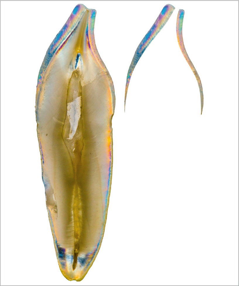
Fig 4-12
Schematic division of the enamel.
Also known as an adamantine substance, it covers the dentin, offering protection to the dentin–pulp complex. It is the hardest tissue on the human body and is constituted by millions of mineralized prisms – hydroxyapatite crystals. It is acellular, avascular, and has no innervation, so it cannot self-repair. Its thickness varies in the different regions of the tooth.
Its major thickness is located at the incisal edge and the minimum in the cervical, where it ends in a sharp edge. It is translucent, and its color is dependent on the subjacent dentin. In areas of greater thickness, it has a white-grayish color, and where it is thinner, it presents a yellowish-white color.
Approximate composition:
- 95% inorganic matter (hydroxyapatite crystals)
- 1–2% organic matter (protein origin with no collagen)
- 3–5% water
DENTAL MORPHOLOGY
It is essential tohave a basic notion about the structure of a tooth to understand its morphology. As we have seen before, dentin plays a major role in defining the final shape and color. Now, we are ready to go for the study of the basic morphological characteristics of each one of the anterior human teeth. To facilitate the comprehension of a tooth, the crown and root are normally divided into thirds to precisely define the location of its characteristics14 (Fig 4-13).

Fig 4-13
Total wax-up in a 1:1 ratio of a superior canine vestibular and proximal views. (left) Division in thirds of the tooth crown and root viewed from vestibular, in horizontal and vertical directions. (right) Division in thirds of the tooth crown viewed from proximal, in a vertical direction.
BASIC CHARACTERISTICS OF ANTERIOR TEETH MORPHOLOGY
 Anatomical cervical line: Continuous and sinuous line that divides the tooth into crown and root. It is concave in vestibular and palatal and convex in mesial and distal.
 Surface: Each anterior tooth presents five surfaces: facial, lingual or palatal, mesial, distal, and incisal.
 Border/Edge: These delimitate the transition between two surfaces, taking note of the ones that they’re in, forming a dihedral angle.
 Angle: When the different surfaces of a tooth meet, there is a formation of angles between them.
 Bulges: They are enamel elevations that protrude on dental surfaces. Four humps are common in every anterior tooth, and their location is:
- Cervical third in the buccal surface.
- Incisal third in the mesial surface.
- Incisal third, slightly going to the medium third on the distal surface.
- Cervical third, in the lingual surface.
 Marginal crest: Cylindrical enamel projections located on the proximal portions of the lingual surface, going from the incisal edge to the cingulum on the anterior teeth. They act as a reinforcement pilar and allow the maintenance of the vertical occlusion dimension of the anterior sector
 Cingulum: Lingual lobe of anterior teeth, which determines the width of the cervical third of the lingual surface. It is important in the masticatory process to protect the soft tissue from the friction produced.
 Lingual fossa: A concavity or irregular depression delimited by the incisal edge, marginal ridges, and the cingulum. It is located on the palatal/lingual surface of the anterior teeth. It is less evident on the mandibular canines and incisors.
 Scape groove: A small sulcus located between the coronal portion of the termination of the marginal crests and the incisal edge. It runs from the lingual fossa into the proximal surface and provides the function of escape for food during the masticatory process.
 Pit: A sharp, pointed depression point between the cingulum and the lingual fossa. It can be found in both superior incisors but is more frequent in the lateral one.
 Development grooves: Two linear depressions (mesiovestibular and distovestibular), parallel to the major axis of the tooth, located on the vestibular surface of anterior teeth. They have variable development and are more frequent in younger teeth. They go from the cervical third (where they are less evident) to the incisal edge.
 Development lobes: Three lobes that are formed by the development grooves: mesial, medial, and distal. They can divide the incisal edge and form the incisal mamelons.
 Equatorial line: The line of the major contour of the tooth crown, resulting from the union of all the humps. It passes through the areas of bigger convexity of the dental surfaces. It divides the crown into two parts: one retentive located cervically to the equatorial line, and one expulsive located occlusally to it.
With the aim of a schematic description of each of the already mentioned characteristics, I have tried to replicate one of each tooth of two anterior sectors, three superior and three inferior. They were made in wax, applying a stratification technique, and then photographed. The morphological characteristics allow the teeth to have a correct function. The primordial function of teeth is to accomplish a good masticatory process; they are formed for cutting, tearing and crushing food, but they also have other functions.
 THE FUNCTIONS OF INCISORS AND CANINES
The functions of incisors, situated anteriorly, are to cut the food, enable articulated speech, support the lip, maintain an esthetic appearance, and aid mandible guidance during the final closing phase. They all have some common characteristics that can help us identify them: a relatively straight incisal edge with a curvature in distal, a relatively rectangular crown, three labial lobes (form the vestibular surface) and one palatal/lingual (form the cingulum), a crown outline more convex on the distal surface (except the inferior central incisors, which are symmetrical), the mesioincisal angles are more acute than the distal ones (except on the inferior central incisors) and the mesial contact areas located on the incisal third, while the distal ones are in between the incisal third and the middle third. They all have a wedge shape when viewed from proximal and an S-shaped lingual outline.
Canines have the function of cutting, piercing, or shearing food, supporting the lip and facial muscles, and functional protection due to their occlusal lateral guidance. They also have some common characteristics, which are: the presence of two incisal cusp ridges, being the mesial shorter than the distal one; the presence of a vertical labial ridge; they are the only teeth with just one cusp; the mesial contact areas are also located on the incisal third while the distal ones are in between the third and middle third; the crown outline is also more convex in distal than in mesial.
When viewed from proximal, they also have a wedge shape and an S-shaped lingual outline.14
 TIP
TIP
The morphological characteristics allow the teeth to have a correct function. The primordial function of teeth is to accomplish a good masticatory process; they are formed for cutting, tearing, and crushing food, but they also have other functions. Anatomy is a way to understand internal teeth characteristics.
ANTEROSUPERIOR TEETH (Fig 4-14)

Fig 4-14
Photographs of anterosuperior teeth (11, 12, and 13) made of wax, applying a stratification technique.
 Superior central incisor (Figs 4-15 to 4-17)
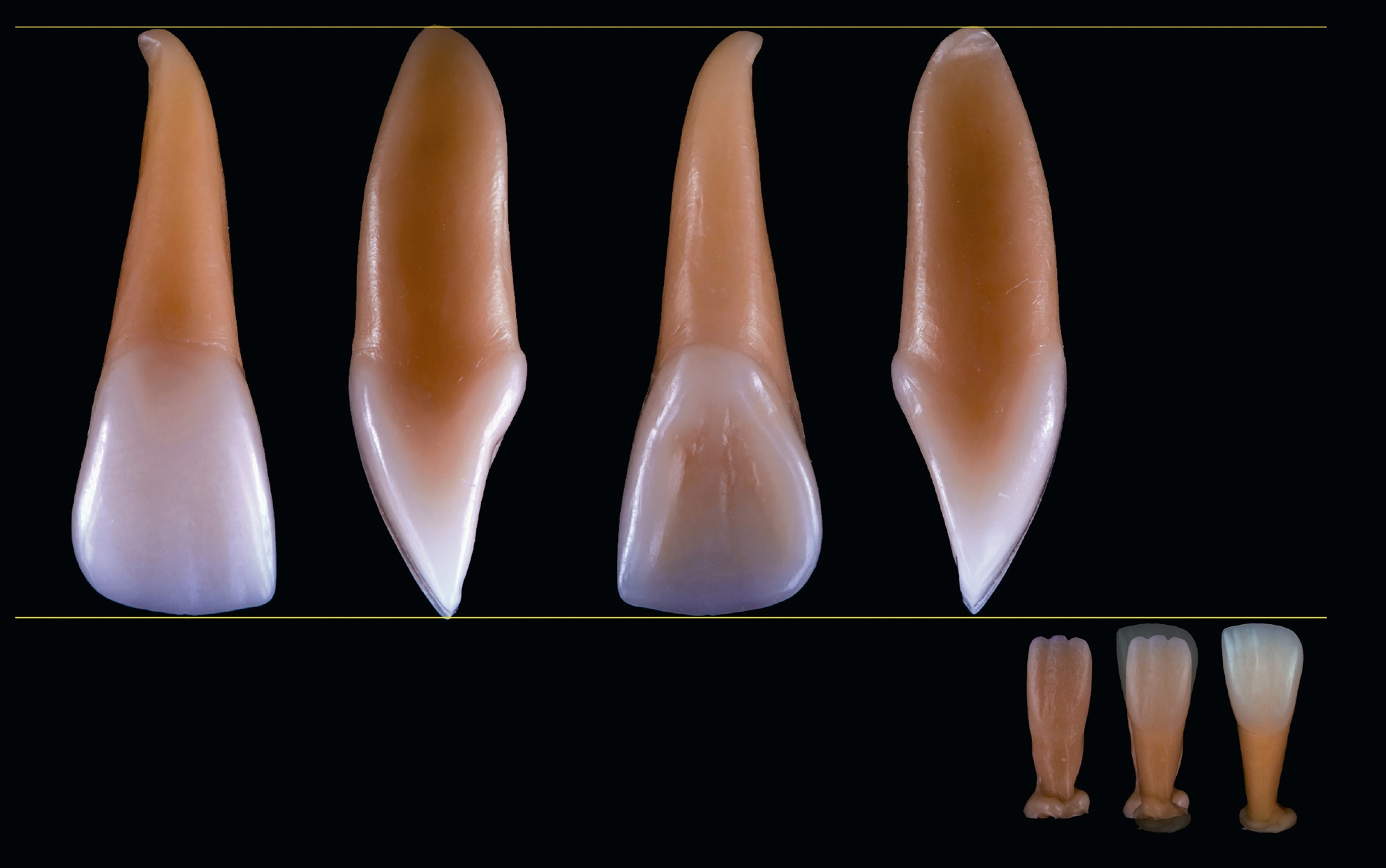
Fig 4-15
Photographs of tooth 11 (vestibular, mesial, palatal, and distal views). Stratification technique.
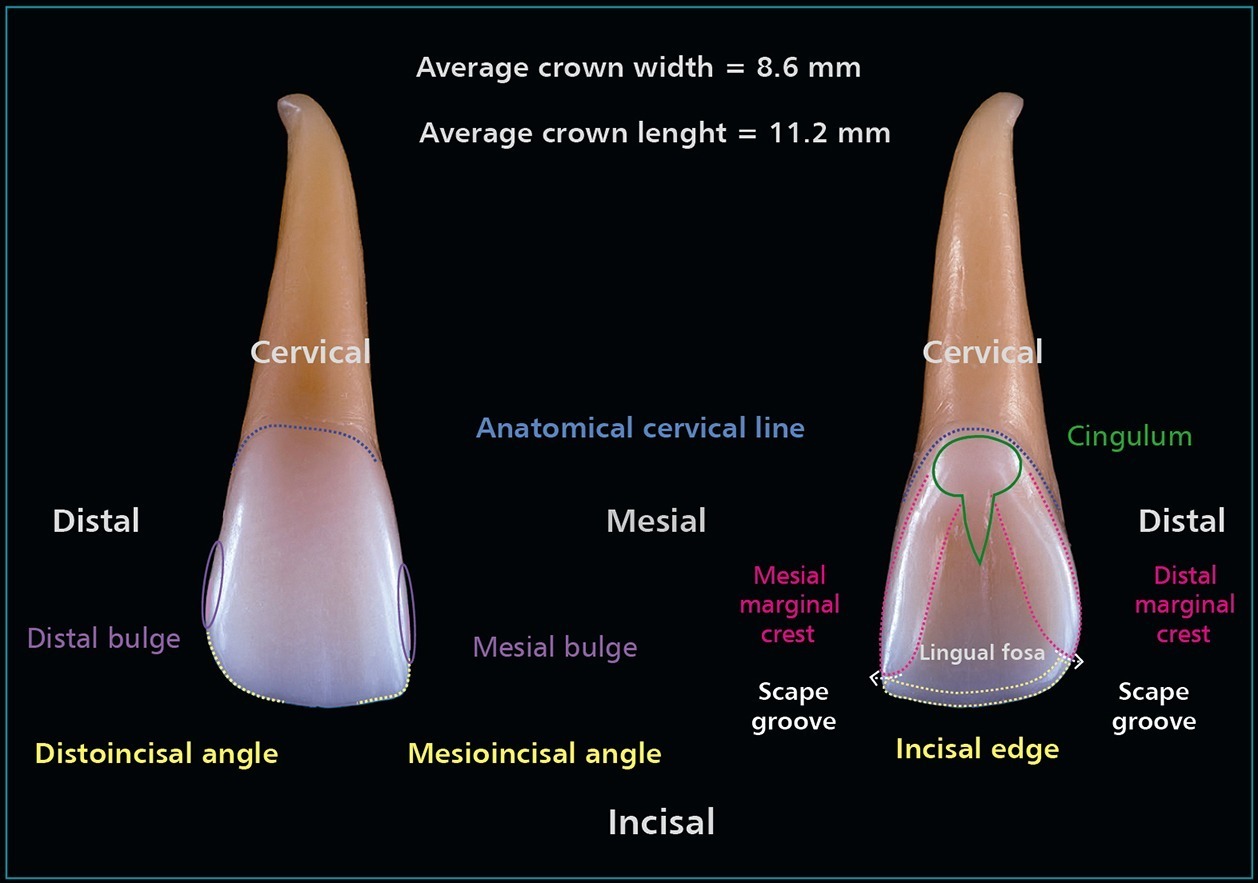
Fig 4-16
Tooth 11 (vestibular and palatal views). Scheme with basic tooth characteristics.
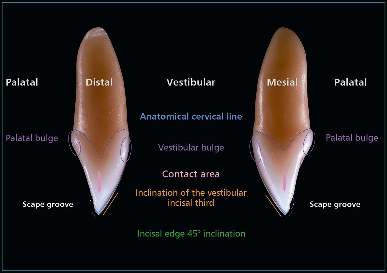
Fig 4-17
Tooth 11 (mesial and distal views). Scheme with basic tooth characteristics.
- Mesioincisal angle is a right angle
- Incisal edge closer to horizontal
- Mesial outline is flat
- Distal outline is more rounded
- Distal contact is more cervical than mesial one
- Larger shallow lingual fossa
- Distal marginal ridge more curved than mesial one
 Superior lateral incisor (Figs 4-18 to 4-20)
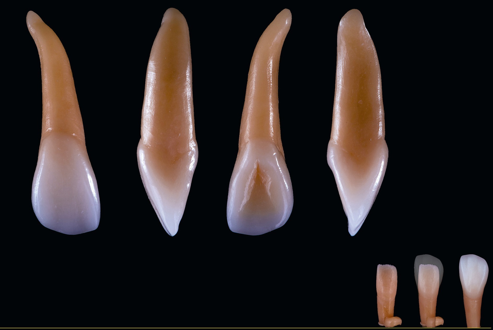
Fig 4-18
Tooth 12 (vestibular, mesial, palatal, and distal views). Stratification technique.
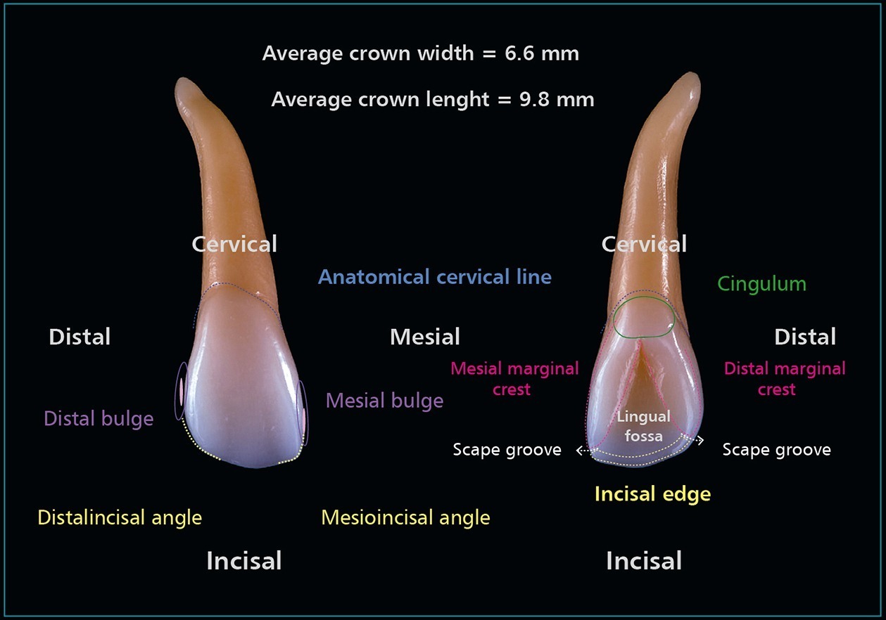
Fig 4-19
Tooth 12 (vestibular and palatal views). Scheme with basic tooth characteristics.

Fig 4-20
Tooth 12 (mesial and distal views). Scheme with basic tooth characteristics.
- Smaller crown, narrower cervically
- Mesioincisal angle is rounder than in the central incisor, but still straighter than the distal angle
- Distal outline is round
- Distal contact is near the middle third incisal edge toward cervical in distal
- Deep but small fossa
 Superior canine (Figs 4-21 to 4-23)

Fig 4-21
Tooth 13 (vestibular, mesial, palatal, and distal views). Stratification technique.

Fig 4-22
Tooth 13 (vestibular and palatal views). Scheme with basic tooth characteristics.
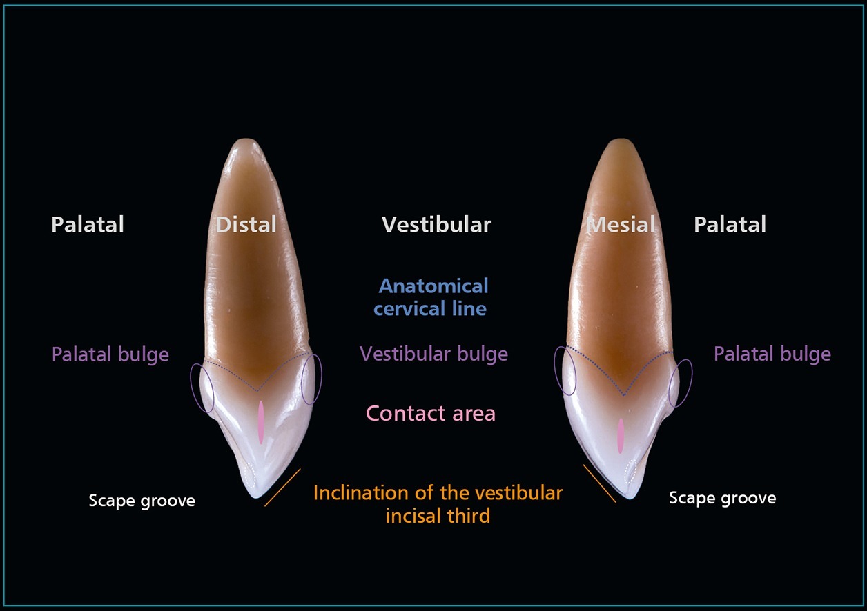
Fig 4-23
Tooth 13 (mesial and distal views). Scheme with basic tooth characteristics.
- They do not have mamelons, but they can have a notch on either cusp ridge
- The labial ridge is always vertical
- The distal contact area is more cervically positioned than the mesial one
- The crown outline is more convex on the distal than on the mesial
- The anatomy is more prominent in lingual (marginal crests and lingual ridge)
ANTERIO-INFERIOR TEETH (Fig 4-24)
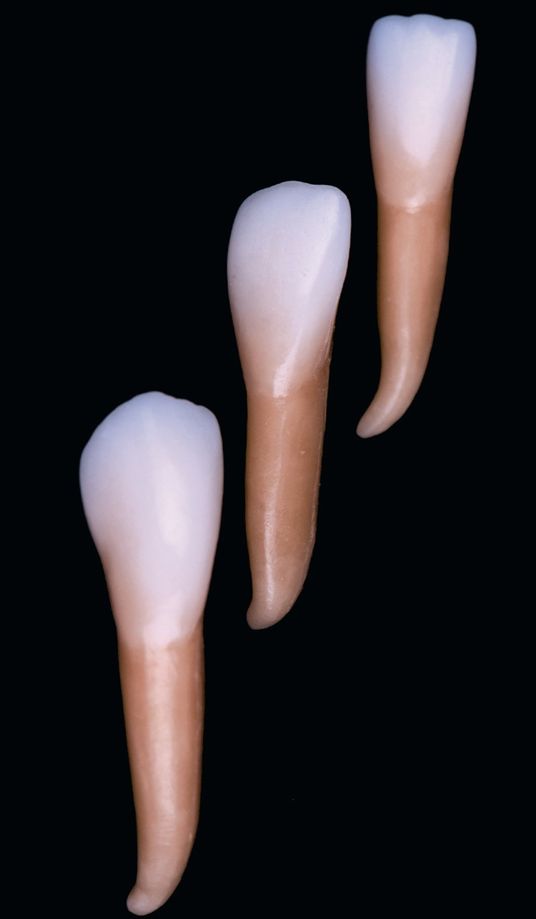
Fig 4-24
Teeth 41, 42, and 43 wax-ups, applying a stratification technique.
 Inferior central incisor (Figs 4-25 to 4-27)
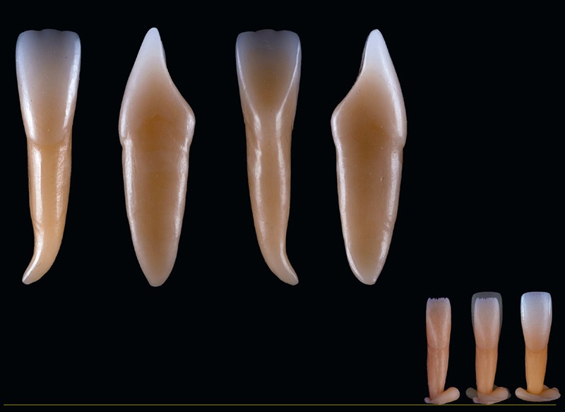
Fig 4-25
Tooth 41 (vestibular, mesial, palatal, and distal views). Stratification technique.

Fig 4-26
Tooth 41 (vestibular and palatal views). Scheme with basic tooth characteristics.

Fig 4-27
Tooth 41 (mesial and distal views). Scheme with basic tooth characteristics.
- It is the narrowest crown in the mouth
- It is the only symmetrical tooth in the mouth
- Mesial and distal contacts are at the same level (incisal third)
- Mesioincisal and distoincisal angles are right angles
- Straight mesial and distal outlines, becoming narrower toward cervical
- Facial surface is smooth but can have two shallow development depressions
- The lingual fossa is barely visible, smooth, without grooves, and shallow, slightly concave in the middle and incisal thirds
 Inferior lateral incisor (Figs 4-28 to 4-30)
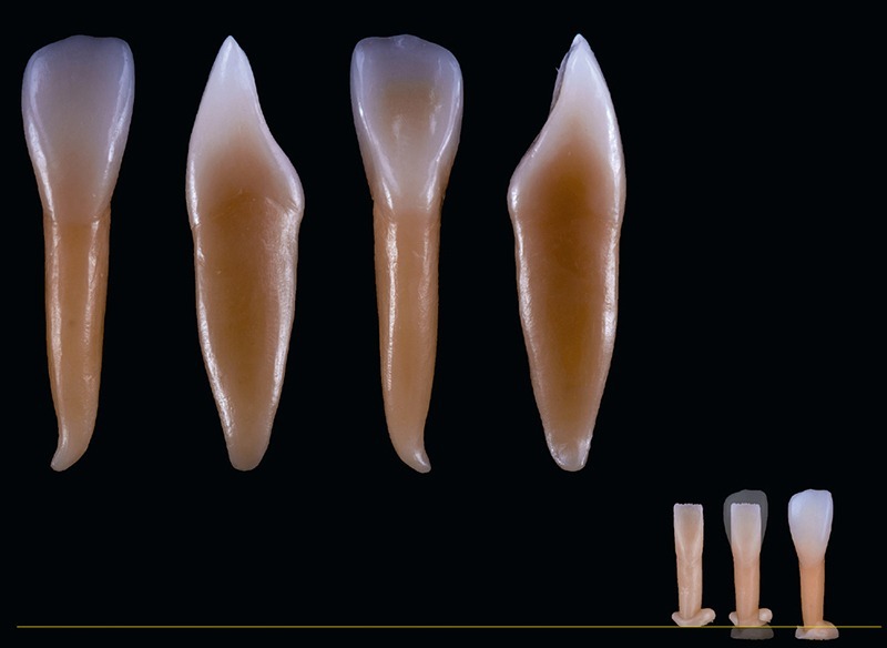
Fig 4-28
Tooth 42 (vestibular, mesial, palatal, and distal views). Stratification technique.

Fig 4-29
Tooth 42 (vestibular and palatal views). Scheme with basic tooth characteristics.

Fig 4-30
Tooth 42 (mesial and distal views). Scheme with basic tooth characteristics.
- The crown is slightly wider than the mandibular central incisor
- Distal crown outline is rounded, and the mesial one is straight
- Mesial proximal contact is more incisal
- Distal proximal contact is in a more cervical position
- Larger than mandibular central incisor
- Facial surface is smooth but can have two shallow development depressions in the incisal third
- The lingual fossa is barely visible, smooth, without grooves, and shallow, slightly concave in the middle and incisal thirds
 Inferior canine (Figs 4-31 to 4-33)
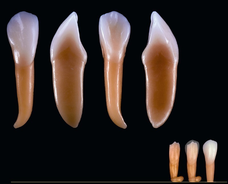
Fig 4-31
Tooth 43 (vestibular, mesial, palatal, and distal views). Stratification technique.

Fig 4-32
Tooth 43 (vestibular and palatal views). Scheme with basic tooth characteristics.
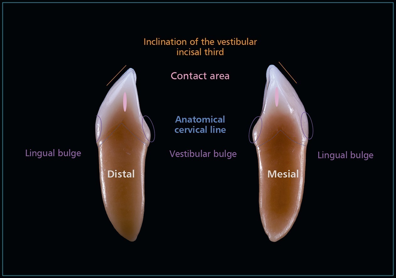
Fig 4-33
Tooth 43 (mesial and distal views). Scheme with basic tooth characteristics.
- Crown appears narrower mesiodistally and longer than the maxillary canine
- Cusp angle is obtuse, so less pronounced
- Distal outline is more convex on the distal than on the mesial surface, where it is almost flat
- Mesial cusp ridge is shorter than the distal one and is almost horizontal
- The mesial proximal contact is more incisal than the distal one
- Less pronounced labial ridge than the maxillary canine
- Lingual ridge, marginal ridges, and fossa are less prominent than in the maxillary canine
Stay updated, free dental videos. Join our Telegram channel
VIDEdental - Online dental courses


