CHAPTER 4 Anatomy and Physiology
4.1 General Anatomy and Physiology
The structure of the body
The Cells
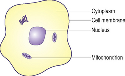
There are many different cell types, but the basic design is the same for all cells (Figure 4.1.1):
The Tissues
The Organs
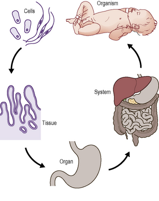
An organ is a structure that contains at least two different types of tissue that work together for a common purpose (Figure 4.1.2). Organs include the brain, heart, liver, kidneys, skin and others.
Terms used in anatomy
The special terms used in anatomy to describe the relationship of one part of the body to another are shown in Table 4.1.1. To help describe the position of structures in the body relative to each other and also the movement of various parts of the body in relation to each other, the body can be divided into anatomical planes (Figure 4.1.3) that correspond to the vertical and horizontal planes of space (Table 4.1.2).
| Term | Definition |
|---|---|
| Superficial | Closer to the surface |
| Deep | Further from the surface |
| Anterior | Closer to the front of the body |
| Posterior | Closer to the back of the body |
| Superior | Closer to the top of the head |
| Inferior | Closer to the soles of the feet |
| Medial | Closer to the midline of the body |
| Lateral | Away from the midline of the body |
| Proximal | Closer to the point of origin of a structure |
| Distal | Further from the point of origin of a structure |
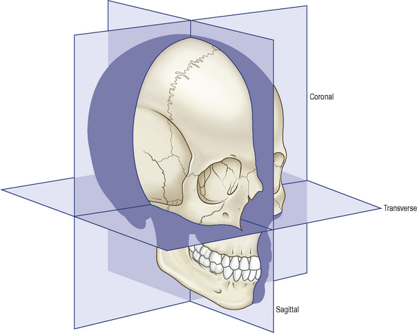
| Anatomical Plane | Spatial Plane | Body is Divided by Plane into Portions |
|---|---|---|
| Coronal | Vertical | Anterior and posterior |
| Transverse | Horizontal | Superior and inferior |
| Sagittal | Vertical | Right and left |
The circulatory system
Blood
Blood cells
There are three types of blood cell:
BOX 4.1.1 Haemostasis and Wound Healing
Haemostasis
Production of blood cells
Blood cell production also requires a healthy bone marrow. People whose bone marrow is damaged (e.g. because they have had radiotherapy or chemotherapy for a cancer) may lack all types of blood cells. They can then have anaemia and they also have a tendency to catch infections due to a lack of white blood cells for defence (see Box 4.1.2 below) and they can also have a tendency to bleed (since platelets are also damaged).
BOX 4.1.2 Immunity and Inflammation
The other white blood cells involved in defence are the lymphocytes:
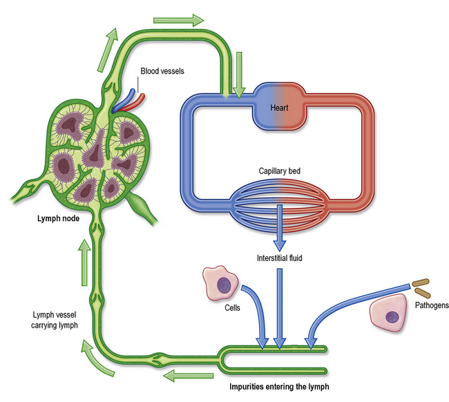
Central to the immune response are also organs such as the spleen and lymph nodes, which are together called the lympho-reticular or reticulo-endothelial system (RES)). The lymph nodes are basically collections of lymphocytes and macrophages. These cells catch and deal with pathogens or other foreign materials that have escaped from the blood into the tissues and then entered the lymph (Figure 4.1.7).
The Blood Vessels
There are three types of blood vessel:
The pulmonary artery and vein are the opposite of the rest of the arteries and veins, since the pulmonary artery carries de-oxygenated blood from the heart to the lungs and the pulmonary vein carries the oxygenated blood from the lungs to the heart (see Figure 4.1.5 below).
The Heart and the Flow of Blood around the Body
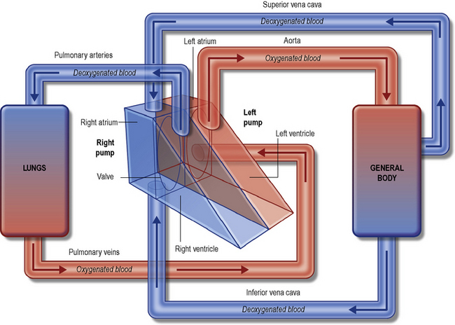
The heart is the organ that is found in the centre of the chest (the thorax). It has four chambers: two large ventricles and two smaller atria (Figure 4.1.4).
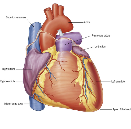
The heart pumps de-oxygenated blood to the lungs via the pulmonary arteries. In the lungs the blood releases the carbon dioxide and becomes oxygenated. It then travels in the pulmonary veins back to the heart entering it at the left atrium (Figure 4.1.5). From the left atrium blood is pumped into the left ventricle. The opening between these two chambers is controlled by a valve called the mitral valve.
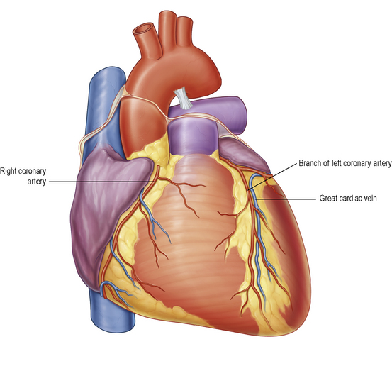
Oxygenated blood reaches the tissues of the heart itself by way of branches of the aorta called the coronary arteries (Figure 4.1.6). If the coronary arteries get blocked, this stops the oxygen supply to the heart and causes the condition called angina or a heart attack (also called myocardial infarction or coronary thrombosis; see Chapter 2 and Chapter 17.1).
From the arteries, the blood enters the capillaries. It is in the capillaries that the oxygen is released to the tissues and the carbon dioxide collected as a waste product. The space between the capillaries and the cells of a tissue is filled with a substance called the interstitial fluid and the gases and nutrients travel through this (see Figure 4.1.7 below).
The Lymphatic System
The lymphatic system (Figure 4.1.7) is the part of the circulatory system that cleanses it of impurities. It also forms an important part of the immune system (Box 4.1.2). The lymphatic system consists of the following parts:
The respiratory system
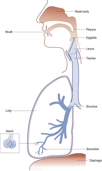
The respiratory system (Figure 4.1.8) has two parts:
Defence Mechanisms in the Respiratory Tract
The defence mechanisms in the respiratory tract include those discussed in Box 4.1.2 as well as:
The Nose
Air enters the nose through the openings in the face called the anterior nares. It exits the nose into the pharynx which connects to the lungs via the larynx, trachea and bronchi (Figure 4.1.8). The mucosal lining of the nose is rich in blood vessels and glands that produce protective mucus. The plentiful blood warms the inspired air. The nose also has special cells that enable us to smell. The functions of the nose thus include:
The Larynx
The larynx has three important functions:
The larynx is also close to a very important gland called the thyroid gland (see p. 116). The larynx can be visualised in the neck as the ‘Adam’s apple’. The larynx leads into the trachea (the windpipe).
The Trachea and Bronchi
The right main bronchus is shorter, wider and more vertical than the left. Thus, if, for example, a small dental instrument or a part of a tooth or a filling is inhaled, it tends to enter the right bronchus because it is wider and more directly continuous with the trachea. The use of rubber dam (p. 217) should prevent such catastrophic accidents.
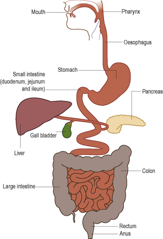
The digestive system
The digestive system (Figure 4.1.9) starts at the mouth and ends at the anus. It is responsible for:
Swallowing
The Small Intestine
The Large Intestine
The large intestine has two functions:
Lying between the small intestine and the anus, the large intestine consists of:
The large intestine is not crucial to life, and it is a common site for cancer.
Liver
The liver is located in the right part of the upper abdomen. Its functions are:
Stay updated, free dental videos. Join our Telegram channel
VIDEdental - Online dental courses


