39
Oral Pathology in the Newborn
Serious oral pathology in newborns and young infants is fortunately very uncommon and many of the “abnormalities” that are observed by the parent or our medical and nursing colleagues are in fact variations of normal. With the advent of more specialised antenatal ultrasound imaging techniques, many congenital abnormalities such as clefts of the lip and palate and cardiac defects can be detected in utero.
Developmental Cysts of Newborns
Epstein’s Pearls
It is estimated Epstein’s pearls may be found in up to 80% of all newborns. These small pale white well circumscribed papules appear in the midline of the palate, usually at the junction of the hard and soft palates, and are true keratinised cysts that have arisen from epithelial cells left in areas along the lines of fusion of the palatal shelves. They are sometimes termed inclusion cysts. Over time they sequestrate and resolve without any intervention.
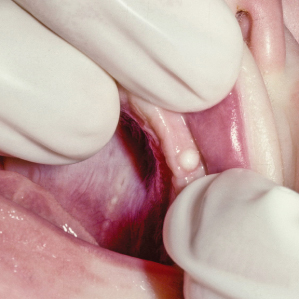
Bohn’s Nodules (Fig. 39.1)
Similar to Epstein’s pearls, these pale white or yellow papules measuring 1–4 mm in size are sometimes termed dental lamina cysts. They are remnants of the dental epithelium that form keratinised cysts and usually occur anywhere on the alveolar ridges, although they are observed predominately on the maxillary arches. They resolve spontaneously.
Figure 39.1 A single large Bohn nodule in a neonate. Commonly, multiple lesions are present and vanish within a few months.

Granular Cell Tumours
Two neoplasms present in newborns and infants that are histologically identical. The granular cell tumour of infancy, sometimes termed the congenital epulis or Neumann tumour, is an uncommon lesion that arises from the alveolar processes of either arch. The granular cell myoblastoma is found predominately on the tongue in older individuals.
Congenital Epulis
Unlike the granular cell myoblastoma, this lesion is negative for S100 stain. Over 85% of the lesions are found in girls and in many cases, small lesions resolve spontaneously. Only large lesions require treatment an/>
Stay updated, free dental videos. Join our Telegram channel

VIDEdental - Online dental courses


