37: The Medically Compromised Patient

chapter 37 The Medically Compromised Patient

When we previously discussed the management of stress related to medical and dental treatment, it was stated that many patients are unable to tolerate the “usual” degree of stress associated with dental therapy and, in some cases, with everyday existence. These persons usually have an underlying medical problem that limits their ability to handle stress in a normal manner. When such a patient is faced with a situation in which increased stress is present, there is an increased likelihood of this patient experiencing an acute exacerbation of the underlying disease process. Examples might include the patient with a history of coronary artery disease suffering from chest pain, the epileptic patient having a tonic-clonic seizure, or an asthmatic patient having an acute episode of bronchospasm during periods of increased stress, as might occur during dental treatment.

Advances in medicine and pharmacotherapeutics have made it possible for more medically compromised patients to become increasingly functional to the degree that their existence is no longer limited to their home. Partially or fully ambulatory, they may now be gainfully employed and seek dental and medical care as would any other patient. These persons, however, do represent a greater degree of risk during stressful times. How then may the medically compromised patient be better managed? Reduction of stress associated with treatment is the primary goal in the successful dental management of these patients. Awareness and knowledge of the underlying disease process are essential. In this chapter, disease entities that are more commonly seen in an ambulatory setting are reviewed. Following a brief description of each disease process, we list factors that tend to exacerbate it and review the methods available to successfully minimize perioperative stress in this patient.

CARDIOVASCULAR DISEASE

Cardiovascular disease ranks as the number one cause of death in the United States, United Kingdom, and other industrialized nations. It is estimated that the number of persons in the United States with signs and symptoms of cardiovascular disease exceeds 80.7 million. In 2005, 869,724 persons died from cardiovascular disease in the United States. Cancer, the second leading cause, was responsible for 553,888 deaths.1

Significant advances have occurred in both the surgical and pharmacologic management of many cardiovascular disorders. Patients who once suffered from extremely severe, almost debilitating anginal pains caused by coronary artery disease are today asymptomatic as a result of coronary artery bypass and graft procedures. Newer drugs, such as β-adrenergic blockers (e.g., propranolol), calcium channel blockers (e.g., verapamil, nifedipine), and angiotensin-converting enzyme (ACE) inhibitors (e.g., captopril), have enabled patients to lead more normal lives despite the continued presence of a serious cardiovascular disorder.

In view of the fact that more than 10% of the American population has signs and symptoms of clinically significant cardiovascular disease, it stands to reason that the dentist will be called on to manage the oral health needs of many cardiovascular-risk patients. Although there are many causes for the various cardiovascular diseases, there is one factor which, when present, is responsible for dramatically increasing the risk of an acute exacerbation. This is a myocardial oxygen (O2) requirement that exceeds the supply capability of the coronary arteries. When myocardial O2 requirements are not satisfactorily met, the patient responds with an acute exacerbation of the underlying disorder. For example, chest pain and/or dysrhythmias may develop in the patient with angina, and dyspnea will occur in the patient with heart failure (HF).

Patients at cardiovascular risk are usually able to tolerate elective and emergency dental care. Modifications in the planned dental treatment are based on the severity (American Society of Anesthesiologists [ASA] 2, 3, or 4) of the disease process as determined through physical evaluation of the patient (see Chapter 4). Sedation and pain control are of much greater importance in these patients than in ASA 1 patients. Specific details relating to the management of cardiovascular-risk patients are discussed in the following section.

Angina Pectoris

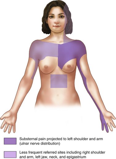
Angina pectoris is usually a result of arteriosclerotic heart disease, but may occasionally occur in the absence of significant disease through coronary artery spasm, severe aortic stenosis, or aortic insufficiency. The basic mechanism involved in angina pectoris is a discrepancy between myocardial O2 demand and O2 delivery through the coronary arteries. Anginal pain is described as a squeezing or pressurelike pain, retrosternal or slightly to the left of the sternum, that appears suddenly during exertion and may radiate in a set pattern (Figure 37-1); it typically resolves with rest or the administration of nitrates. Patients with angina may be taking long-acting nitrates, such as isosorbide dinitrate (Isordil, Sorbitrate), to prevent the occurrence of acute episodes. Nitroglycerin is available for administration in several forms, including an intravenous (IV) form, translingual spray (Nitrolingual), transmucosal tablet, oral sustained-release form, topical ointment, transdermal patch, and the traditional sublingual tablet.2

The patient with stable angina represents an ASA 3 risk. Persons with easily managed and less frequent episodes might be classified as ASA 2, and persons with daily (or more frequent) episodes or episodes that are increasing in frequency or severity (unstable angina) are categorized as ASA 4.

Factors increasing the likelihood of an acute exacerbation of anginal pain include the following:

As with all patients, those with a history of anginal pain may exhibit heightened anxiety before dental or surgical treatment. Sedation is especially indicated in these patients because of the effects of anxiety on the cardiovascular system. Increased blood levels of the catecholamines epinephrine and norepinephrine lead to an increase in the heart rate and an increase in the strength of each contraction. The net result is an increase in the O2 requirement of the myocardium. In the presence of coronary artery disease, this increased demand may not be met, resulting in an acute anginal episode. Dysrhythmias may also occur at this time. Minimizing stress and maximizing oxygenation of the patient are desired goals in preventing anginal episodes.

Oral sedation is indicated. Minimal to moderate levels of sedation minimize the risk of developing possible clinically significant respiratory depression, which could induce myocardial ischemia. Though not essential, the continuous delivery of O2 through a nasal cannula or nasal hood during treatment is recommended, especially in the ASA 3 or 4 patient with a history of angina.

Intramuscular (IM) and intranasal (IN) sedation are not usually indicated. The use of IM or IN sedation in adult patients is rarely indicated in general; these routes are used primarily in pediatric patients (IM, IN) or adults with disabilities (IM). Should an occasion necessitate the administration of IM sedation to a patient with angina, the level of sedation should be kept minimal to moderate. O2 should be delivered via nasal cannula or nasal hood throughout the procedure and recovery period.

Inhalation sedation is highly recommended. Nitrous oxide-oxygen (N2O-O2) inhalation sedation is the preferred technique of sedation for patients with angina. Because anginal episodes are provoked by an unmet myocardial O2 requirement, the administration of N2O-O2 serves the following beneficial purposes: (1) relaxes the patient, (2) increases the pain reaction threshold, and (3) increases oxygenation of the patient, including the myocardium. Not only does N2O-O2 minimize any increase in myocardial activity as a result of its sedative and analgesic properties, but also the typical patient will receive more than 50% or at the least approximately 30% O2. This represents a significant increase in O2 delivery to the cells of the patient’s body. N2O-O2 is the most nearly ideal sedative technique for the management of patients with angina.

IV sedation is recommended for the more fearful patient with a history of angina. Levels of sedation should be kept minimal to moderate (e.g., diazepam or midazolam) to lessen the potential for respiratory depression, hypoxia, or myocardial ischemia. In addition, the patient should receive O2 via nasal cannula or nasal hood throughout the procedure.

Elective general anesthesia is not recommended for all patients with angina. Outpatient general anesthesia in these patients is generally not indicated because of the increased risk of hypoxia during anesthesia and the inherent increased stress of general anesthesia and of the surgical procedure. Patients with angina who require general anesthesia are usually hospitalized, undergoing treatment as described earlier for inpatient procedures.

Unstable Angina

Unstable angina, also known as intermediate coronary syndrome, preinfarction angina, premature or impending myocardial infarction (MI), or coronary insufficiency, is a syndrome intermediate in severity between angina pectoris and acute MI.

Because mortality from acute MI is greatest within the first hour, recognition of a syndrome that has an increased likelihood of impending MI mandates immediate hospitalization and monitoring of the patient in an intensive care unit to prevent sudden dysrhythmias and death. Many, if not most, unstable anginal patients are candidates for reperfusion therapy with either thrombolytics or primary balloon angioplasty and cardiac stent.

Unstable angina is recognized by the appearance of pain that is different in character, duration, radiation, and severity from the typical stable anginal episode or pain that, over a period of hours or days, demonstrates progressive ease of induction (decreased exercise tolerance) or that develops at rest or during sleep.

Patients with unstable angina who do not develop signs and symptoms of acute MI are considered to be in precarious balance between coronary artery supply and myocardial demand and should be managed as though they had suffered an MI.

Patients with unstable angina are considered ASA 4 patients and therefore are not candidates for elective dental or surgical care. Immediate medical consultation is recommended. In the event that emergency care is required, hospitalization of the patient should be seriously considered. O2 delivery via nasal cannula or nasal hood is recommended throughout the procedure, and N2O-O2 inhalation sedation is the only sedative technique recommended for this patient. Medical consultation is definitely indicated before any treatment is carried out on this very high-risk patient.

Oral sedation is indicated if absolutely necessary. Only minimal levels of central nervous system (CNS) depression should be sought. The administration of O2 to this patient throughout the procedure is recommended.

IM sedation is not recommended in patients with unstable angina because of the possibility of hypotension, which further compromises coronary blood flow and respiratory depression. Inhalation sedation is recommended because of the increased O2 delivery to the patient throughout the procedure. IV sedation is not recommended unless the occasion absolutely demands it. The possibility of hypotension and respiratory depression, although minimal, could further aggravate the precarious balance between coronary O2 supply and demand. Moderate levels of sedation, such as that seen with benzodiazepines, plus supplemental O2 would best serve this patient.

Myocardial Infarction

MI is a clinical syndrome resulting from a deficient coronary arterial blood supply to a region of the myocardium, resulting in cellular death and necrosis. Synonyms for MI include coronary occlusion, coronary thrombosis, and heart attack.

More than 1.39 million Americans suffer acute MI annually.1 In 2004, ischemic heart disease and acute MI were responsible for 607,000 deaths in the United States.3 Coronary heart disease is the leading cause of death in the United States, responsible for 20% of all deaths occurring in the United States in 2004.3

Patients who have suffered an acute MI and survived represent a definite risk during dental and surgical treatment. Immediately after an MI, the incidence of reinfarction is high (36% reinfarction rate on surgical patients within 3 months of first MI).4,5 With time and the formation of a myocardial scar, the incidence of reinfarction declines. Reinfarction rates fall to 16% at 5 months for post-MI patients undergoing surgical procedures and to 5% at 6 months after infarction. Reinfarction rates then level off at 5% and remain at that level indefinitely. By comparison, the risk of infarction during surgical procedures for a patient who has not had an MI is less than 0.1%.

Patients who have had an MI may be receiving a number of drugs to manage complications, such as HF, angina, and dysrhythmias. Drug categories include anticoagulants, antidysrhythmics, digitalis, vasodilators (e.g., nitroglycerin), and various categories of drugs to manage high blood pressure.

The patient who has had an MI is considered an ASA 3 patient if more than 6 months have passed since the initial MI and no further cardiovascular complications have developed. In the event that cardiovascular complications have developed after the MI, medical consultation should be obtained. This patient is classified as either an ASA 3 or 4 patient depending on the severity of the prior MI and the degree of cardiovascular dysfunction still present. For the 6 months immediately following the MI, the patient is considered an ASA 4 patient, with all elective care deferred until a full 6 months after the infarction. In the event that emergency care is required, hospitalization should be given serious consideration.

An acute MI may be precipitated when the patient undergoes unusual stress, whether physical (pain) or emotional (anxiety). Unfortunately, the patient need not be undergoing any physical activity at the time of onset of the MI. Alpert and Braunwald reported that 51% of patients were at rest and 8% were asleep when the signs and symptoms of MI initially developed.6 Of the patients, 18% were performing moderate or usual exertion, whereas only 13% were physically exerting themselves. It therefore appears to be more a matter of (bad) timing than a result of dental treatment when an acute MI develops in the dental office. Stress, however, does increase the risk to the patient who has had an MI and must be considered.

Sedation in the patient who has had an MI is extremely valuable because these patients are usually stress intolerant. Any unmet increase in myocardial O2 demand can lead to serious complications, including anginal pain, increased severity of HF, serious dysrhythmias, and reinfarction.

Oral sedation is recommended for minimal levels (see Chapter 7). More profound (deep) sedation increases the risk of hypotension and respiratory depression with hypoxia. In the event that this does occur, airway management with supplemental O2 administration is essential. IM sedation is not recommended unless other techniques are unavailable or ineffective, and then only minimal to moderate sedation is indicated, with the administration of O2 via nasal cannula or nasal hood encouraged.

Inhalation sedation is highly recommended. N2O-O2 inhalation sedation provides the myocardium with additional O2 throughout the procedure. N2O-O2 has been used by paramedical and medical personnel for pain management during acute MI and has proven valuable in decreasing or eliminating the pain of MI.7

IV sedation is recommended in the patient who has experienced an MI when inhalation sedation has proven ineffective. Only minimal to moderate levels of sedation are recommended in the ASA 3 patient, with supplemental O2 administered. Hospitalization is highly recommended for the ASA 4 patient with angina for whom only emergency care is recommended and then only in a controlled environment.

Both outpatient and inpatient general anesthesia for elective dental or medical procedures are relatively contraindicated in the patient who has had an MI. The risk of reinfarction in this patient during general anesthesia is such that other (conscious) techniques should be attempted before considering the use of general anesthesia.

High Blood Pressure

Elevated blood pressure is not uncommon within the dental office because the stress associated with treatment leads to increased catecholamine release and subsequent elevations in heart rate and blood pressure. In Chapter 4, ASA classifications for blood pressure were presented. The two categories that must be reviewed are ASA 3 and 4. ASA 3 patients have a blood pressure of 160 to 199 mm Hg systolic and/or 95 to 115 mm Hg diastolic. ASA 3 patients may receive elective dental care; however, steps should be taken to prevent any further elevation of blood pressure. Two of the most important steps are the management of pain through the effective use of local anesthesia (vasopressors are not contraindicated in the hypertensive ASA 3 patient) and the management of fear and anxiety. Hypertensive ASA 4 patients have a systolic blood pressure above 200 mm Hg and/or a diastolic blood pressure in excess of 115 mm Hg. Elective dental care is postponed until the blood pressure is better controlled. Emergency procedures may be performed; however, sedation and effective pain control are absolutely mandatory to prevent any further elevation in blood pressure. Hospitalization of the ASA 4 patient who requires emergency dental care should receive serious consideration.

Further elevation of the hypertensive patient’s blood pressure may lead to a number of acute cardiovascular crises, including cerebrovascular accident (CVA, stroke, “brain attack”), acute MI, acute renal failure, and acute HF (pulmonary edema). Most patients with high blood pressure are taking antihypertensive drug therapy to lower their blood pressure. Many drugs, each of which has its own side effects, are used to manage high blood pressure. The dentist must be aware of these side effects and any possible drug-drug interactions and take steps to minimize their occurrence or at least be able to manage them successfully. Table 37-1 lists the major categories of antihypertensive drugs and their more common side effects.

Table 37-1 Side Effects and Drug Interactions of Antihypertensive Medications

Drug Major Side Effects Drug Interactions
ACE inhibitors Hypotension  
Reversible renal insufficiency  
Reversible hyperkalemia  
Clonidine Drowsiness  
Orthostatic hypotension  
Xerostomia  
Guanethidine Orthostatic hypotension Alcohol increases orthostatic hypotension
Hydralazine Tachycardia  
Palpitation  
Increased angina  
Increased CHF  
Loop diuretics Hypokalemia  
αα-Methyldopa Orthostatic hypotension  
Drowsiness  
Depression  
Xerostomia  
Potassium-sparing diuretics Hyperkalemia  
Nausea (triamterene)  
Prazosin Orthostatic hypotension with syncope  
Dizziness  
Weakness  
Blurred vision  
Nausea  
Headache  
Palpitation  
Propranolol Bradycardia
CHF
Increased asthmaWeaknessDepression
Epinephrine may induce bradycardia
Reserpine Drowsiness
Sedation
Weakness
Depression
Bradycardia
Hypotension with general anesthesia
Thiazide diuretics GI upset  
Weakness  
Hypokalemia  
Hyperglycemia  

ACE, Angiotensin-converting enzyme; CHF, congestive heart failure; GI, gastrointestinal.

The primary side effects of antihypertensive drugs of concern during ambulatory patient care are orthostatic (postural) hypotension, CNS depression, and sedation. Prevention of clinically significant orthostatic hypotension requires that alterations in dental chair position occur gradually, allowing the patient to adapt to the increasing effect of gravity as the chair becomes more elevated. Many CNS depressant drugs, especially opioids, enhance this effect of antihypertensive drugs. The use of CNS-depressant drugs in patients who may be somewhat CNS depressed from their antihypertensive drugs must be managed with extreme care to prevent excessive sedation from occurring. Titratable techniques are, as always, preferred.

Elevations in a patient’s blood pressure can be expected during dental procedures, especially those that are potentially traumatic. The stress-reduction protocols are especially valuable in these patients. Adequate pain control through the use of local anesthetics with vasopressors (if indicated) and anxiety reduction enable these patients to receive dental care with minimal risk.

In the hypertensive patient, increases in stress further elevate blood pressure. Increased blood pressure can precipitate acute medical crises. The use of sedation should minimize or eliminate blood pressure elevations and thereby decrease patient risk during treatment.

Oral minimal or moderate sedation is recommended. IM sedation is recommended. Opioid analgesics may enhance orthostatic hypotension associated with some antihypertensive drugs.

Inhalation sedation is recommended. Inhalation sedation may be used in patients in whom blood pressure readings are slightly above the ASA 4 level (i.e., 206/112 mm Hg). N2O-O2 is titrated to the point at which the patient is comfortably relaxed. The patient’s blood pressure is rechecked at this time. If it has fallen into the ASA 3 range, the planned treatment may proceed; however, when the blood pressure remains in the ASA 4 range (above 200/115 mm Hg), treatment should be postponed, the patient unsedated and dismissed.

IV moderate sedation is recommended. Opioid analgesics may enhance orthostatic hypotension produced by some antihypertensive drugs.

General anesthesia is indicated in ASA 2 or 3 hypertensive patients for traumatic procedures in which sedative techniques are not indicated or have proven ineffective. The ASA 2 patient may be managed as an outpatient, whereas the ASA 3 and 4 patients might be better managed as an inpatient.

Dysrhythmias

Myocardial rhythm disturbances are not uncommon. Fortunately, most dysrhythmias are of relatively benign nature in that the myocardium still functions effectively, pumping blood. However, some dysrhythmias are potentially more dangerous, requiring immediate treatment or referral to a physician.

Patients with clinically significant dysrhythmias will be receiving antidysrhythmic drugs. These drugs include quinidine, procainamide, disopyramide, flecainide, propafenone, sotalol, and many others.8

In the absence of an electrocardiogram (ECG) and of training in its interpretation, the dentist is frequently unable to determine the precise nature of any dysrhythmia that might develop. Termination of dental treatment, administration of O2, and consideration for immediate medical consultation are indicated in such cases.

Dysrhythmias may develop in patients with organic heart disease (e.g., in the patient who has had an MI) and in those with a “normal, healthy” heart. Stress, the ingestion (by the patient) of certain drugs (e.g., cocaine) and chemicals, or the administration (by the dentist) of certain drugs can precipitate or exacerbate cardiac dysrhythmias. Caffeine and nicotine are examples of two substances that may precipitate dysrhythmias, and several inhalation anesthetics, including halothane, sensitize the myocardium to catecholamines. Stress causes the release of significant amounts of the catecholamines epinephrine and norepinephrine into the cardiovascular system (CVS), increasing myocardial work and inducing dysrhythmias.

Sedation is indicated in most patients with rhythm disturbances. Although any technique may be used, it is important that hypoxia and hypotension be prevented because of their dysrhythmogenic characteristics.

Oral sedation is recommended. IM sedation is recommended when other sedative techniques have proven ineffective. The use of supplemental O2 delivered via nasal cannula or nasal hood is recommended when the IM route is used.

Inhalation sedation is recommended. N2O-O2 increases oxygenation, thereby eliminating a common cause of dysrhythmias.

IV moderate sedation is recommended. O2 supplementation is suggested to minimize the risk of hypoxia.

In patients in whom dysrhythmias are well controlled through the use of drugs and no other cardiovascular disease is evident, IV outpatient general anesthesia (e.g., propofol, methohexital) is a consideration, although this patient should be considered a candidate for hospitalization. ECG monitoring throughout the procedure is strongly recommended, as is administration of supplemental O2. The administration of inhalation anesthetics, such as halothane, which sensitize the myocardium to catecholamines, should be avoided, if possible. A commonly used general anesthesia technique for patients with dysrhythmias is N2O-O2 and IV opioids.

Local anesthesia is recommended for intraoperative and postoperative pain management. The use of vasopressor-containing local anesthetics is not contraindicated in most ASA 2 and 3 patients. Epinephrine-impregnated gingival retraction cord should be avoided in these patients. Box 37-1 lists contraindications (absolute and relative) to the inclusion of vasopressors in local anesthetic solutions.

Heart Failure

HF is a pathophysiologic state in which an abnormality in cardiac function is responsible for the failure of the heart to pump blood in a volume adequate to meet the requirements of the metabolizing tissues of the body. Left-sided HF is associated with signs and symptoms associated with pulmonary vascular congestion; right-sided HF commonly exhibits signs and symptoms of systemic venous and capillary engorgement. Left- and right-sided HF can develop independently, or they may coexist. The term CHF (congestive heart failure) refers to the combination of left- and right-sided HF in which there is evidence of both pulmonary and systemic congestion.

Pulmonary edema is usually an acute condition marked by excess serous fluid in the alveolar spaces or interstitial tissues of the lungs accompanied by extreme difficulty in breathing.

HF may be produced by a number of causes, including coronary artery disease; myocarditis; hypertension; aortic or pulmonary valve stenosis; hypertrophic cardiomyopathy; aortic, mitral, or tricuspid valve insufficiency; thyrotoxicosis; anemia; pregnancy; and congenital left-to-right shunts. HF is a common sequela of MI.

An estimated 4.8 million Americans have HF with an estimated 400,000 new cases occurring each year. The incidence of HF is equally frequent in men and women, and annual incidence approaches 10 per 1000 population after 65 years of age. Incidence is twice as common in persons with hypertension compared with normotensive persons and five times greater in persons who have had a myocardial infarction compared with persons who have not. High blood pressure is a common precursor, with more than 75% of patients with CHF having a history of preexisting high blood pressure.9

Patients with HF commonly take drugs to control their high blood pressure in addition to preparations of digitalis. Digitalis increases cardiac output, decreases right atrial pressure, decreases venous pressure, and increases the excretion of sodium and water.

There is considerable variation in the severity of HF. A commonly used method of classification of HF is called the functional reserve category. Four classes are recognized, based on a patient’s ability to climb a normal flight of stairs (Figure 37-2). The functional reserve classification is defined as follows:

image

Figure 37-2 ASA classification for HF.

(Courtesy Dr. Lawrence Day.)

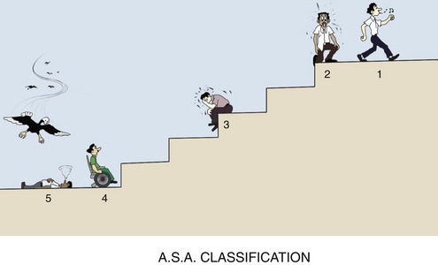
These numbers can be considered the ASA physical status classification for HF.

Hypoxia or stress may increase the degree of HF by increasing the workload of the myocardium thereby increasing its O2 requirement. The stress-reduction protocol is of considerable importance in the management of patients with HF. Scheduling appointments early in the morning, when the patient is well rested; limiting the length of the appointment so as not to exceed the limit of the patient’s tolerance; and monitoring vital signs preoperatively are recommended. If the weather becomes extremely warm or humid or if the patient appears somewhat fatigued before the start of treatment, it may be prudent to postpone the planned treatment to another day. Intraoperatively, the need for effective pain and anxiety control is quite important because increased stress produces an increased myocardial workload and an increase in the degree of HF.

Chair positioning of patients with HF may require modification from that recommended for the sedated patient. Although the supine or semisupine position is strongly recommended for patients during sedative procedures, the patient with HF may exhibit orthopnea that precludes the use of this position. Should this occur, the patient should be placed in the most recumbent position in which he or she can still breathe comfortably.

Local anesthetics containing vasopressors are indicated for pain control in the patient with HF. The recommended concentration of epinephrine is 1 : 200,000 or 1 : 100,000.

There are no contraindications to using any of the techniques of sedation in the patient with HF. It is important to remember that the primary problem in this patient is the failure of the heart to deliver an adequate volume of blood (and O2) to the tissues of the body. Because all sedative drugs are CNS, respiratory, and potentially cardiovascular depressants, it is essential that any additional hypoxia be prevented during the procedure. The ASA 2 patient with coronary heart disease is an excellent candidate for sedation with any technique, whereas the ASA 3 patient with HF should be restricted to minimal to moderate levels of sedation by the oral or inhalation route.

Oral sedation is quite appropriate for the patient with HF (ASA 2 or 3) for preoperative anxiety control. Only light levels of sedation should be sought (minimal to moderate), such as is obtained with the benzodiazepines.

IM sedation should be reserved for the patient in whom other more controllable techniques have proven ineffective. Minimal to moderate levels of sedation may be sought, with O2 supplementation provided throughout the procedure for the ASA 2 patient only. Potent respiratory-depressant drugs, such as opioids and barbiturates, should be used with extreme care, if at all.

N2O-O2 inhalation sedation is an appropriate technique for the ASA 2 or 3 patient with HF because it provides sedation and analgesia, and additional O2 for the patient.

Minimal to moderate levels of IV sedation are recommended for the ASA 2 patient with HF. Supplemental O2 is recommended for all IV sedative procedures. The use of IV sedation is not recommended for use in the ASA 3 patient with HF unless it is considered essential by the dentist, in which case only minimal to moderate sedation levels are recommended (with O2 supplementation).

Outpatient general anesthesia is not usually recommended for the ASA 2, 3, or 4 patient with HF. In-hospital general anesthesia should be considered when other sedative techniques have proven inadequate in patient management.

Congenital Heart Disease

The incidence of congenital heart disease is 9 per 1000 live births. Some defects develop as a result of genetic abnormalities; however, most congenital heart lesions occur in the absence of any detectable chromosomal abnormality. Although there are a large number of congenital lesions, those listed in Box 37-2 account for more than 80% of those seen in children with congenital heart disease. Ventricular septal defects account for approximately one third of all lesions, and atrial septal defects and patent ductus arteriosus account for 10% each; other relatively common defects include pulmonary stenosis and coarctation of the aorta. Less common are tetralogy of Fallot, aortic stenosis, and transposition of the great arteries.

Because great variation in clinical signs and symptoms and relevance toward dental care exists with congenital heart lesions, a thorough medical history, dialog history, and clinical evaluation are absolutely essential. When a history of congenital heart disease is obtained, medical consultation with the patient’s (child or adult) physician is recommended.

Primary concerns associated with dental management of this patient include the exacerbation of HF and cardiac dysrhythmias secondary to the stresses associated with dental treatment and the possibility of infection producing bacterial endocarditis. Consulting the most recent American Heart Association (AHA), American Medical Association (AMA), and American Dental Association (ADA) guidelines for prophylaxis along with possible medical consultation with the patient’s primary care physician will aid in determining the need for prophylactic antibiotics.10 In many patients with surgically repaired defects, the need for antibiotic coverage during dental care exists for life.

As specifically relates to the management of pain and anxiety in patients with congenital heart disease, pain control through the use of local anesthetics is vitally important as a means of minimizing stress. The administration of vasopressor-containing local anesthetics is not contraindicated in these patients.

Sedative techniques are indicated as a means of minimizing intraoperative stress in this patient. The primary goal during the procedure is to provide adequate sedation without inducing hypoxia. The myocardium of the patient with congenital heart disease may be less able to tolerate hypoxic episodes than healthy heart muscle.

The oral route is indicated for minimal to moderate levels of sedation. Deep sedation via the oral route cannot be recommended.

IM sedation should be relegated to a last-choice technique for patients in whom other sedative procedures are unavailable or have proven ineffective. Only minimal to moderate sedation levels are recommended by the IM route, along with administration of supplemental O2 throughout the procedure.

Inhalation sedation is an excellent technique for these patients primarily because of the additional levels of O2 supplied throughout the procedure.

IV sedation is also recommended provided the level of sedation remains minimal to moderate. Deep sedation is not recommended because of the increased likelihood of hypoxia and depression of respiratory and cardiovascular function. Supplemental O2 should be administered when IV sedation is used.

Outpatient general anesthesia is not recommended in patients with congenital heart lesions, repaired or not. General anesthesia should be reserved for patients in whom sedative procedures have been ineffective. Because of the nature of the underlying disease, the patient is admitted to the hospital before the procedure to receive a more in-depth medical evaluation.

Valvular Heart Disease

Valvular heart disease is a possible sequela of rheumatic fever. The incidence of valvular heart disease secondary to rheumatic fever has diminished over the past 4 decades; however, congenital valvular lesions are diagnosed with increasing regularity. It is estimated that more than 18,000 cardiac valvular replacements are performed annually in the United States.11

Life expectancy is prolonged for most patients receiving valvular replacements. Along with this benefit, however, is the ever-present prospect of bacterial endocarditis. The reader is referred to the guidelines for prophylaxis, which present detailed antibiotic regimens for these patients.10 The patient’s primary care physician may be consulted before dental treatment.

The primary concern during the dental management of the patient with valvular heart disease is the prevention of bacterial endocarditis. In addition, stress should be minimized through the effective use of local anesthesia and sedation, as indicated. Hypoxia should be prevented.

The administration of local anesthetics with vasopressors is indicated in patients with valvular replacement.

Oral sedation is indicated in the management of lesser degrees of preoperative anxiety. Minimal levels of sedation only are recommended.

IM sedation is recommended when other sedative techniques are unavailable or have proven ineffective. Intraoperative O2 administration is recommended for the minimal to moderate sedation recommended by the IM route.

Inhalation sedation with N2O-O2 is highly recommended for anxiety control in patients with valvular prostheses.

IV sedation is also recommended with minimal to moderate levels only suggested. Intraoperative O2 administration is suggested.

Outpatient general anesthesia is not recommended for the patient with a valvular prosthesis. Hospitalization and thorough work-up are strongly suggested.

RENAL DISEASE

An estimated 20 million adults in the United States have chronic kidney disease (CKD)—about one in nine adults.12 Glomerulonephritis, pyelonephritis, nephrotic syndrome, chronic renal insufficiency, and chronic renal failure are the most common disorders of renal function. Renal dialysis and transplantation are used in the management of chronic renal failure. In 2006 it was estimated that more than 200,000 persons were undergoing dialysis for end-stage renal disease.12 In 2006, 17,092 patients received renal transplants in the United States. There were more than 95,000 patients awaiting kidney transplant as of April 2007. In 2006, 3916 kidney patients died while awaiting their kidney transplant.12

Most renal failure patients may be safely managed in an outpatient setting, representing ASA 2, 3, or 4 risks. Specific questioning and examination can determine the degree of risk.

All patients with altered renal function, especially those undergoing dialysis in the days just preceding their dialysis appointments, must be managed carefully because their blood chemistries may be in disarray. It is recommended that dental appointments be scheduled on the day following dialysis so that the patient’s metabolic status is more optimal and the effects of systemic anticoagulation are minimal.

Prophylactic antibiotics may be required before dental care in the patient with renal disease, especially the renal transplant patient. Consultation with the patient’s physician (nephrologists) is strongly recommended to determine an appropriate antibiotic regimen.

Many patients with chronic renal disease, especially patients having undergone renal transplantation, receive long-term corticosteroid and antirejection drug therapy. Such therapies diminish the patient’s capacity to respond appropriately to increased stress. The administration of supplemental corticosteroid may be recommended before particularly traumatic (emotionally or physically) procedures. Medical consultation is recommended.

Patients undergoing renal dialysis and renal transplant are considered at high risk for contracting hepatitis B and should be evaluated before the start of dental care. Patients who are surface-antigen negative and surface-antibody positive may be treated in the usual manner, whereas those who are surface-antigen positive should be treated using current recommendations to minimize transmission of hepatitis B. There is also an increased risk of human immunodeficiency virus (HIV) and acquired immunodeficiency syndrome (AIDS) infection. This increased risk should be considered before the use of parenteral techniques of sedation.

Most drugs are excreted through the kidneys, a percentage of the drug unchanged along with its major metabolites. Drugs, such as cocaine and gallamine, that are excreted entirely unchanged in the urine should not be administered to patients undergoing renal dialysis. Blood levels of these drugs would become overly high, increasing the risk of overdose (toxic reaction). Approximately 10% to 15% of most amide local anesthetics are excreted unchanged in the urine, whereas virtually no ester local anesthetic is found unchanged in/>

Stay updated, free dental videos. Join our Telegram channel

Jan 5, 2015 | Posted by in General Dentistry | Comments Off on 37: The Medically Compromised Patient

VIDEdental - Online dental courses

Get VIDEdental app for watching clinical videos