32
Root Fractures
Root Fractures
Root fractures tend to occur in older children and adolescents. In young children the bone is softer, so teeth tend to be displaced and luxated; however, as the bone becomes harder and teeth more brittle with age, then root fractures are more common.
Diagnosis
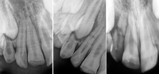
- Take several radiographs at different angulations (Fig. 32.1).
- Check for both vertical and horizontal root fractures.
- Fractures may not be evident initially, it is only with inflammation and swelling that the fragments separate and are visible.
- Suspect a vertical root fracture when an isolated periodontal defect is present or there is inability to resolve a periapical infection.
Figure 32.1 When a root fracture is suspected it is essential to take radiographs at different horizontal and vertical angulations. The fracture is not evident on the first radiograph and with elongation, the fracture appears faintly as an ellipse. It is only in the final film that the true extent of the fracture is shown.
Aim: to align fragments, provide stability and achieve healing.
Management
- Reposition coronal fragment and splint rigidly for up to 12 weeks with composite resin and wire or orthodontic appliances.
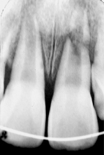
- High apical fractures usually require no treatment (Fig. 32.2).
- Review.
Figure 32.2 High apical root fractures have a very good prognosis especially if the tooth is immature. A rigid stainless steel wire has been used here for splinting.
Healing
The apical portion of the fracture almost always retains its vitality.
- Hard tissue union between fragments (very uncommon).
- Interposition of bone (Fig. 32.3).
- Interposition of fibrous connective tissue.
- Granulation tissue between fragments – coronal pulp necrosis.
Stay updated, free dental videos. Join our Telegram channel
VIDEdental - Online dental courses


