DEFINITION OF OPERATIVE DENTISTRY
CLASSES OF HUMAN TEETH
• Incisors
• Canines
• Premolars
• Molars
SETS OF TEETH
TOOTH NUMBERING SYSTEMS
• Zsigmondy-Palmer System
• Universal System
• FDI-Federation Dentaire Internationale
• Comparison of Tooth Numbering Systems
NOMENCLATURE OF TOOTH SURFACES
• Buccal
• Labial
• Facial
• Mesial
• Distal
• Lingual
• Occlusal
• Incisal
• Gingival
• Cervical
• Anatomic Crown
• Clinical Crown
NOMENCLATURE RELATED TO DENTAL CARIES
• Dental Caries
• Primary Caries
• Recurrent Caries
• Residual Caries
• Active Carious Lesion
• Inactive/Arrested Carious Lesion
• Pits and Fissure Caries
• Smooth Surface Caries
• Root Caries
• Acute Dental Caries
• Rampant Caries
• Chronic Dental Caries
NOMENCLATURE RELATED TO NONCARIOUS DEFECTS OF TEETH
• Attrition
• Abrasion
• Erosion
• Abfraction
• Resorption
• Localized Non-hereditary Enamel Hypoplasia
•Localized Non-hereditary Enamel Hypocalcification
Operative dentistry plays an important role in enhancing dental health and now branched into dental specialties. Today operative dentistry continues to be the most active component of most dental practice. Epidemiologically, demand for operative dentistry will not decrease in the future.
DEFINITION OF OPERATIVE DENTISTRY
According to Mosbys dental dictionary. “Operative dentistry deals with the functional and esthetic restoration of the hard tissues of individual teeth.”
According to Sturdvent, “Operative dentistry is defined as science and art of dentistry which deals with diagnosis, treatment and prognosis of defects of the teeth which do not require full coverage restorations for correction.”
Such corrections and restorations result in the restoration of proper tooth form, function and esthetics while maintaining the physiological integrity of the teeth in harmonious relationship with the adjacent hard and soft tissues. Such restorations enhance the dental and general health of the patient.
According to Gilmore, “Operative dentistry is a subject which includes diagnosis, prevention and treatment of problems and conditions of natural teeth vital or nonvital so as to preserve natural dentition and restore it to the best state of health, function and esthetics.”
Definition of dental anatomy or anatomy of teeth is the branch of anatomy which deals with the study of human teeth structures. It includes development, appearance and classification of teeth. Dental anatomy is also a taxonomical science; it is concerned with the naming of teeth and the structures of which they are made.
For convenience human dentition is divided into four quadrants viz; upper (maxillary) right, upper (maxillary) left, lower (mandibular) right and lower (mandibular) left (Fig. 2.1). Right and left here relate to patient’s right and left side.
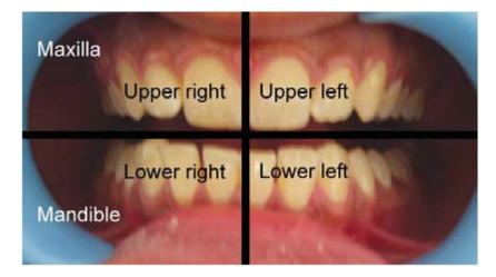
Fig. 2.1: Photograph showing division of whole dentition into four quadrants, i.e. upper right, upper left, lower right and lower left. Here right and left relate to patient’s right and left
It is important to understand anatomy of teeth because of following reasons:
• For maintenance of supporting tissues in the healthy state.
• For restoration of damaged tooth to its original form.
0 For optimal functions of teeth.
CLASSES OF HUMAN TEETH
Depending upon their form and function human teeth can be divided into following classes:
Incisors
The square-shaped teeth located in front of the mouth, with four on the upper and four on the lower are called incisors. Incisors are important teeth for phonetics and esthetics. They help in cutting the food (Fig. 2.2).
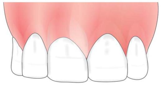
Fig. 2.2: Maxillary central and lateral incisors
Canines
The sharp teeth located near the corner of the mouth. Because of their anatomy and long root, they are strong teeth. They help in tearing and cutting of food (Fig. 2.3).
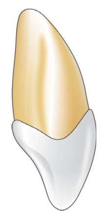
Fig. 2.3: Maxillary canine showing sharp tip and long root
Premolars
There are a total of eight premolars inside the mouth present after the canines. Four premolars are present in upper and lower arch, two on each side of the canine. Facially they resemble canines and lingually as molars (Fig. 2.4). They help in tearing and grinding of the food.
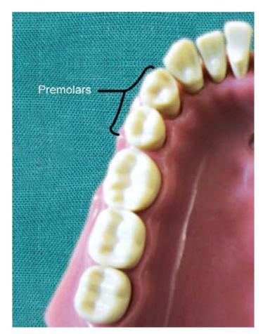
Fig. 2.4: Photograph showing premolars
Molars (Fig. 2.5)
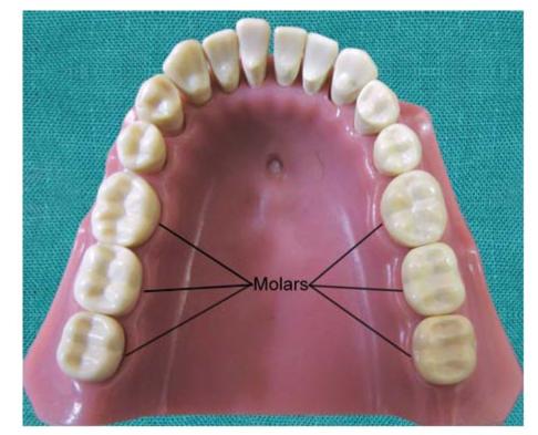
Fig. 2.5: Photograph showing molars
Distal to premolars are the molars. There are six molars on each arch (three on each side), therefore a total of 12 inside the mouth. They have multiple cusps which help in crushing and grinding the food. These teeth also help in maintenance of vertical height of the face.
SETS OF TEETH
There are two sets of teeth that develop in a person’s mouth (Fig. 2.6). The first set of teeth is called as “milk or baby” or “primary teeth”. The total number of teeth in this set are twenty. Primary teeth erupt at the age between 6 months and 2 years. Most children develop all their primary teeth at the age of three.
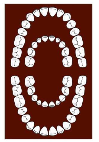
Fig. 2.6: Figure showing two sets of teeth. The outer ring represents the permanent teeth. The inner ring represents the deciduous teeth
The second set of teeth, i.e. the permanent teeth erupt at the age of six. There are a total of 32 permanent teeth in an adult mouth. Total number of teeth are divided into two arches, i.e. an upper and a lower arch each found on the upper and lower jaws respectively. Normally, a total of 16 teeth may be found on each complete arch.
TOOTH NUMBERING SYSTEMS
There are different tooth numbering systems for naming a specific tooth. The three most commons systems are the FDI World Dental Federation notation, Universal numbering system and Zsigmondy-Palmer notation method. The FDI system is used worldwide and the universal is used widely in the USA.
Most commonly used tooth numbering systems
1. Zsigmondy-Palmer system
2. Universal system (ADA system)
3. FDI system (Federation Dentaire Internationale)
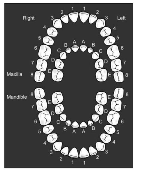
Zsigmondy-Palmer System (Fig. 2.7)
It was originally termed as “Zsigmondy system” after the Austrian dentist Adolf Zsigmondy who developed the idea in 1861, using a Zsigmondy cross to record quadrants of tooth positions.
Adult teeth were numbered 1 to 8 and the primary dentition as Roman numerals I, II, III, IV, V from the midline. Palmer changed this to A, B, C, D, E. This makes it less confusing and less prone to errors in interpretation.
Stay updated, free dental videos. Join our Telegram channel
VIDEdental - Online dental courses


