INTRODUCTION
MAXILLARY CENTRAL INCISOR
• Labial Aspect
• Lingual Aspect
• Mesial Aspect
• Distal Aspect
• Incisal Aspect
MAXILLARY LATERAL INCISOR
• Labial Aspect
• Lingual/Palatal Aspect
• Mesial Aspect
• Distal Aspect
• Incisal Aspect
CANINE
• Labial Aspect
• Lingual/Palatal Aspect
• Mesial Aspect
• Distal Aspect
• Incisal Aspect
FIRST PREMOLAR
• Buccal Aspect
• Palatal Aspect
• Mesial Aspect
• Distal Aspect
• Occlusal Aspect
SECOND PREMOLAR
• Buccal Aspect
• Palatal Aspect
• Mesial Aspect
• Distal Aspect
• Occlusal Aspect
FIRST MOLAR
• Buccal Aspect
• Palatal Aspect
• Mesial Aspect
• Distal Aspect
• Occlusal Aspect
SECOND MOLAR
• Buccal Aspect
• Palatal Aspect
• Mesial Aspect
• Distal Aspect
• Occlusal Aspect
CENTRAL INCISOR
• Labial Aspect
• Lingual Aspect
Mesial Aspect
• Distal Aspect
• Incisal Aspect
MANDIBULAR LATERAL INCISOR
• Labial Aspect
• Lingual Aspect
• Mesial Aspect
• Distal Aspect
• Incisal Aspect
CANINE
• Labial Aspect
• Lingual Aspect
• Mesial Aspect
• Distal Aspect
• Incisal Aspect
FIRST PREMOLAR
• Buccal Aspect
• Lingual Aspect
• Mesial Aspect
• Distal Aspect
• Occlusal Aspect
SECOND PREMOLAR
• Buccal Aspect
• Lingual Aspect
• Mesial Aspect
• Distal Aspect
• Occlusal Aspect
MANIBULAR FIRST MOLAR
• Buccal Aspect
• Lingual Aspect
• Mesial Aspect
• Distal Aspect
• Occlusal Aspect
SECOND MOLAR
• Buccal Aspect
• Lingual Aspect
• Mesial Aspect
• Distal Aspect
• Occlusal Aspect
DIFFERENTIATION BETWEEN RIGHT AND LEFT MAXILLARY CENTRAL INCISOR
DIFFERENTIATION BETWEEN RIGHT AND LEFT MAXILLARY LATERAL INCISOR
DIFFERENTIATION BETWEEN RIGHT AND LEFT MAXILLARY CANINE
DIFFERENTIATION BETWEEN RIGHT AND LEFT FIRST MAXILLARY PREMOLAR
DIFFERENTIATION BETWEEN RIGHT AND LEFT SECOND PREMOLAR
DIFFERENTIATION BETWEEN RIGHT AND LEFT MAXILLARY FIRST MOLAR
DIFFERENTIATION BETWEEN RIGHT AND LEFT MAXILLARY SECOND MOLAR
DIFFERENTIATION BETWEEN RIGHT AND LEFT MANDIBULAR LATERAL INCISOR
DIFFERENTIATION BETWEEN RIGHT AND LEFT MANDIBULAR CANINE
DIFFERENTIATION BETWEEN RIGHT AND LEFT MANDIBULAR FIRST PREMOLAR
DIFFERENTIATION BETWEEN RIGHT AND LEFT SECOND PREMOLAR
DIFFERENTIATION BETWEEN RIGHT AND LEFT MANDIBULAR FIRST MOLAR
DIFFERENTIATION BETWEEN RIGHT AND LEFT MANDIBULAR SECOND MOLAR
INTRODUCTION
As we know there are 32 teeth in permanent dentition and 20 teeth in deciduous dentition. A tooth has crown and a root portion. Crown part of the tooth is covered with enamel and root portion of tooth is covered by cementum. The crown and root join at cementoenamel junction (CEJ).
Before discussing the individual tooth, one should be familiar with few terms. These are:
Cervical line: Each tooth has a crown and root portion. The crown is covered with enamel and the root portion is covered with cementum. The crown and root join at the cementoenamel junction (CEJ). This junction is also called the cervical line.
Cingulum: It is lingual lobe of an anterior tooth and makes up the bulk of the cervical third of the lingual surface.
Ridge: It is linear elevation on the surface of a tooth.
Marginal ridges: These are rounded borders of enamel that form the mesial and distal margins of occlusal surfaces of posterior teeth (premolars and molars) and mesial and distal margins of the lingual surfaces of anterior teeth (incisors and canines).
Triangular ridges: These descend from the tips of the cusps of molars and premolars toward the central part of occlusal surfaces.
Transverse ridge: When both buccal and lingual triangular ridges join, they combine to form a transverse ridge.
Oblique ridge: It is a ridge obliquely crossing the occlusal surfaces of maxillary molars. It is usually formed by the union of triangular ridge of distobuccal cusp and distal cusp ridge of the mesiolingual cusp.
Fossa: It is an irregular depression or concavity.
Lingual fossae: Occur on lingual surface of incisors.
Central fossae: Occur on occlusal surface of molars.
Sulcus: It is a long depression on the surface of tooth ridges and cusps.
Developmental groove: It is shallow groove between the primary parts of the crown or root.
Pits: These are small pinpoint depressions located at the junction of developmental grooves or at ending of those grooves.
Lobe: It is one of the primary sections of formation in the development of the crown.
Mamelons: These are three rounded protuberances found on the incisal edges of newly erupted incisor teeth.
Cusp: It is elevation on the crown portion of a tooth making up a divisional part of the occlusal surface.
Tubercle: Smaller elevation on some portion of crown produced by an extra formation of enamel.
DESCRIPTION OF PERMANENT TOOTH
MAXILLARY CENTRAL INCISOR
Labial Aspect (Fig. 1.1)
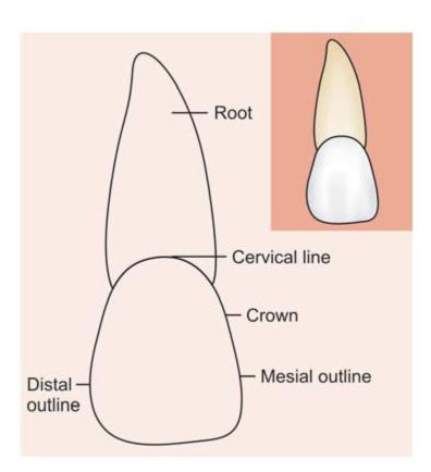
Fig. 1.1: Labial aspect
1. Widest mesiodistally (10-11 mm) of any anterior teeth, with almost square or rectangle shape.
2. Medial outline is straight or slightly convex; whereas the distal outline is more convex.
3. Distoincisal angle is not as sharp as mesioincisal angle.
4. Incisal outline is almost straight, but newly erupted teeth may show mamelons.
5. Cervical outline follows a semicircular shape with convexity towards root surface.
Lingual Aspect (Fig. 1.2)
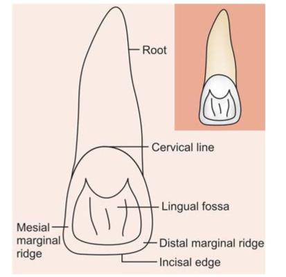
Fig. 1.2: Lingual aspect
1. Mesial and distal outlines converge palatally.
2. Below cervical line, a smooth convexity is present called cingulum.
3. Below cingulum there is a shallow concavity which is bordered by mesial and distal marginal ridge, incisal ridge and cingulum.
Mesial Aspect (Fig. 1.3)
Fig. 1.3: Mesial aspect
1. Wedge triangular shaped crown with base towards cervix and apex towards incisal ridge.
2. Incisal edge of crown is in line with center of the root.
3. Labial outline is convex from cervix till incisal edge.
4. Lingual outline is convex at the point where it joins crest of curvature at cingulum. After this, it becomes concave and then slightly convex again when it approaches linguoincisal ridge.
5. Cervical line curves incisially. This curve is more on the mesial surface then on distal surface.
Distal Aspect (Fig. 1.4)
Distal aspect is almost similar to that of mesial aspect except in following:
1. Crown appears thicker at mesial.
2. Curvature of cervical line is less than on the mesial surface.
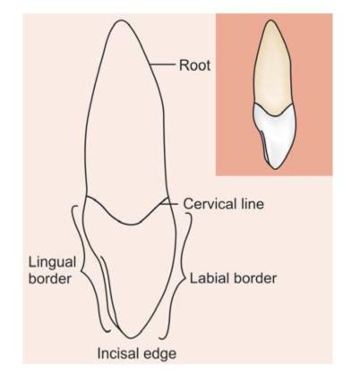
Fig. 1.4: Distal aspect
Incisal Aspect (Fig. 1.5)
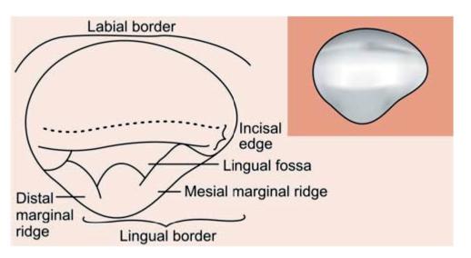
Fig. 1.5: Incisal aspect
1. Incisal edge is centered over the root.
2. Labial surface appears broad and flat, whereas the lingual portion tapers lingually towards the cingulum.
MAXILLARY LATERAL INCISOR
Special features
• Most commonly found missing tooth.
• Peg shaped lateral-common finding.
• Palatogingival groove.
Labial Aspect (Fig. 1.6)
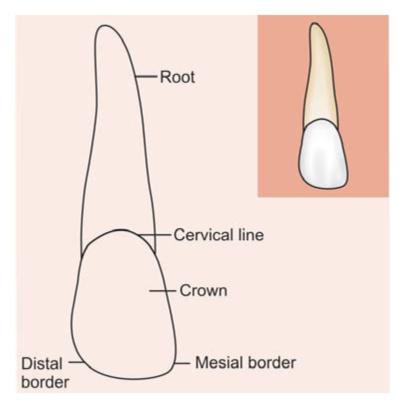
Fig. 1.6: Labial aspect
1. When compared to central incisor, it has more curvature, rounded incisal edge and rounded incisal angles mesially and distally.
2. Mesioincisal angle can be as sharp as that of central incisor.
3. Distal outline is more rounded than central incisor.
4. Crest of contour mesially is at point of junction of middle and incisal third and on distal side, it lies more towards cervical aspect.
5. Lateral incisor is narrower mesiodistally and shorter cervicoincisally than central incisor.
Lingual/Palatal Aspect (Fig. 1.7)
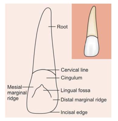
Fig. 1.7: Lingual aspect
1. Palatal aspect is narrower than labial.
2. Cingulum is prominent with affinity towards deep developmental grooves within lingual fossa.
3. Marginal ridges are more prominent than that of central incisor.
4. When compared to central incisor, lingual fossa is more concave and circumscribed.
Mesial Aspect (Fig. 1.8)
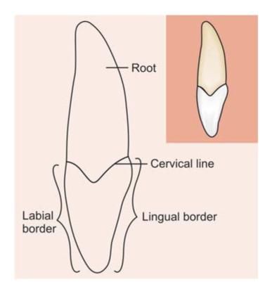
Fig. 1.8: Mesial aspect
1. Almost similar to that of central incisor.
2. Curvature of cervical line more on mesial surface than on distal surface.
Distal Aspect (Fig. 1.9)
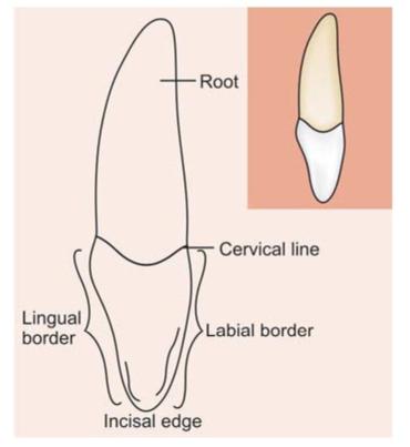
Fig. 1.9: Distal aspect
1. Width of crown appears more than on mesial surface because of placement of crown on the root.
2. Curvature of cervical line is usually less than that of mesial surface.
Incisal Aspect (Fig. 1.10)
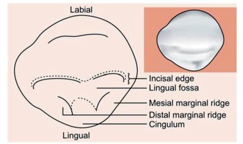
Fig. 1.10: Incisal aspect
1. May resemble central incisor or canine.
2. Labial surface is more convex with prominent cingulum.
3. Labiolingual dimensions may be greater than mesiodistal dimensions.
CANINE
Labial Aspect (Fig. 1.11)
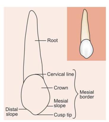
Fig. 1.11: Labial aspect
1. Mesiodistal dimensions are shorter than central incisor.
2. Labial surface is smooth with slight shallow depressions.
3. Due to more development of middle lobe, labial ridge is seen.
4. Mesial outline is convex from cervix to mesial contact area.
5. Distal outline is usually concave from cervical line to distal contact area.
6. Incisal edge comes to a distinct point in form of cusp. It has mesial and distal slopes. The mesial slope is shorter than distal slope.
7. Root appears slender from the labial aspect. Canine has the longest root.
Lingual/Palatal Aspect (Fig. 1.12)
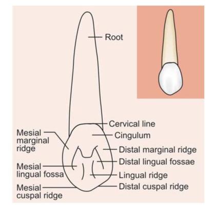
Fig. 1.12: Lingual aspect
1. Crown is narrower on lingual side.
2. Cingulum is prominent sometimes pointed and may appear as a small cusp.
3. Marginal ridges are prominent. Lingual ridge is found below cingulum and between marginal ridges. It divides lingual fossa in mesial and distal lingual fossa.
Mesial Aspect (Fig. 1.13)
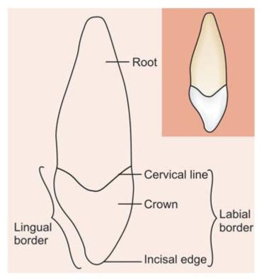
Fig. 1.13: Mesial aspect
1. Outline is wedge shaped with greatest measurements towards cervical third.
2. Labial surface appears convex from cervical line towards cusp tip.
3. Lingual outline shows convexity at cervical area, straight at middle third and again convex at incisal third.
4. A line bisecting the cusp is labial to the line bisecting root.
Distal Aspect (Fig. 1.14)
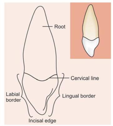
Fig. 1.14: Distal aspect
It is almost similar to that of mesial aspect except that:
1. Cervical line shows less curvature.
2. Distal marginal ridge is heavier and more irregular in outline.
Incisal Aspect (Fig. 1.15)
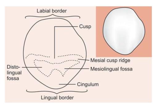
Fig. 1.15: Incisal aspect
1. Labiolingual dimensions are more than mesiodistal.
2. Labial ridge appears on labial surface.
3. On lingual surface, cingulum makes up the cervical third of the crown.
4. Cusp tip and cusp slopes lie labial to long axis of the root.
5. Distal cusp ridge is longer than the mesial cusp ridge.
FIRST PREMOLAR
Buccal Aspect (Fig. 1.16)
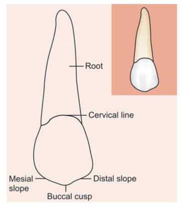
Fig. 1.16: Buccal aspect
1. Crown is roughly trapezoidal with convex buccal surface and buccal ridge.
2. Mesial outline is slightly concave from cervical line to the mesial contact area.
3. Distal outline is straighter than mesial outline.
4. Buccal cusp is long and sharp, and thus resembles canine.
5. Mesial cusp slope is longer than distal cusp slope. This difference places buccal cusp tip distal to long axis of the tooth.
Palatal Aspect (Fig. 1.17)
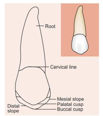
Fig. 1.17: Palatal aspect
1. Due to palatal convergence, crown is narrower on palatal side than on buccal side.
2. Palatal cusp is smooth, short and blunt as compared to buccal cusp.
3. Mesial and distal slopes of palatal cusp make rounded angle at cusp tip.
4. Since palatal cusp is shorter than buccal cusp, the tips of both cusps with their mesial and distal slopes can be seen from palatal aspect.
Mesial Aspect (Fig. 1.18)
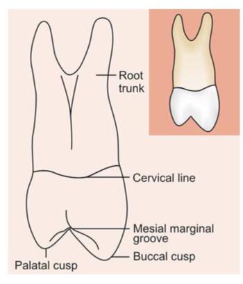
Fig. 1.18: Mesial aspect
1. Roughly trapezoidal in shape with longest side towards cervical portion, and shortest towards occlusal portion.
2. Buccal and palatal outlines are smoothly curved from the cervical line till the tips of buccal and palatal cusps.
3. Mesial developmental depression is found cervical to mesial contact area bordered by mesiobuccal and mesiolingual line angles. This depression continues apically beyond cervical line and joins the deep developmental depressions of roots.
4. Developmental groove is found in enamel of mesial marginal ridge. This groove is usually continuous with central groove of occlusal surface of crown.
5. Two roots; one buccal and one palatal are clearly visible from mesial aspect.
Distal Aspect (Fig. 1.19)
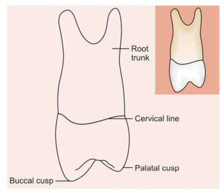
Fig. 1.19: Distal aspect
It is almost similar to mesial aspect except that:
• Convex distal surface with no depression at cervical third.
• Absence of deep developmental groove.
• Curvature of cervical line is less than on mesial aspect.
Occlusal Aspect (Fig. 1.20)
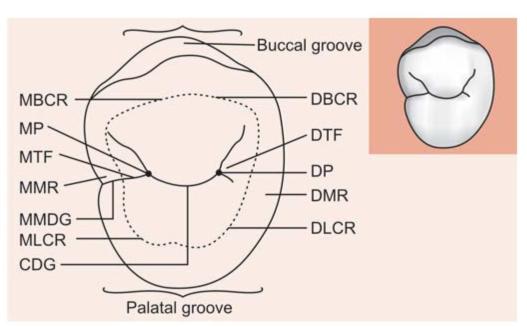
Fig. 1.20: Occlusal aspect: MBCR: Mesiobuccal cusp ridge; MP: Mesial pit; MTF: Mesial triangularfossa; MMDG: Mesial marginal development groove; CDG: Central development groove; DBCR: Distobuccal cusp ridge; DTF: Distal triangular fossa; DMR: Distal ma/>
Stay updated, free dental videos. Join our Telegram channel
VIDEdental - Online dental courses


