CHAPTER 3 Anatomy, Biochemistry, and Physiology
GENERAL ANATOMY
 •
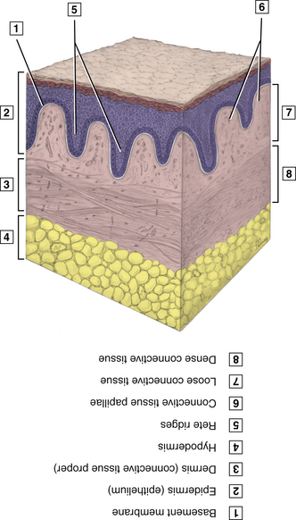
• Integumentary System
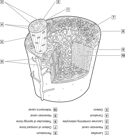
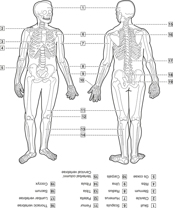
Skeletal System
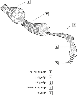
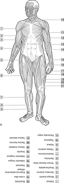
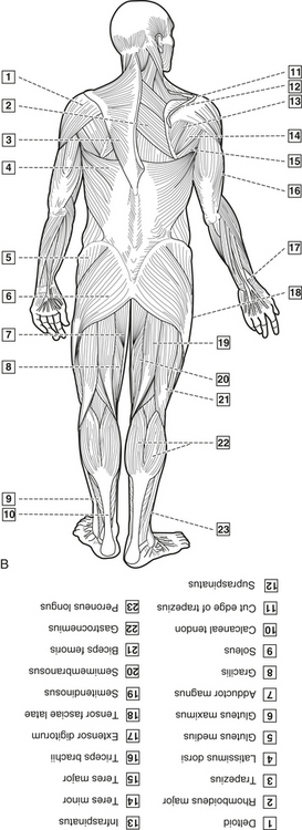
Muscular System
Nervous System
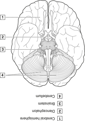
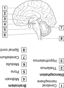
Nervous system includes all neural tissue (neurons and neuroglia). The synapse is in the region of communication between neurons; communication occurs through the effects of neurotransmitters. Sensory nerves (dorsal root) transmit information to the brain and spinal cord. Motor nerves (ventral root) transmit information from the brain and spinal cord to effectors, such as muscles and glands. Integrative functions interpret the sensory information and act by stimulating motor pathways. Divided into central nervous system (CNS) and peripheral nervous system (PNS), which function together to integrate and coordinate body activities, assimilate experiences, and assist in memory and learning.
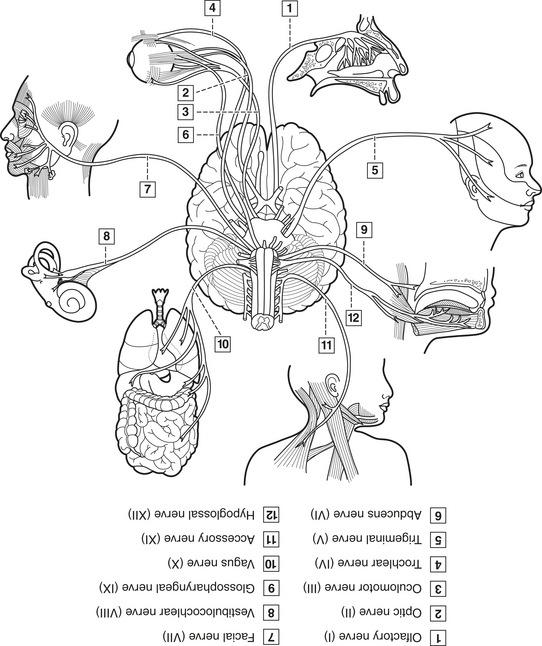
Cranial Nerves
Cranial nerves are part of the PNS; designated by both number and Roman numerals I through XII, as well as specific name (Figure 3-8). Include 12 pairs that are connected to the brain at its base and pass through the skull by way of fissures and foramina; may be either afferent or efferent or have both types.
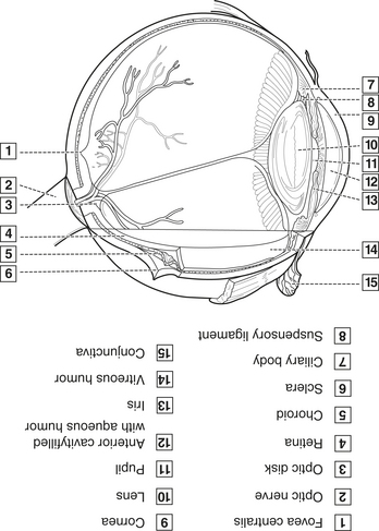
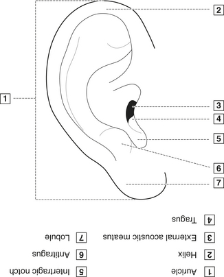
Sense Organs
CLINICAL STUDY
| Age | 68 YRS | SCENARIO |
| Sex |  Male Male  Female Female |
The dental hygienist calls to remind the patient that she is due for her 3-month recall appointment. The patient says that she has just been diagnosed with an eye disease that has permanently narrowed her fi eld of vision. She does not like the drops prescribed and wonders why she should take them. |
| Height | 5′5″ | |
| Weight | 145 LBS | |
| BP | 118/74 | |
| Chief Complaint | “How am I going to get to the appointment?” | |
| Medical History |
Endocrine System
Endocrine system consists of endocrine glands (release product into bloodstream) and hormones produced (Figure 3-11). Acts with nervous system to regulate body activities to maintain homeostasis. Nervous system acts through electrical impulses and uses a neurotransmitter, and effects are MORE localized and of LESS duration; endocrine system acts through a chemical messenger called a hormone, and effects are MORE generalized and of MORE duration.
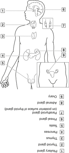
Figure 3-11 Endocrine glands.
(From Fehrenbach MJ, ed: Dental anatomy coloring book, St. Louis, 2008, Saunders/Elsevier.)
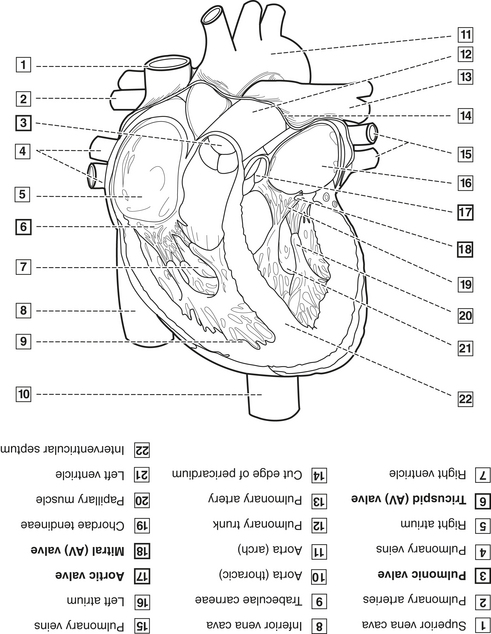
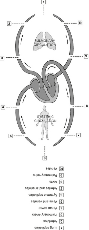
Cardiovascular System
Cardiovascular system (CVS) includes blood, heart, and blood vessels.
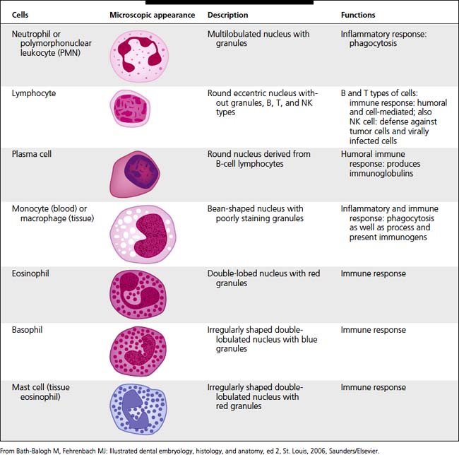
| Immunoglobulin | Description |
|---|---|
| IgA | Has two subgroups: serous in blood and secretory in saliva and other secretions; aids in defense against proliferation of microorganisms in body fluids |
| IgE | Involved in hypersensitivity reactions, since can bind to mast cells and basophils and bring about release of bioactive substances such as histamine |
| IgD | Functions in activation of B-cell lymphocytes. |
| IgG | Major antibody in blood serum; can pass placental barrier and forms first passive immunity for newborn |
| IgM | Involved in early immune responses owing to involvement with IgD in activation of B-cell lymphocytes |
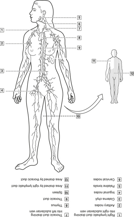
Lymphatic System
CLINICAL STUDY
| Age | 23 YRS | SCENARIO |
| Sex |  Male Male  Female Female |
During an extraoral examination, a hard fi xed mass, approximately 6 mm wide by 36 mm long by 6 mm deep, is noted on the right side of the neck; enlargement is clearly visible but no discomfort is noted when it is palpated. Other enlarged areas have been detected by the patient in his armpit and groin areas on same side. He states that he feels fi ne, although he tires easily. Intraoral exam shows poor oral hygiene and moderate gingivitis but no evidence of caries, only decalcifi cation. |
| Height | 6′2″ | |
| Weight | 185 LBS | |
| BP | 112/69 | |
| Chief Complaint | “There is a swelling on my neck that has been there for 6 months and is getting larger.” | |
| Medical History | Broken kneecap at age 8 | |
| Current Medications | None | |
| Social History |
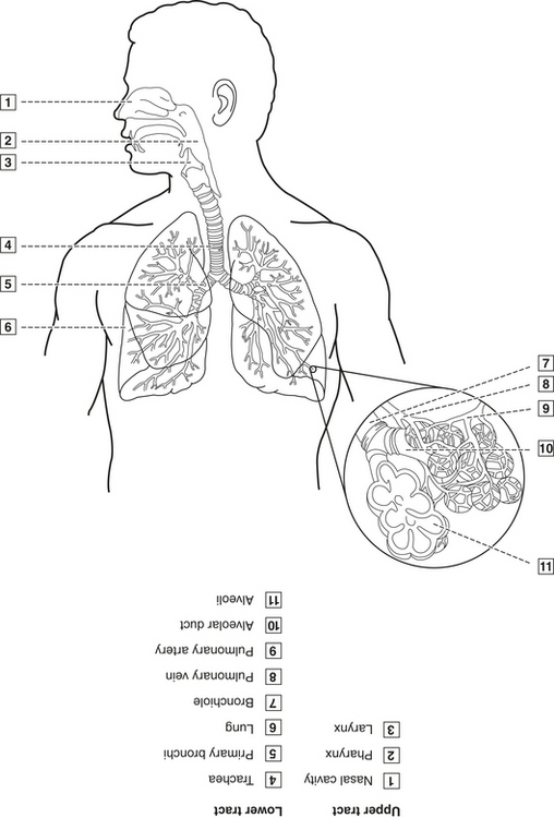
Respiratory System
Respiratory system includes the upper and lower respiratory tracts and the lungs (Figure 3-15). It functions to move gases to and from exchange surfaces, where diffusion can occur. Defends body against pathogens, permits speech, helps regulate acid-base balance in body.
Digestive System
Digestive system includes gastrointestinal (alimentary) tract (GIT; includes oral cavity, pharynx, esophagus, stomach, small intestine, large intestine, rectum, anus) and accessory organs (liver, gallbladder, pancreas) (Figure 3-16). Embryologically and physiologically, the mouth is the beginning of GIT. Functions include ingestion, mechanical digestion, chemical digestion, secretion, mixing and propelling movements, absorption, elimination of waste products, immunological functions (e.g., secretory IgA).

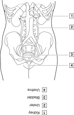
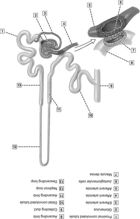
Urinary System
Urinary system consists of kidneys, ureters, urinary bladder, urethra (Figure 3-17). Functions to rid wastes, regulate fluid volume, maintain electrolyte concentrations, control blood pH, secrete renin and erythropoietin responsible for long-term maintenance of blood pressure.
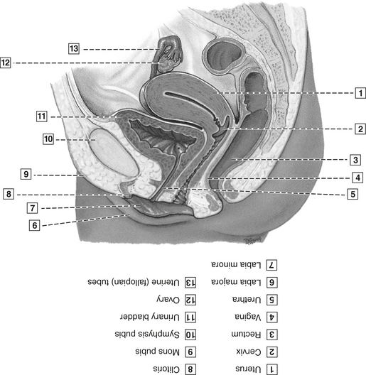
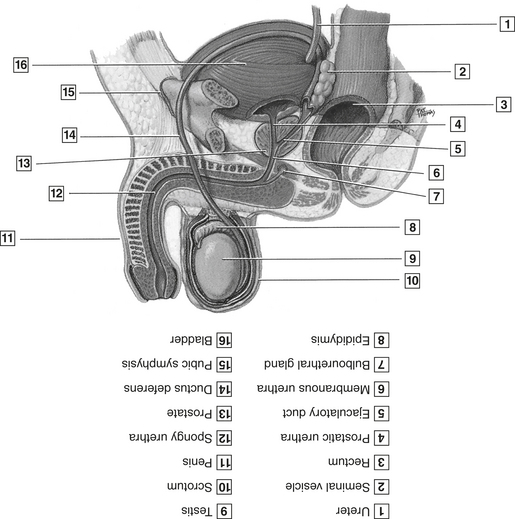
Reproductive System
BIOCHEMISTRY
Biochemistry is the study of living matter or organisms at a molecular level. Requires understanding of how molecules are structured, bonding to form MORE complex structures and thus affect living matter. Hydrocarbons are carbon- and hydrogen-based molecules that exist in several different forms and have various properties. Found within several products used daily (including in the dental office) and also form major components of the body.
 •
• Stay updated, free dental videos. Join our Telegram channel
VIDEdental - Online dental courses


