27: Bone Defects and Furcation Lesions

27

Bone defects and furcation lesions

Figure 27.1 Anatomy of bone defects.

ch27_054_img0271.gif


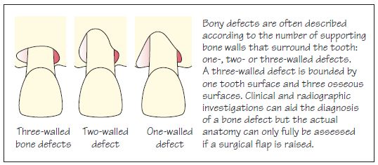
Figure 27.2 Treatment of bone defects.

ch27_054_img0272.gif


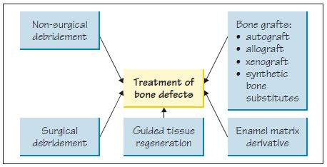
Figure 27.3 Use of synthetic bone graft (PerioGlas®). (a) Synthetic bone graft material on an instrument prior to placement. (b) Placement of the synthetic bone graft material. Courtesy of Mr P. J. Nixon.

ch27_054_img0273.gif


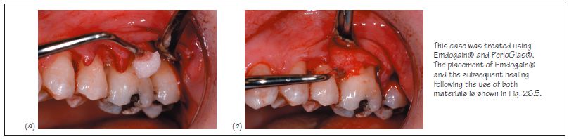
Figure 27.4 Classification of furcation involvement.

ch27_054_img0274.gif


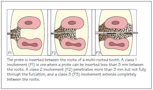
Figure 27.5 Treatment of furcation lesions.

ch27_054_img0275.gif


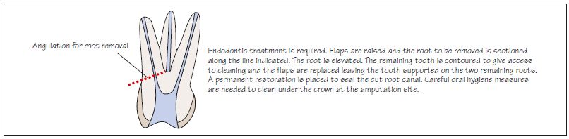
Figure 27.6 Root resection.

ch27_054_img0276.gif


Bone grafts

Alveolar bone resorption leads to the creation of bony defects (infrabony or intrabony), which differ in anatomy from one tooth site to another (Fig. 27.1).


Treatment of bone defects

The different methods of treatment are depicted in Fig. 27.2.


Non-surgical debridement

• Bone defects can be treated as at any other site by non-surgical debridement.
• Healing is with a long junctional epithelium and potentially some bony infill, particularly where there are several walls to the defect.


Surgical access for debridement

• Raising a surgical flap can improve access to the site and enable thorough removal of the inflammatory tissue.
• Healing is as for non-surgical debridement.
• The bony defect can be reshaped during the procedure.

– May result in further bone loss so use with caution to create an architecture that allows better flap adaptation.

• Bone fill may be obtained in two- and three-walled defects but great variability in the degree of fill has been found.

Simp/>

Only gold members can continue reading. Log In or Register to continue

Stay updated, free dental videos. Join our Telegram channel

Jan 14, 2015 | Posted by in Periodontics | Comments Off on 27: Bone Defects and Furcation Lesions

VIDEdental - Online dental courses

Get VIDEdental app for watching clinical videos