Potentially malignant disorders
 Potentially malignant (sometimes termed premalignant) clinically obvious disorders include actinic cheilitis, erythroplakia (erythroplasia – red patch), leukoplakia (white patch), submucous fibrosis and lichenoid lesions (Ch. 29)
 Risk factors for erythoplakia and leukoplakia may include tobacco use, alcohol use, betel nut chewing and sunlight exposure
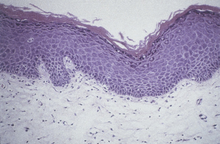
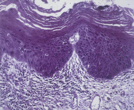
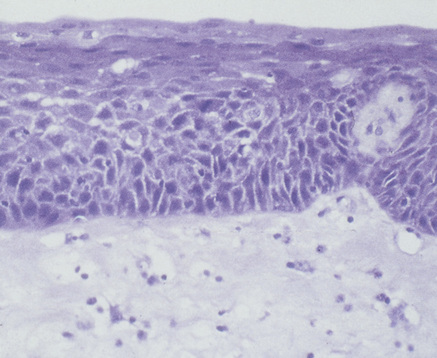
 Diagnosis and prognosis are aided by biopsy
 Factors predictive of future malignant transformation may include dysplasia, history of cancer in the upper aerodigestive tract, and changes in expression of P53 tumour suppressor protein, DNA and chromosomes 3p or 9p
 Treatment involves removing risk factors, and excision of dysplastic lesions
INTRODUCTION
 have had previous oral malignancy
 have had previous malignancy in the upper aerodigestive tract (nose, pharynx, trachea, lungs, oesophagus)
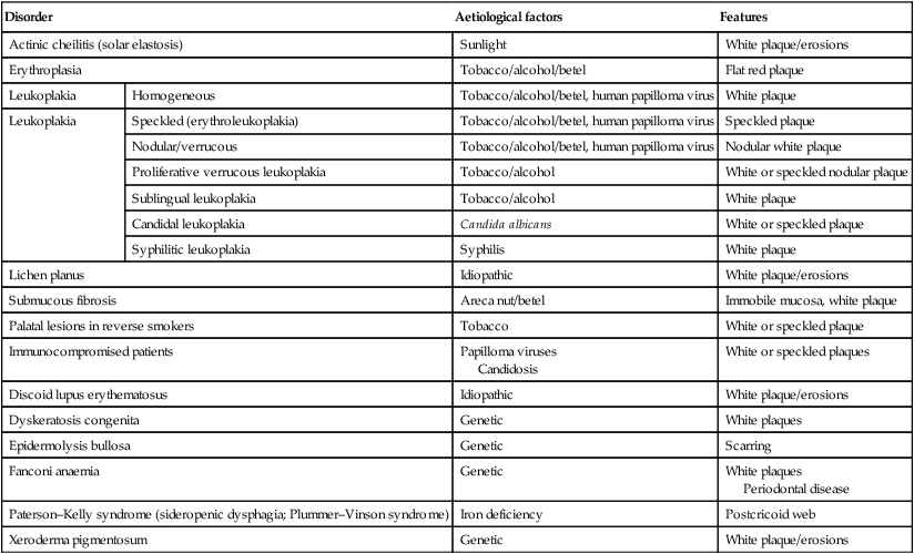
PMD include (Table 25.1) mainly:
Table 25.1
Potentially malignant oral disorders
| Disorder | Aetiological factors | Features | |
| Actinic cheilitis (solar elastosis) | Sunlight | White plaque/erosions | |
| Erythroplasia | Tobacco/alcohol/betel | Flat red plaque | |
| Leukoplakia | Homogeneous | Tobacco/alcohol/betel, human papilloma virus | White plaque |
| Leukoplakia | Speckled (erythroleukoplakia) | Tobacco/alcohol/betel, human papilloma virus | Speckled plaque |
| Nodular/verrucous | Tobacco/alcohol/betel, human papilloma virus | Nodular white plaque | |
| Proliferative verrucous leukoplakia | Tobacco/alcohol | White or speckled nodular plaque | |
| Sublingual leukoplakia | Tobacco/alcohol | White plaque | |
| Candidal leukoplakia | Candida albicans | White or speckled plaque | |
| Syphilitic leukoplakia | Syphilis | White plaque | |
| Lichen planus | Idiopathic | White plaque/erosions | |
| Submucous fibrosis | Areca nut/betel | Immobile mucosa, white plaque | |
| Palatal lesions in reverse smokers | Tobacco | White or speckled plaque | |
| Immunocompromised patients | Papilloma viruses Candidosis |
White or speckled plaques | |
| Discoid lupus erythematosus | Idiopathic | White plaque/erosions | |
| Dyskeratosis congenita | Genetic | White plaques | |
| Epidermolysis bullosa | Genetic | Scarring | |
| Fanconi anaemia | Genetic | White plaques Periodontal disease |
|
| Paterson–Kelly syndrome (sideropenic dysphagia; Plummer–Vinson syndrome) | Iron deficiency | Postcricoid web | |
| Xeroderma pigmentosum | Genetic | White plaque/erosions | |
 Actinic cheilitis (solar elastosis) (Ch. 26).
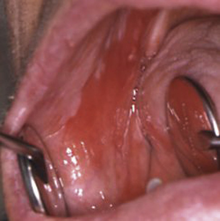
 Erythroplakia (erythroplasia – red patch; Fig. 25.1) (Ch. 27): defined by the World Health Organization (WHO) as ‘any lesion of the oral mucosa that presents as bright red velvety plaques which cannot be characterized clinically or pathologically as any other recognizable condition’. This is rare but has a very high malignant potential and many cases are already a carcinoma on microscopic examination.
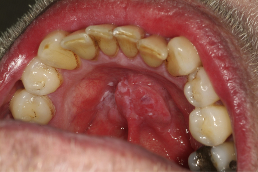
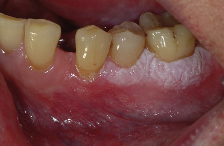
 Leukoplakia (white patch; Figs 25.2–25.6) (Ch. 28): defined by the WHO as ‘clinical white patches that cannot be wiped off the mucosa and cannot be classified clinically or microscopically as another specific disease entity (such as lichen planus)’. Leukoplakia is, thus, a clinical diagnosis only and can only be made by exclusion. A workshop coordinated by the WHO Collaborating Centre for Oral Cancer and Precancer agreed that the term leukoplakia should be used to recognize ‘white plaques of questionable risk having excluded (other) known diseases or disorders that carry no increased risk for cancer’.
 Lichen planus/lichenoid lesions (Ch. 29): in many geographic areas these are the most common PMDs.
Apart from the conditions noted above, rare conditions that predispose to OSCC include:
 discoid lupus erythematosus (Ch. 57)
 dyskeratosis congenita (Ch. 57)
 epidermolysis bullosa (Ch. 57)
 Paterson–Kelly syndrome (sideropenic dysphagia; Plummer–Vinson syndrome) (Ch. 56)
There are also occasional associations of OSCC with:
Erythroplakia has a very high malignant potential. Leukoplakia can have a malignant potential – particularly high in the following types (Table 25.2):
Table 25.2
Malignant potential in the most important oral potentially malignant disorders
| Malignant potential | |||
| High (> 60%) | Medium (< 30%) | Low (< 10%) | |
| Main entities | Erythroplakia | Leukoplakia (non-homogeneous) Candidal leukoplakia |
Leukoplakia (homogeneous) Lichen planus/lichenoid lesions |
| Uncommon entities | Actinic cheilitis Submucous fibrosis |
Discoid lupus erythematosus | |
| Rare entities | Dyskeratosis congenita* | Fanconi anaemia* | |

*Malignant potential unclear but involves OSCC (mainly tongue) and other neoplasms, especially acute myeloid leukaemia.
Stay updated, free dental videos. Join our Telegram channel

VIDEdental - Online dental courses


