Maxillary Arch Implant Considerations
Treatment Plans for Partial and Complete Edentulous Fixed and Overdenture Prostheses
Carl E. Misch
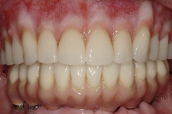
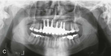
More than 18 million people in the United States, or 10.5% of the adult population, are completely edentulous.1 Maxillary dentures usually are tolerated better by patients than their mandibular complete denture. As such, many treatment plans initially concentrate on the problems associated with the mandibular prosthesis (Figure 25-1). However, after the patient obtains an implant prosthesis in the mandible that is a stable and retentive (and perhaps a fixed mandibular prosthesis), often the patient’s attention is brought to the inadequacies of the maxillary restoration.
In addition to the edentulous segment of the population without any teeth, 7% of the employed adult population wears a maxillary denture opposing some remaining mandibular teeth.2 These people routinely have more problems with their upper denture because their opposing arch is fixed and the plane of occlusion is often compromised. This means a total of 17% of the U.S. adult population (30 million people) have no natural maxillary teeth.
The first chapter of this book addressed the esthetic and psychological consequences of the loss of maxillary teeth. After patients become aware of the anatomical and esthetic consequences of multiple missing anterior teeth (as a result of continued bone loss), there is an increased desire for implant restorations. In addition, traditional fixed or removable prostheses accelerate the loss of additional teeth. As a result of continued patient and doctor education related to the consequences of the loss of several adjacent teeth, implant restoration of the edentulous maxilla will become even more prevalent in the future.
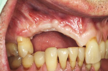
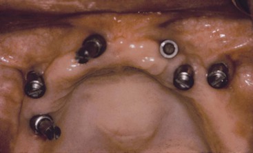
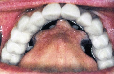
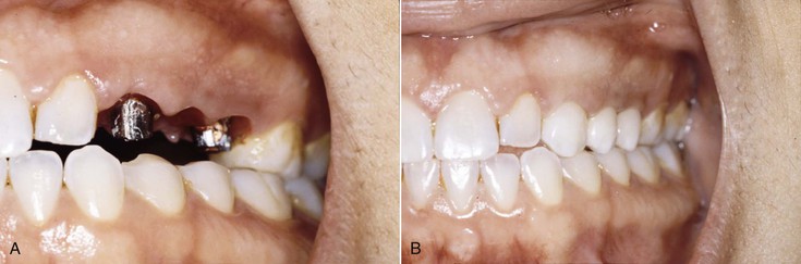
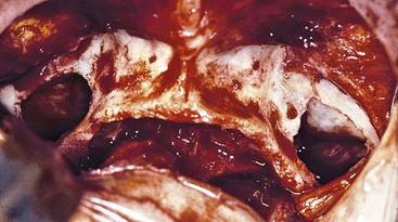
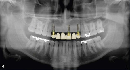
Partially edentulous patients missing multiple maxillary anterior teeth are not unusual. Failed fixed partial dentures (FPDs) often result in additional tooth loss. Car accidents and other sources of trauma also result in multiple anterior teeth missing (Figure 25-2). Less often are the effects of periodontal disease affecting only anterior teeth. Most of the partially edentulous patients prefer a fixed prosthesis to restore their dentition. There are many advantages to restore missing teeth with a fixed implant restoration, independent from the remaining natural teeth.
Edentulous Anterior Maxilla
Treatment Limitations
In a 20-year review of the literature compiled by Goodacre et al., restorations associated with the edentulous maxilla have the highest early loading implant failure rate compared with any other dental prostheses.3 For example, overdentures in the maxillary arch averaged 19% implant failure, and complete arch fixed prostheses in the edentulous maxilla have an early implant failure of 10%. In comparison, mandibular overdentures and partial or mandibular full-arch fixed restorations demonstrated a 3% implant failure rate. When 10% to 20% of implants fail in an edentulous maxillary prosthesis, the number of restorations affected may be more than half of the patients. For example, if four implants are used to support a prosthesis (fixed or removable) and 25% of the implants fail (one per patient), all of the final restorations would be affected because three remaining implants cannot predictably support a maxillary full-arch restoration.
Anatomic Limitations
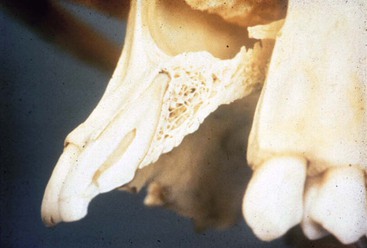
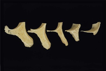
Several factors affect the condition of the edentulous maxilla and may result in a decrease in implant survival or an increase in prosthetic complications. The facial cortical plate of the premaxilla is thin over the roots of the teeth and may be resorbed from periodontal disease or is often fractured during the extraction of these teeth (Figure 25-3). In addition, the facial cortical plate rapidly resorbs during initial bone remodeling, and the anterior ridge loses more than 25% of its width within the first year after tooth loss and 40% to 50% over 1 year, mostly at the expense of the labial plate. As a result, the residual available bone migrates to a more palatal position.4–7
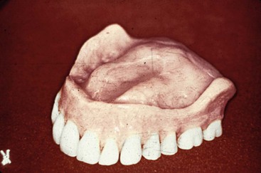
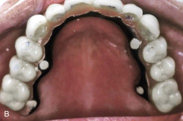
The patient is more likely to wear and functionally accommodate to a maxillary complete denture compared with its mandibular counterpart. The greater retention, support, and stability compared with the lower denture are well documented. As such, the patient often is able to wear the maxillary removable prosthesis for many years before complications arise. During this time, from a patient’s perspective, the need to replace the maxillary denture is more related to a desire to improve esthetics or for a fixed restoration as the motivating factor. By the time the patient notices problems of stability and retention caused by resorption of the premaxilla, the maxillary bone often has advanced atrophy and may be division C–h or D in volume (Figure 25-4). Therefore, unlike the anterior mandible (in which denture complications occur before advanced bone atrophy), in the completely edentulous maxilla, the anterior bony ridge is often inadequate for ideal endosteal implant insertion even though few denture complications exist. It is the doctor’s responsibility to inform the patient about the continued bone loss in the maxilla before complications arise.
To achieve predictable esthetics for a maxillary anterior or full-arch fixed prosthesis, the hard and soft tissue, volume, and character should be adequate in most aspects. Available bone should be evaluated closely for implant insertion in esthetic regions because of its influence on the soft tissue drape, implant size, implant insertion (angulation and depth), and the final prosthetic result. Bone loss after maxillary anterior tooth loss is rapid and has considerable consequences. Therefore, most multiple maxillary anterior edentulous sites in the esthetic zone require at least some bone and soft tissue augmentation before or during implant insertion and/or at implant uncovery.
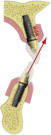
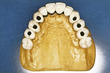
As the bone resorbs from division B to C–w in the anterior edentulous mandible, the cross-section of the residual ridge is triangular (with a wide base). As a consequence, an osteoplasty removes the narrower crestal bone and the residual ridge becomes wider, often converted to a division A bone volume. In the maxilla, however, the division B to C–w crest often remains narrow almost to the floor of the nose. An osteoplasty to gain bone width results in a division C–h to D ridge (Figure 25-5). The implant dentist often has difficulty inserting implants in the correct position when augmentation does not restore the region prior to implant insertion (Figure 25-6). Therefore, bone augmentation is more often required to increase bone width in the anterior maxilla compared with the anterior mandible.
In general, the premaxilla requires the most varied surgical approaches to improve success and is the most critical region for esthetics and phonetics. Initially, the anterior maxilla has less bone height than the anterior mandible and may compose only one third of the vertical dimension. Bone grafting is much more predictable for width gains rather than increases in height. Surgical options for division B and C–w bone more often require bone augmentation rather than osteoplasty, as often advocated in the mandibular anterior region. Division B bone grafting may use a synthetic bone component for the graft; division C–w often requires at least some autologous bone as a donor.
In some C–h premaxillae, implants may be inserted for a fixed prosthesis FP-3 or overdenture prosthesis. It should be noted that the opposing landmark is the floor of the nose, and this structure may be modified slightly by nasal elevation of 1 to 2 mm to improve implant support. However, when the premaxilla is less than 7 mm in height and when a D edentulous maxilla is present, this condition requires height augmentation before implant insertion. As a result, the dentist often must resort to the iliac crest or other extraoral donor sites for large volumes of bone. As such, the maxillary completely edentulous patient should understand that the surgical rehabilitation is much more complex and extensive because the volume of bone needed to reconstruct the atrophic maxilla increases. Therefore, notifying patients of their continued maxillary bone loss is even more important than in the anterior mandible rather than waiting until problems with their removable restoration develop.
Biomechanical Limitations
From a biomechanical perspective, the implant-restored anterior maxilla is often the weakest region of the mouth compared with other sections of the mouth. Compromised biomechanical conditions and their consequences include the following (Box 25-1):
2. In the premaxilla, esthetics and phonetics dictate that the replacement teeth be placed at or near their original position. The teeth are often cantilevered from the implants and the residual ridge, which usually is resorbed palatally and superiorly. The use of facial cantilevers results in increased moment loads at the implant crest and often leads to localized crestal remodeling bone loss and soft tissue recession. The cantilevered forces are also applied to the cement or screw that retains the restoration and the abutment screws that connect the implant components. This increases the risk of partially unretained restorations (Figure 25-7).
The farther forward the maxillary anterior crowns are positioned from the implants, the greater the moment force leverage on the implants, bone–implant interface, abutment screws, and prosthetic components. Yet many dentists attempt to do plastic surgery with plastic, hoping to eliminate vertical lines in the lip by bulking out the labial flange of an overdenture and positioning the teeth farther forward than the natural tooth position. Patients who desire to eliminate wrinkles in the lips from bone loss should have plastic surgery and bone augmentation, not the teeth positioned more labially for the maxillary prosthesis.
The facial position of the lip relative to esthetics is an important criterion to evaluate at the onset of treatment before the placement of the implants. This is even more important when the patient desires a fixed prosthesis. Bone and soft tissue augmentation is usually required to restore the natural appearance of the face without the help of a labial denture flange when a fixed restoration is planned. This criterion alone may indicate an overdenture rather than a fixed prosthesis or an onlay graft to position the implants more labial.
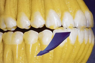
3. The arc of closure of the mandible is anterior to the maxillary residual ridge and is usually at an angle of 15 degrees or more. An angled load to an implant crown increases the force by 25.9% when it is 15 degrees off axis. As a consequence, the moment force is greater against the maxillary anterior crowns supported by implants compared with any other position in the mouth. Oblique centric contacts result in potentially harmful, off-axis load components. The force is also directed against the thinner facial bone (Figure 25-8).
4. All mandibular excursions place lateral forces on the maxillary anterior teeth, with resulting increased stress on the implant system, including the prosthesis and crestal bone of the supporting implants, especially on the labial aspect. These lateral loads in excursion further increase the moment loads applied to the implant system (Figure 25-9).
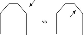
5. A mandibular arch receives a load from the outside of the arch toward the center. An arch is constructed for this force direction. A maxillary arch receives a force from within the arch to the outside of the structure, especially in mandibular excursions. An arch is not as effective to resist this type of force (Figure 25-10).
6. In most patients with available bone, the bone is less dense in the anterior maxilla than in the anterior mandible, where a dense cortical layer surrounds coarse trabeculae of adequate bone strength to provide implant support. In contrast, the maxilla presents thin porous bone on the labial aspect, very thin porous cortical bone on the floor of the nasal and sinus region, and more dense cortical bone on the palatal aspect.8,9 The trabecular bone of the maxilla is usually fine and is less dense than the anterior region of the mandible. The trabecular bone of D3, often found in the maxilla, is 45% to 65% weaker than the trabecular bone of D2, usually found in the anterior mandible.10 Reduced trabecular bone density of the maxilla results in compromised bone strength and a weaker implant–bone interface.
As a consequence of these biomechanical factors, not only is implant failure more common, but prosthetic complications are also more often found in maxillary restorations.
Treatment Options
The treatment options for the restoration of a complete or partially edentulous patient with multiple maxillary anterior teeth missing include a removable partial or complete denture, an implant-supported overdenture, or an implant-supported fixed prosthesis. Most traditional maxillary dentures have adequate retention, stability, and function, and soft tissue sore spots are rarely a problem. Therefore, less benefit is perceived with an implant overdenture (IOD) compared with the situation in an edentulous mandible.
A major disadvantage of a maxillary complete denture is most often the psychological aspect of removable teeth, especially when the mandibular arch is natural teeth or a fixed prosthesis. In contrast, a fixed prosthesis presents significant benefits for maxillary denture patients. In fact, after 3 years of function, most patients feel the maxillary fixed prosthesis is as good as or better than their natural teeth. On the other hand, an IOD is always considered by the patient as a removable prosthesis.
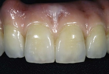
An independent, fixed implant-supported restoration has become the treatment of choice for most patients with complete or partial edentulism. A fixed prosthesis presents several advantages over a removable partial denture or an overdenture for a maxillary edentulous patient. However, when one or two canine teeth or implants are missing in addition to two or more adjacent teeth, it is contraindicated for a fixed prosthesis, regardless of how many teeth (or implants) are splinted together, unless two or more implants are used to replace the teeth11,12 (Figure 25-11). Additional contraindications for a FPD include long edentulous spans, poor abutment support, and inadequate edentulous bone for proper prosthetic contour.
The primary reason for a conventional maxillary removable denture is economic reasons or because a patient is unwilling to undergo bone grafting or implant surgery. However, the easiest interim treatment prosthesis for the replacement of several anterior teeth during implant-submerged healing is a removable restoration. If bone augmentation is necessary, this prosthesis may need to be used for longer than 1 year before delivery of the final implant restoration.
Sequence of Treatment Planning
Maxillary Labial Lip Position
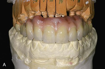
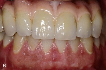
The maxillary anterior region with multiple adjacent teeth missing often is restored with an overdenture or a fixed restoration that replaces teeth and the soft tissue drape (FP-3 prosthesis) (Figure 25-12). Whether a denture, an overdenture, or a fixed prosthesis is being fabricated, a full-arch or anterior edentulous maxillary reconstruction begins with the determination of the facial position of the maxillary incisal edge. Its modification at a later step may alter all other determinants of a reconstruction. A baseplate and wax rim (or the patient’s existing denture) may determine the facial support necessary for the labial contour of the maxillary lip. Most often the facial surfaces of the central incisors are 12.5 mm from the most posterior aspect of the incisive papilla.13,14 The wax rim is initially positioned with this in mind. The farther forward the labial flange and teeth position, the higher the resting position of the lip and the greater the incisal edge exposure. The philtrum of the lip should have a visible depression in the midline under the nose. If the philtrum is too flat, the lip is extended too far, and wax should be removed from the labial aspect of the wax rim.
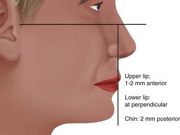
The position of the maxillary lip also may be determined by the position of the lower lip and chin with the face at the proper vertical dimension. A horizontal line, represented by the Frankfort plane, may be drawn from the highest point of the auditory meatus (top of the tragus) to the lowest point on the margin of the orbit, with the patient’s head in a vertical position. Ideally, a vertical perpendicular line drawn from the Frankfort plane to the lower lip should have the maxillary lip anterior to this landmark 1 to 2 mm and the chin 2 mm posterior to this line15 (Figure 25-13).
The labial position of the lip in relationship to the premaxillary bone is the primary criterion to determine whether a fixed restoration, a bone graft and fixed restoration, or a maxillary overdenture is indicated. When the labial position of the wax rim is forward of the residual ridge more than 5 mm, a bone graft before implants or a hydroxylapatite graft on the labial plate is required to support the lip for a fixed restoration, or a maxillary overdenture with a labial flange is considered (Figure 25-14).
Key Implant Positions
After the prosthesis type and labial tooth position are determined, the key implant positions are then determined for the maxillary restoration.16,17 An important parameter in treatment planning is to provide adequate biomechanical position and surface area of support for the load transmitted to the prosthesis. Four guidelines are presented in Chapter 9 for key implant positions in an implant prosthesis. For the edentulous maxilla, these four guidelines may be slightly modified and summarized as no posterior cantilever, no posterior three adjacent pontics, the canine rule, and the first molar sites.
In addition to these four key implant position guidelines, the complete edentulous maxilla should further reduce the increased biomechanical risks by adding a fifth guideline, the five-sided arch12 (Box 25-2).
Guideline 1: No Posterior Cantilever
The premaxillary teeth may be cantilevered forward from the implants for esthetics and phonetics. Hence, it is more important to place posterior implants connected to anterior implants to increase the anterior-posterior (A-P) distance and counter this affect. There should be little to no posterior cantilever in a complete edentulous maxilla (Figure 25-15).
Guideline 2: No Posterior Three Adjacent Pontics
When the posterior teeth are included in a prosthesis, there should not be three (or more) adjacent pontics.11 Under those conditions, the adjacent abutments must support five or more adjacent teeth, the amount of the force is greater in the posterior regions, and the metal of the restoration flexes 27 times more than a one pontic prosthesis. In addition, the bone density to support the implants is often less in the posterior maxilla, hence the strength of the bone is reduced (Figure 25-16). This further increases the overload risk to the implants.
When all six anterior teeth are missing, at least one implant should usually be positioned between the canine implants in the maxillary arch. However, the “no three pontic” rule may be modified in the front of the mouth because the force is less in the anterior region compared with the posterior region In addition, a lateral incisor is the smallest maxillary tooth, so the length of the span is reduced (compared with the posterior regions).
Guideline 3: The Canine Sites
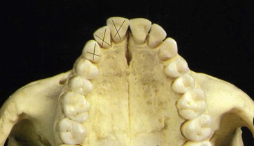
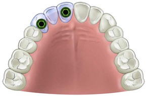
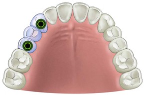
A fixed prosthesis replacing a canine tooth is at greater risk than almost any other tooth in the mouth. The adjacent maxillary lateral incisor is the weakest anterior tooth, and the first premolar is often the weakest posterior tooth. A traditional prosthodontic axiom indicates that a fixed prosthesis is contraindicated when a canine and two or more adjacent teeth are missing.11 Therefore, if a patient desires a fixed restoration, implants are required whenever the following adjacent teeth are missing: (1) the first premolar, canine, and lateral incisor; (2) the canine, lateral incisor, and central incisor (Figure 25-17); and (3) the canine, first premolar, and second premolar (Figure 25-18).
When any of these three missing teeth combinations are present, a fixed restoration is contraindicated because of the length of the span (three pontics), the amount of force on the abutments (forces greater in the canine region compared with the anterior and the force on the abutments includes the three missing teeth), the direction of the force on the abutments (angled forces to the canine region), the proprioawareness of the canine site, and the need to establish posterior disocclusion of the teeth in excursions (Box 25-3).
A tooth-supported prosthesis is less at biomechanical risk than an implant-supported restoration when the canine and two adjacent teeth are missing. Teeth are more mobile than implants; therefore, the stress relief mechanism of the periodontal–ligament complex reduces the flexure, force, and effect of an angled force. Despite this, it is contraindicated for patients with natural teeth to use three pontics in a fixed prosthesis whenever the natural canine and two adjacent teeth are missing. Therefore, under these conditions with implant treatment plans, at least two implants are indicated to support an independent fixed restoration (usually in the terminal positions of the span to eliminate cantilever forces) (Figure 25-19).
Using the missing canine and two adjacent natural teeth guideline, a fixed prosthesis is obviously contraindicated when either (or both) canine(s) and anterior four incisors are missing. For example, when a right canine, right lateral incisor, right central incisor, left central incisor, left lateral incisor, and left canine are missing, the condition is contraindicated for a fixed restoration. Yet in some underengineered treatment plans, implants are placed in each posterior maxillary quadrant, and a fixed restoration with five or six pontics is fabricated to replace the anterior teeth (Figure 25-20). The anterior cantilever from the first premolar sites in this treatment option is more detrimental than a posterior cantilever because of all of the biomechanical issues of the premaxilla. Apparently, the rationale for violating the prosthetic guidelines established in the literature for teeth are the following:
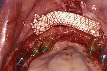
1. To augment a complete premaxilla, autologous bone grafts are often necessary, but synthetic materials can be used to predictably graft in the posterior maxillary sinus. The large volumes of autograft required for the complete premaxilla may require advanced bone graft procedures (which patients do not want, and few doctors are properly trained for) (Figure 25-21).
Guideline 4: The First Molar Site
The first molar is an important abutment position in an edentulous maxilla. The bite force in this region increases to 200 lb compared with half this amount in the premolar sites. As a consequence, the first molar natural tooth surface area is more than two times greater than the premolars. In addition, the bone density in the molar region is often poorer than the premolar regions of the jaws. As a result, larger diameter implants or more implants are suggested, not a cantilevered force applied in the molar regions.
An anatomical problem for implant treatment in the posterior maxilla is the rapid expansion of the maxillary sinus after tooth loss. As a result, the edentulous posterior maxilla rarely has enough bone height without sinus grafting. Therefore, a treatment option to cantilever the posterior missing teeth from anterior implants is used to eliminate sinus grafting. Posterior cantilevers from anterior maxillary implants are less predictable than cantilevers from anterior mandibular implants for all the reasons addressed previously in this chapter. In addition, when first molar implants are not present, the A-P distance of the splinted implants is reduced, and the anterior cantilever from the most anterior implants is more of a biomechanical risk (Figure 25-22). Instead of a posterior cantilever, sinus grafting and larger diameter implants (or two smaller diameter implants instead of one larger diameter) are indicated in the first molar region and render improved overall implant success rates and, as important, fewer prosthetic-related complications.
Guideline 5: Five-Sided Arch
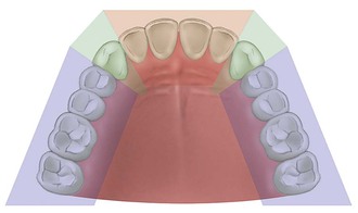
The dental arch may be divided into five different components related to their direction of movement (Figure 25-23). The posterior regions (first premolar, second premolar, first and second molars) each move in lateral directions to the midline. The canine positions move in two different oblique directions, and the anterior teeth (laterals and central incisors) move in an A-P direction. When three or more different components of an arch are splinted together, the different force directions are blended together, and the splint has less movement. In addition, when the three or more sections are splinted together, an A-P dimension is present, which also resists lateral forces. The more sections of the arch splinted together, the greater the A-P distance and the more resistant the splint is to any lateral force or cantilever.
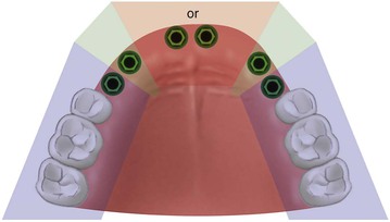
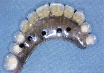
In the maxilla, most often at least one implant should be placed in each of the five sections missing teeth and then splinted together when replacing multiple adjacent teeth. This means at least three implants usually are required to replace the anterior six teeth in the premaxilla: one in each canine position and one in any of the four incisor positions16,17 (Figure 25-24). Previous studies by Bidez and Misch have shown that the force distributed over three abutments results in less localized stress to the crestal bone than two abutments.17,18 When these three implants are splinted around an arch, connecting at least three segments creates a tripod effect and provides an A-P distance (A-P spread) with mechanical properties superior to a straight line and with greater resistance to lateral forces.
When anterior and posterior teeth are missing, additional posterior implants are usually required (Figure 25-25). Because the premaxillary prosthesis has many biomechanical conditions that increase forces (both in centric and excursions), it may be considered a cantilever from the implant support system. When the implants in all five sections are splinted together, they act as one side of the class I lever with the most anterior implants in the position of the fulcrum, and the incisal edge of the prosthesis as the cantilever length of the lever. The A-P distance of the implants for the anterior cantilever in the premaxillary restoration corresponds to the distance between the center of the most distal implants on each side (in the splint) and the anterior aspect of the most anterior implant. Hence, an implant in the most distal missing tooth position greatly improves the A-P spread and reduces the forces of the premaxilla to the implant system (Figure 25-26).
Premaxilla Arch Form
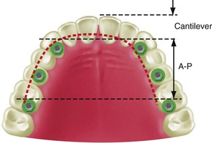
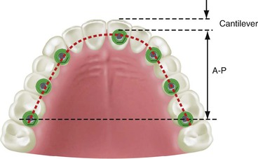
The arch form of the maxilla influences the fixed prosthesis treatment plan of the edentulous premaxilla. Three typical dentate arch forms for the maxilla are square, ovoid, and tapering. The dental arch form of the patient is determined by the final teeth position in the premaxilla and not the arch shape of the residual ridge. There are also three edentulous bone arch forms. As a consequence of bone resorption, the edentulous ridge arch form may be different from the dentate arch form. A residual ridge may appear square because of resorption or trauma. However, the final teeth position may need to be cantilevered facially with the final prosthesis. In other words, a dental ovoid or tapering arch form may be needed to restore a residual edentulous square arch form (Figure 25-27). The number and position of implants are related to the arch form of the final dentition (restoration), not only the existing edentulous arch form.
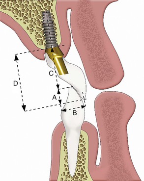
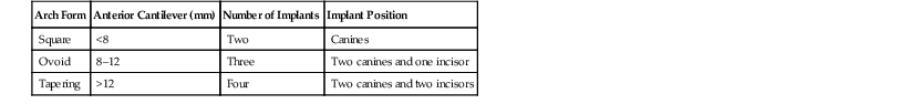
The dental arch form in the anterior maxilla is determined by the distance from two horizontal lines. The first line is drawn from one canine incisal edge tip to the other. This line most often bisects the incisive papilla regardless of the dentate arch form.13,14 The second line is drawn parallel to the first line along the facial position of the anterior teeth16 (Figure 25-28). When the distance between these two lines is less than 8 mm, a square dental arch form is present. When the distance between these two lines is 8 to 12 mm, an ovoid dentate arch form is present—the most commonly observed. When the distance between the two horizontal lines is greater than 12 mm, the dentate arch form is tapering12,17 (Table 25-1).
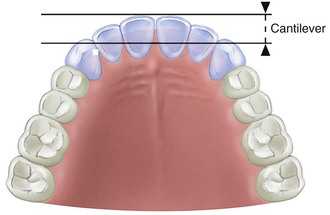
TABLE 25-1
Treatment Plan for Edentulous Premaxilla
| Arch Form | Anterior Cantilever (mm) | Number of Implants | Implant Position |
| Square | <8 | Two | Canines |
| Ovoid | 8–12 | Three | Two canines and one incisor |
| Tapering | >12 | Four | Two canines and two incisors |
In a dental square arch form, lateral and central incisors are not cantilevered very much facially from the canine position. Therefore, mandibular excursions and occlusal forces exert less stress on the canine implants. As a result, implants in the canine position to replace the six anterior teeth may suffice when the force factors are low (parafunction, masticatory dynamics, CHS) and if they are splinted to additional posterior implants (Figure 25-29). The four pontics between the canines may counter rule 2 of key implant positions (no three adjacent pontics) because (1) the forces are lowest in the incisor region and (2) in a dentate square arch form in the maxilla, minimal cantilevers are placed on the canines.
If the final teeth position is an ovoid arch form, at least three implants should be inserted into the premaxilla: one in each canine and one between the canines (preferably one in a central incisor position) (Figure 25-30). The central incisor position increases the A-P distance from the canine to central and provides improved biomechanical support to the prosthesis. In long-term edentulous maxillae, this most likely will require bone augmentation before implant insertion. When patient force factors are low to moderate, the anterior implant may be positioned in a lateral incisor site, when anterior implants are connected to molar implants and the A-P distance is increased. These three anterior implant positions resist the additional forces created in this arch form, enhance prosthesis retention, and reduce the risk of abutment screw loosening.
The restoration of a tapered dental arch form places the greatest forces on anterior implants, especially during mandibular excursions when the residual bone is an ovoid or square ridge form. The anterior teeth create a significant facial cantilever from the canine position. As such, four implants should be considered to replace />
Stay updated, free dental videos. Join our Telegram channel
VIDEdental - Online dental courses


