23 DESCRIPTION OF COLOR, COLOR-REPLICATION PROCESS, AND ESTHETICS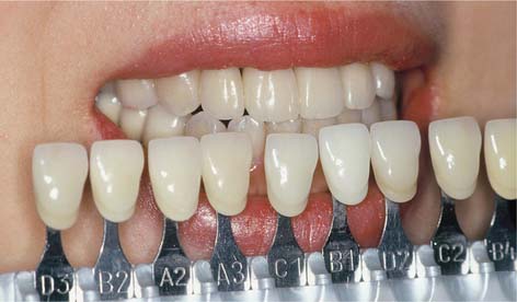
DESCRIPTION OF COLOR
Munsell Color Order System1
This system was widely used in the dental literature and also used in the past to quantify color.2,3 It is still a popular method of visually describing color. The three attributes of color in this system are called Hue, Chroma, and Value.*
Hue
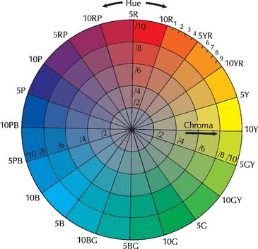
Hue is defined as the particular variety of a color. The hue of an object can be red, green, yellow, and so on, and is determined by the wavelength of the reflected and/or transmitted light observed. The place of that wavelength (or wavelengths) in the visible range of the spectrum determines the hue of the color. The shorter the wavelength, the closer the hue is to the violet portion of the spectrum; the longer the wavelength, the closer it is to the red portion. In the Munsell color system, Hues are arranged around the wheel (Fig. 23-1).
Chroma
Chroma is defined as the intensity of a hue. The terms saturation and chroma are used interchangeably in the dental literature; both mean the strength of a given hue or the concentration of pigment. A simple way to visualize differences in chroma is to imagine a bucket of water. When one drop of ink is added, a solution of low chroma results. Adding a second drop of ink increases the chroma, and so on, until a solution is obtained that is almost all ink and consequently of high chroma. In the Munsell color system, the intensity of Chroma of a particular Hue is more intense on the outer rim than near the hub of the wheel (Fig. 23-2).
CIELAB Color System
The CIELAB color system is used almost exclusively for color research in dentistry around the world.4–7 It was introduced in 1976 and recommended by the International Commission on Illumination. The strength of this system, unlike that of the Munsell system, is its ability for clinical interpretation, as equal distances across the CIELAB color space (color differences or ΔE) represent approximately uniform steps in human color perception, improving the interpretation of color measurements. This means that the magnitude of perceptible and/or acceptable color difference can be defined between, for example, a porcelain crown and the adjacent natural dentition.
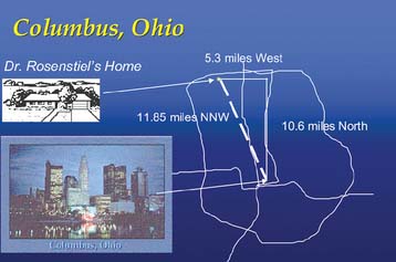
The CIELAB color order system defines color space by three coordinates: L*, a*, and b*. L* is similar to the Munsell system’s Value and represents the lightness, brightness, or black/white character of the color. The coordinates a* and b* describe the chromatic characteristics of the color. L* describes the achromatic character of the color. Colors with high value or L* (such as tooth colors) are located near the top of the color space, as depicted in Figure 23-3. The chromatic, or non–black/white, characteristics of a color are represented in the Munsell system by Hue and Chroma and in the CIELAB system by a* and b*. In each system, these two coordinates define the location of color on a plane of given lightness, such as the one depicting color B in Figure 23-3. In the Munsell system, the color is identified by one polar coordinate (Hue) and one linear, or Cartesian,† coordinate (Chroma); in the CIELAB system, both coordinates (a* and b*) are Cartesian. For an analogy, consider how the location of a house in a city might be described. It could be said that someone lived a distance of 11.85 miles (linear coordinate) in the north-northwest direction (polar coordinate) from downtown. This is analogous to describing a color in the Munsell system. The identical location could also be defined as being 10.6 miles north and 5.3 miles west of downtown (two Cartesian coordinates) (Fig. 23-4). This is analogous to describing a color in CIELAB. They represent the same location in space. However, unlike the Munsell coordinates, the CIELAB coordinates define the color space in approximately uniform steps of human color perception. This means that equal distances across the CIELAB color space (color differences, or ΔE) represent approximately equally perceived shade gradations, an arrangement that makes interpretation of color measurements more meaningful.
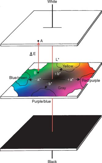
Fig. 23-3 La*b* color space. Any color can be defined in terms of these coordinates. L* (the vertical axis) defines the lightness or darkness of the color and corresponds to Value in the Munsell system; a* and b* define the chromatic characteristic. The color difference (ΔE) between two colors (A and B) can be calculated from the sum of the squares of the differences among the three coordinates. The system is arranged so that a color difference of 1 is perceivable by 50% of observers with normal color vision.60
(From Rosenstiel SF, Johnston WM: The effect of manipulative variables on the color of ceramic metal restorations. J Prosthet Dent 60:297, 1988.)
a* and b*
The a* and b* coordinates describe the chromatic characteristics of the color. Although they do not correspond directly to Munsell’s Hue and Chroma, they can be converted by numerical parameters8 (see Fig. 23-3). The a* coordinate corresponds to the red-purple/blue-green axis in the Munsell color space. A positive a* relates to a predominantly red-purple color, whereas a negative a* denotes a color that is more blue-green. Similarly, the b* coordinate corresponds to the yellow/purple-blue axis.
COLOR REPLICATION PROCESS
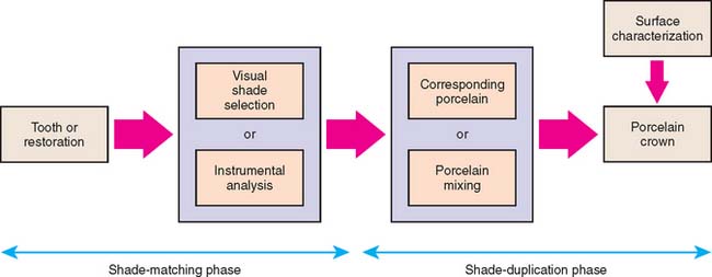
The process in which the color of adjacent teeth is replicated in a metal-ceramic or all-ceramic crown is termed in this chapter the color replication process. The color replication process for fixed restorations (Fig. 23-5) consists of the shade-matching phase followed by a shade-duplication phase. Shade matching can be accomplished through either the more common visual shade matching or the increasingly popular instrumental analysis. The shade duplication takes place in the dental laboratory, in which either the use of corresponding porcelain selected in the shade-duplication phase or the use of more sophisticated porcelain mixtures is used to fabricate the fixed restoration. If visually perceptible differences can be observed between the final restoration and the originally matched restoration, it is possible for the clinician to apply surface characterization porcelains to the restoration to adjust any color discrepancy.
SHADE-MATCHING PHASE
Visual Shade Matching
Visual assessment of the shade and translucency is the method most frequently applied in dentistry.9 Studies have shown that this often-used method is difficult to apply with accuracy and often yields unreliable and inconsistent results.10,11 Fortunately, a lifelike and successful restoration does not have to be an exact duplicate of the color and translucency of the adjacent teeth. It should, however, blend with the teeth as a result of the distribution of ceramic materials in the restoration. Not only is the apparent color of an object influenced by its physical properties, the nature of the light to which the object is exposed, and the subjective assessment of the observer; the variability of two of the three factors (e.g., lighting and subjectivity of the observer) can cause the same object (e.g., tooth) to look very different. Understanding the three main factors (lighting, subjectivity of human vision, and the object) that influence the outcome of visual shade matching can improve the accuracy and reliability of this process.
Lighting
Although daylight was initially thought to be the ideal light source for color matching,10 its use is not recommended, in view of inconstant color characteristics. The color of daylight can vary from red-orange at sunset to blue when the sky is clear. The relative intensity of daylight also fluctuates during the day with cloud cover.12 An ideal light source for visual shade matching is one that is diffuse and comfortable for the eyes, allowing observers to assess the color accurately and comfortably.12 In one study, evaluators obtained better visual shade matching in controlled stable, constant, and standard full-spectrum lighting than in daylight.13
Description of light
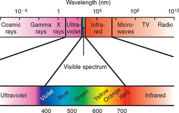
Scientifically, light is described as visible electromagnetic energy whose wavelength is measured in nanometers (nm), or billionths of a meter. The eye is sensitive only to the visible part of the electromagnetic spectrum, a narrow band with wavelengths from 380 to 750 nm. At the shorter wavelengths lie ultraviolet, x, and gamma rays; at the longer wavelengths are infrared radiation, microwaves, and television and radio transmission waves (Fig. 23-6).
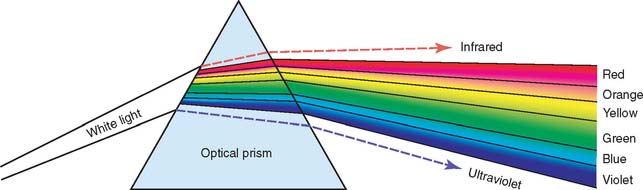
Pure white light consists of relatively equal quantities of electromagnetic energy over the visible range. When it is passed through a prism (Fig. 23-7), it is split into its component colors because the longer wavelengths are bent (refracted) less than the shorter ones.
Quality of light source
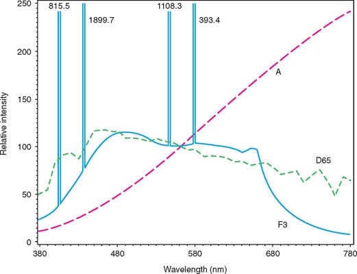
A light source with a color temperature close to 5500° K (D55) that is spectrally balanced throughout the visible spectrum is ideal for color matching. Color temperature is related to the color of a standard black body when heated and is reported in degrees Kelvin (K), or absolute (0° K = −273° C). Accordingly, 1000° K is red; 2000° K is yellow; 5555° K is white; 8000° K is pale blue. D65 (Fig. 23-8) is considered to be the true color temperature of white light as perceived by human observers.14 D65 is very commonly used in dental shade matching as the standard lighting for visual shade matching. A light source with a CRI greater than 90 is recommended for shade matching.15 The CRI, on a scale of 1 to 100, indicates how well a particular light source renders color in comparison with a specific standard source. Dental personnel’s shade-matching ability on a designed color test16 was significantly better with a full-spectrum light source of 5700° K (CRI = 91) than with the following light sources: 6000° K (CRI = 93), 4200° K (CRI = 65) and 7500° K (CRI = 94).17
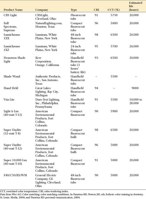
Unfortunately, the most common light sources in dental operatories are incandescent and fluorescent, neither of which is ideal for shade matching. An ordinary incandescent light bulb emits relatively higher concentrations of yellow light waves than of blue and blue-green, whereas fluorescent ceiling fixtures give off relatively high concentrations of blue waves. Color-corrected fluorescent lighting is recommended because it approaches the necessary type of balance. Recommended commercial color-corrected ambient lighting, ideal for shade matching, for the dental operatory can be found in Table 23-1.
Quantity of light source
Appropriate intensity of the ambient lighting in the dental operatory provides the dentist with visual comfort, particularly in terms of contrast. It is recommended that the light intensity for the dental operatory be between 18 to 28 lux* and 28 lux for the dental laboratory.18 The intensity of the dental operatory lighting has not been found to be crucial for color matching when the light intensity ranged from 1.5 to 28 lux.19
Auxiliary light sources
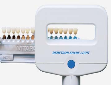
If ambient lighting in the dental operatory is not ideal in terms of quality and quantity for visual shade matching, the use of auxiliary lighting is recommended. The auxiliary light source for shade matching should be intense enough to overcome the influence of the ambient light. It has been recommended that the ratio of task (shade matching) to ambient light should not exceed 3:1; too much intensity does not allow discrimination of small color differences.18 Commercial auxiliary lighting, such as the Demetron Shade Light* (Fig. 23-9) or the Shade Wand,† is recommended for shade matching (see Table 23-1).
Shade-matching environment
The ambient and direct lighting used for shade matching scatters and reflects from surfaces before reaching the structure that it illuminates. The colors of the dental operatory, clothing of the dentist and dental assistants, the patient’s clothing, and the dental drape may influence the perceived color of the patient’s teeth and shade guide.20 To maintain the necessary lighting quality for shade matching, the chroma of the environment should be carefully controlled. It is recommended that the walls, staff clothing, patient drape, and shade-matching environment have a Chroma of four Munsell units or less, which are the pastel18 or the ideal neutral gray tones.21 Further recommendations include that the ceiling have a Munsell Value of 9. All other major reflectors (e.g., walls, cabinets) should present at least a Munsell Value of 7 and a Chroma of no more than 4. Countertops not within the working area can have a Chroma of up to 6 but a Munsell Value retained at 7 or greater.22
Human vision
The eye
Under low lighting conditions, only the rods are used (scotopic vision). These receptors allow an interpretation of the brightness (but not the color) of objects to be made. The rods are most sensitive to blue-green objects. Color vision is dependent on the cones, which are active under higher lighting conditions (photopic vision). The change from photopic to scotopic vision is called dark adaptation and takes about 40 minutes.23
The area with the most cones is in the center of the retina, which is free of rods. The rods begin to predominate toward the periphery. This means that the central field of vision is more color perceptive. Although the exact mechanism of color vision is not known, there are three types of cones—sensitive to red, green, and blue light24—that form an image in much the same way as the additive effect of the pixels in a television picture.
Deceptive color perception
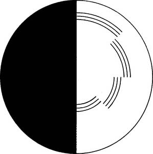
The brain can be tricked in how it perceives color. A classic example of such a trick is the Benham disk (Fig. 23-10). When this black and white disk is illuminated and rotated at an appropriate speed, it appears to be highly colored.
Color is also influenced by surrounding colors, particularly complementary ones (those diametrically opposed in Fig. 23-1). For example, when blue and yellow are placed side by side, their chroma may appear to be increased. The color of teeth can also look different if the patient is wearing brightly colored clothing or lipstick (Fig. 23-11).
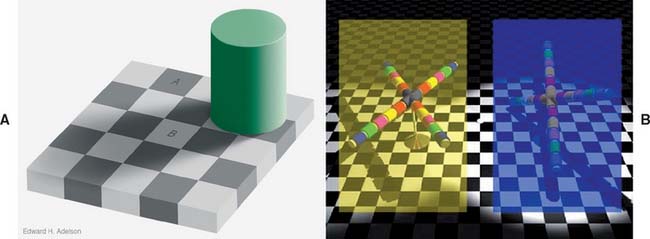
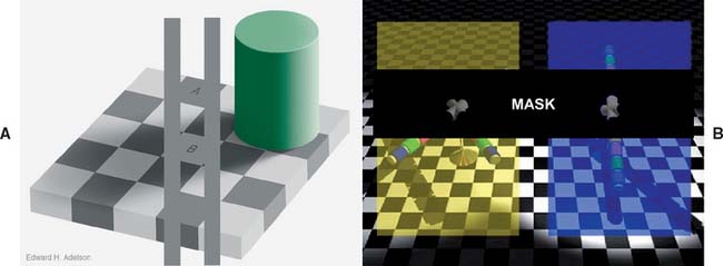
Fig. 23-11a A, The Checker Shadow Illusion. The squares marked A and B are the same shade of gray. For proof, see Fig. 23-11b, A. B, The Colored Cross Illusion. The central element of the two X-shaped objects appear very different in color but are, in fact, exactly the same. For proof, see Fig. 23-11b, B.
(A, Courtesy of Dr. E.H. Adelson; B, Courtesy of Dr. R.B. Lotto.)
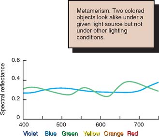
Metamerism
Two colors that appear to be a match under a given lighting condition but have different spectral reflectance (Fig. 23-12) are called metamers, and the phenomenon is known as metamerism. For example, two objects that appear to be an identical shade of yellow may absorb and reflect light differently. Yellow objects normally reflect yellow light, but some may actually absorb yellow light and reflect orange and green. To an observer, the orange and green combination looks yellow, although when the lighting is changed, the metamers no longer match. This means that a sample that appears to match under the operatory light, for example, may no longer be satisfactory in daylight. The problem of metamerism can be avoided by selecting a shade and confirming it under different lighting conditions (e.g., natural daylight and fluorescent light).
Fluorescence
Fluorescent materials, such as tooth enamel, re-emit radiant energy at a lower frequency than it is absorbed.25 For example, ultraviolet radiation is re-emitted as visible light. In theory, a mismatch can occur if the dental restoration has different fluorescence than the natural tooth. In practice, fluorescence does not play a significant role in color matching dental restorations.26
Opalescence
Natural teeth, particularly at their incisal edges, exhibit a light-scattering effect* that creates the appearance of bluish-white colors as the teeth are seen at different angles. This is similar to the bluish-white background seen in opal gemstones (hence the term opalescence). Manufacturers try to match this effect when formulating dental porcelains.27,28
Color blindness
Defects in color vision (color blindness) affect about 8% of the male population and less of the female population.29 Different types exist, such as achromatism (complete lack of hue sensitivity), dichromatism (sensitivity to only two primary hues—usually either red or green is not perceived), and anomalous trichromatism (sensitivity to all three hues with deficiency or abnormality of one of the three primary pigments in the retinal cones). Dentists should therefore have their color perception tested. If any deficiency is detected, the dentist should seek assistance when selecting tooth shades.30
Shade selection systems
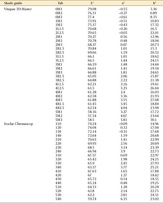
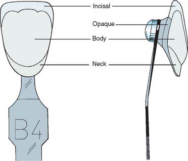
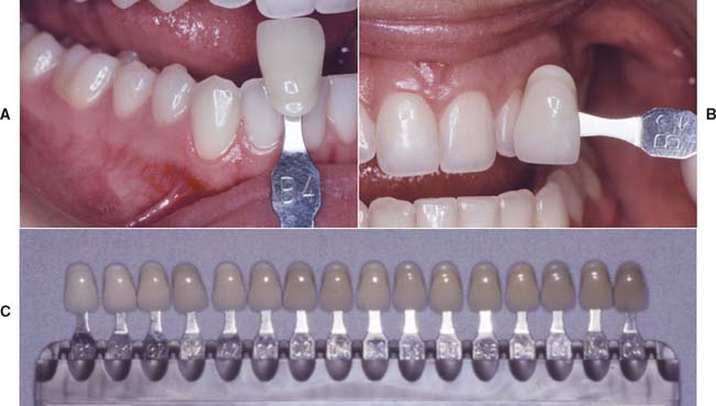
The most convenient method for selecting a shade is a commercially available porcelain shade guide (Fig. 23-13). Table 23-2 presents color measurement values made from Vita Lumin vacuum, Ivoclar Chromascop, and Vitapan 3D-Master shade guides with a spectroradiometer. Each shade tab (Fig. 23-14) has an opaque backing color, neck color, body color, and incisal color. Shade matching consists of picking the shade tab that looks the most natural and reproducing this color in a laboratory with materials and techniques recommended by the manufacturer. The procedure is easier if specimens of the same hue are grouped together in the shade guide. In the past, shade guides were produced in response to the demand for denture teeth rather than on the range of natural tooth color.31 More recently, shade guides have covered the color space occupied by natural teeth,* such as the Vitapan 3D-Master shade guide (see Fig. 23-13C).
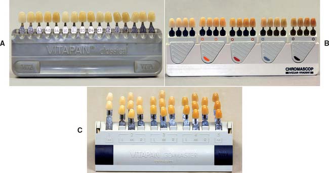
Table 23-2 CIELAB VALUES: VITAPAN 3D-MASTER, IVOCLAR CHROMASCOP, AND VITA LUMIN VACUUM SHADE GUIDES MEASURED WITH SPECTRORADIOMETER WITH 45° ILLUMINATION AND 0° OBSERVER WITHOUT AN APERTURE

Vita Lumin vacuum shade guide: Hue matching
In the popular Vita Lumin vacuum shade guide (see Fig. 23-13A), A1, A2, A3, A3.5, and A4 are similar in hue, as are the B, C, and D shades. Choosing the nearest hue first and then selecting the appropriate match of chroma and value from the tabs available is the recommended technique.
If its chroma or intensity is low, accurately determining a given hue may be difficult. Therefore, the region with the highest chroma (i.e., the cervical region of canines) should be used for initial hue selection (Fig. 23-15A).
Chroma selection
Once the hue is selected, the best chroma match is chosen. For example, if a B hue is determined to be the best match for color variety, there are four available gradations (tabs) of that hue: B1, B2, B3, and B4 (Fig. 23-15B). Several comparisons are usually necessary for determining which sample best represents the hue and its corresponding chroma (saturation) level. Between comparisons, glancing at a gray object rests the operator’s eyes and helps avoid retinal cone fatigue.
Value selection
Finally, value is determined with a second commercial guide whose samples are arranged in order of increasing lightness (Fig. 23-15C). (The lightness readings—L* in Table 23-2—can be used as a guide to the sample sequencing.) By holding the second shade guide close to the patient, the operator should be able to determine whether the value of the tooth is within the shade guide’s range. Attention is then focused on the range of shade that best represents the value of the tooth and how that range relates to the tab matching for hue and saturation. An observer is able to assess the value most effectively by observing from a distance, standing slightly away from the chair, and squinting the eyes. By squinting, the observer can reduce the amount of light that reaches the retina. Stimulation of the cones is reduced, and a greater sensitivity to achromatic conditions may result.32 While squinting, the observer concentrates on which disappears from sight first—the tooth or the shade tab. The one that fades first has the lower value.
When the proper value selection has been made, it is the exception rather than the rule for this to coincide with the determinations for hue and chroma. The operator must decide whether to change the previously selected shade sample. If the independent value determination is lower than the value of the sample selected for hue and chroma, a change is usually necessary, because increasing the value of an object by adding surface stain (which always reduces brightness) is not possible. If the value determination is higher than the hue determination, the operator should decide whether this difference can be bridged through internal or surface characterization of the restoration. The final decisions about hue, chroma, and value are then communicated to the laboratory.
Vitapan 3D-Master shade guide*
The manufacturer of this shade system (Fig. 23-16A) claims that it covers the entire tooth color space. The shade samples are grouped in six lightness levels, each of which has chroma and hue variations in evenly spaced steps (Fig. 23-16B). The shade guide is spaced in steps (ΔE) of four CIELAB units in the lightness dimension and two CIELAB units in the hue and chroma dimensions. The difference between lightness and color steps seems a logical approach to reducing the number of shade samples needed in the guide because of the way the CIELAB units are visually perceived. It seems to match the color difference formula of the Colour Measurement Committee (CMC) of the Society of Dyers and Colourists.33 Because the guide is evenly spaced, intermediate shades can be predictably formulated by combining porcelain powders.34
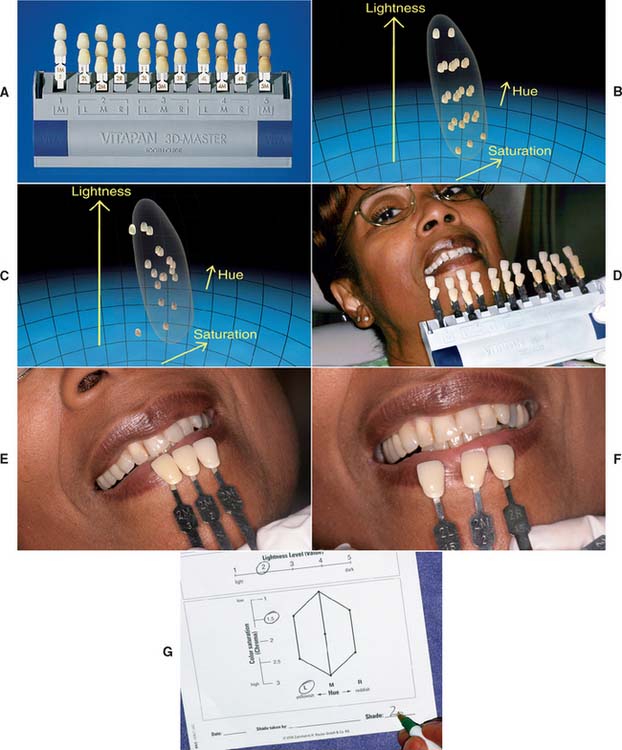
The manufacturer recommends selecting the lightness (Fig. 23-16D) first, then chroma (Fig. 23-16E), and finally the hue (Fig. 23-16F). A form is available to facilitate the laboratory shade prescription, which can include intermediate steps (Fig. 23-16G).
Extended-range shade guides
Most commercial shade systems cover a range more limited than the colors found in natural teeth, and the steps in the guide are greater than can be perceived visually.35 Some porcelain systems are available with extended-range shade guides, and other manufacturers have extended their ranges over the years. The use of two or more shade guides is a practical way to extend the range of commercial guides.
Dentin shade guides
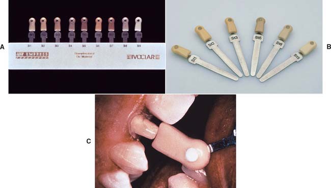
When a translucent all-ceramic system for a crown or veneer is used (see Chapter 25), communicating the shade of the prepared dentin to the dental laboratory is helpful. One system (IPS Empress)* provides specially colored die materials that match the dentin shade guide and enable the technician to judge restoration esthetics (Fig. 23-17).
Stay updated, free dental videos. Join our Telegram channel
VIDEdental - Online dental courses


