22
Hypertension and Target Organ Disease States: Assessment, Analysis, and Associated Dental Management Guidelines
AUTONOMIC NERVOUS SYSTEM
This chapter will begin with a discussion of the autonomic nervous system, which is part of the peripheral nervous system and responsible for regulating involuntary functions such as heartbeat, blood flow, breathing, and digestion. This system has two branches, the sympathetic system and the parasympathetic system. The sympathetic division of the autonomic nervous system regulates the “flight or fight” defense responses: urinary bladder relaxation, increase in heart rate, and dilation of the pupils. The parasympathetic division of the autonomic nervous system helps maintain normal body functions and conserve physical resources.
The heart is innervated by sympathetic and parasympathetic fibers. The sympathetic adrenergic system innervates the SA node, AV node, cardiac conduction pathways, and myocytes in the heart. The parasympathetic cholinergic nerve fibers that come to the heart come from the vagus nerves. Acetylcholine (ACh) released by the vagus nerve fibers binds to the M2 muscarinic receptors in the cardiac muscle, particularly at the SA and AV nodes, which have strong vagal innervations. This produces bradycardia, decreased cardiac conduction, decreased cardiac contractility, and decreased myocyte relaxation in the atria.
Epinephrine and norepinephrine are neurotransmitters associated with the adrenergic system. Epinephrine released by the adrenal medulla in response to the body’s “flight or fight” defense mechanism binds to the alpha and beta-adrenergic receptors on the heart and on the myocytes, causing tachycardia and blood sugar elevation. Norepinephrine (NE) is secreted by most postganglionic neurons and once released binds to specific receptors in the target tissues, producing physiological responses and changes in cardiac function.
The adrenergic system has two types of receptors, alpha and beta-receptors, with each having two subtypes: 1 and 2. The alpha 1 (α-1) and beta 1 (β-1) receptors are stimulatory in action and the alpha 2 (α-2) and beta 2 (β-2) receptors are inhibitory in action. β1- and β2-adrenergic receptors often coexist in the same tissue, sometimes mediating the same physiological effect.
Alpha-adrenergic receptors respond to norepinephrine. The alpha 1 (α-1) receptors are densely located in the vascular smooth muscles of the oral and nasal mucosa; sparsely in the coronary vasculature smooth muscles; around urinary and GI sphincters; in arrector pili muscles of the hair follicles; on apocrine sweat glands of the palms, armpits, and groin; and on dilator muscles of the iris. Contraction of the smooth muscle fibers located in these areas from alpha-1 stimulation causes vasoconstriction, constriction of sphincters, “goose bumps,” nervous sweating, and pupillary dilation. Norepinephrine additionally binds to α1-adrenergic receptors found on myocytes, producing small increases in cardiac contractility.
Alpha-2 receptors are abundantly located in the brain, peripheral smooth muscles of veins, platelets, nerve termini, and pancreatic islets. Alpha-2 receptor stimulation is the negative feedback component that inhibits further secretion of norepinephrine, causing platelet aggregation, vasoconstriction, and inhibition of insulin secretion.
Beta-1 receptors are located in the heart, conduction system, ventricular muscles, cerebral cortex, and eccrine salivary and sweat glands. When β1-receptors are activated by epinephrine or NE tachycardia, increased cardiac conduction, increased cardiac contractility, increased myocyte relaxation, and lipolysis occurs.
Beta-2 receptors are located on the circular smooth muscles of the lungs and some arterioles, the visceral smooth muscles of the GI tract, the urinary bladder, the cerebellum, and the liver. Beta-2 receptors are affected by epinephrine only and the action is inhibitory. Beta-2 receptor stimulation causes bronchodilation, vasodilatation, and an increase in cell energy production and utilization. The β2-adrenergic receptors become functionally more important in heart failure because β1-adrenergic receptors become down regulated. The brain contains both β1 and β2 receptors.
The autonomic nerve terminals also possess prejunctional adrenergic and cholinergic receptors that function to regulate the release of NE. Prejunctional α2-adrenergic receptors inhibit NE release, whereas prejunctional β2-adrenergic receptors increase NE release. Prejunctional M2 receptors inhibit NE release, which is one mechanism by which vagal stimulation overrides sympathetic stimulation in the heart.
Certain diseases can be managed by using drugs that block adrenergic and cholinergic receptors. For example, beta-blockers are used in the treatment of angina, hypertension, arrhythmias, and heart failure. Muscarinic receptor blockers such as atropine are used to treat electrical disturbances (e.g., bradycardia and conduction blocks) associated with excessive vagal stimulation of the heart. Many of these adrenergic and cholinergic blockers are relatively selective for a specific receptor subtype.
HYPERTENSION
Hypertension Classification
Hypertension (Htn) is classified as primary/essential hypertension or secondary hypertension.
Primary/Essential Hypertension
Primary/essential hypertension is the most common type of hypertension and it accounts for 95% of all cases presenting with high blood pressure. This type of hypertension has a genetic link and is often associated with cardiovascular risk factors, such as cigarette smoking, obesity (body mass index ≥30 kg/m2), lipid problems, age (>55 years for men, >65 years for women), family history of premature cardiovascular disease (<55 years for men, >65 years for women), hypertension, microalbuminuria, estimated GFR <60mL/min, physical inactivity, and diabetes. It is insidious in onset and is often asymptomatic in the early stages.
Secondary Hypertension
Secondary hypertension is always due to an underlying cause. Renovascular disease, renal artery stenosis, coarctation of the aorta, pheochromocytoma, Cushing’s syndrome, thyroid or parathyroid disease, heavy alcohol consumption, chronic corticosteroid therapy, chronic NSAIDS therapy, or long-term oral contraceptive use can lead to secondary hypertension. The secondary hypertension patient experiences symptoms relatively early on in the disease process compared to the primary/essential hypertension patient, and the symptoms are also more severe compared with the patient with essential hypertension.
2003 VII JOINT NATIONAL COMMISSION’S HIGH BLOOD PRESSURE GUIDELINES
The following are high blood pressure guidelines from the 2003 VII Joint National Commission (JNC) Report (Table 22.1< ?anchor c22-tbl-0001?>).
Table 22.1 VII Joint National Commission (JNC-2003) Blood Pressure Classification for Adults Aged 18 Years and Older*
| Blood Pressure Classification | ||
| Category/BP Staging | Systolic, mmHg | Diastolic, mmHg |
| Normal | <120 | <80 |
| Pre-Hypertension | 120–139 | 80–89 |
| Stage I | 140–159 | 90–99 |
| Stage II | ≥160 | ≥100 |
| Stage III: Defer Dental Treatment | ≥180 | ≥110 |
|
*Based on the average of two or more readings taken at each of two or more visits after an initial screening, not taking BP medications and not acutely ill. When systolic and diastolic pressures fall into different categories, the higher category should be selected to classify the individual’s blood pressure status. For instance, 154/82mmHg should be classified as stage I, and 164/95mmHg should be classified as stage II. Isolated systolic hypertension is defined as a systolic blood pressure of 140mmHg or more and a diastolic blood pressure of less than 90mmHg plus it is staged appropriately (e.g., 154/82mmHg is defined as stage I isolated systolic hypertension). In addition to staging hypertension on the basis of average blood pressure levels, the clinician should specify presence or absence of target-organ disease and additional risk factors. This specificity is important for risk classification and treatment. |
||
MAJOR RISK FACTORS FOR HYPERTENSION
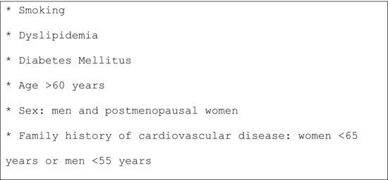
Major risk factors for hypertension are listed in Figure 22.1.
Figure 22.1 Major risk factors for hypertension.

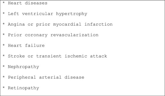
Target Organ Damage/Clinical Cardiovascular Diseases
The target organ damage or clinical cardiovascular diseases that result from hypertension are listed in Figure 22.2.
Figure 22.2 Target organ damage/clinical cardiovascular diseases.

Blood Pressure Measurement Follow-Up Guidelines
Recommendations for follow-up based on the initial set of BP readings for adults are listed in Table 22.2.
Table 22.2 Blood Pressure Monitoring Follow-Up Guidelines
| Systolic | Diastolic | BP Monitoring Follow-Up Guidelines |
| <120mmHg | <80mmHg | Recheck in two years. |
| 120–139 | 80–89 | Recheck in one year. |
| 140–159 | 90–99 | Confirm within two months. |
| ≥160 | ≥100 | Assess or refer to MD in one month. |
| ≥180 | ≥110 | Refer symptomatic patient to MD/ER immediately. Refer asymptomatic patient to MD within one week. |
Blood Pressure Monitoring and Hypertension-Related Facts
When monitoring the BP make sure that the patient has not smoked or had a caffeinated drink 30 minutes prior to measuring the BP, because this will erroneously raise the BP. If the patient has just consumed a heavy meal, it will erroneously lower />
Stay updated, free dental videos. Join our Telegram channel

VIDEdental - Online dental courses


