Variations in Transition of Mandibular Incisors*
The mandible precedes the maxilla in the transition of the incisors. The first deciduous tooth to be lost — the mandibular central incisor — is exfoliated about 1 year earlier than the corresponding maxillary incisor.
The tooth-containing parts of the mandible differ markedly from those in the maxilla in size, shape, and structure. Consequently, the transition of the mandibular incisors differs from that of the maxillary incisors. In the mandible less space is available for the crowns of the not-yet-erupting permanent teeth and the roots of the deciduous and permanent ones than in the maxilla. Further, the mandible does not have an interstitial growth site in the form of a midsagittal suture that can contribute to an increased transverse jaw dimension in the median plane. The aforementioned differences partly explain why the transition of incisors in the mandible varies less than that in the maxilla.
The combination of four features allows the alignment of the larger permanent incisors in the region where previously the smaller deciduous ones were located: (1) the presence of diastemata between the deciduous teeth, (2) the increase in intercanine distance associated with the transition of the incisors, (3) the more labial inclination of the permanent incisors, and (4) the fact that the permanent incisors are more labially positioned than their predecessors. In general, all four features exert a larger effect on the maxilla than the mandible.13, 141, 143, 145, 223
The transition of the mandibular incisors is discussed in relation to the size and shape of the anterior section of the apical area. The variation in location of the permanent incisors and canines prior to emergence and their relation to the adjacent deciduous predecessors are indicated. The eruption is divided into preemergence and postemergence phases. Common features in the transition of the mandibular incisors are explained. Three eruption patterns are presented by means of longitudinal records of dental casts and schematic drawings.
2.2 The anterior section of the apical area* and arrangement of anterior teeth before emergence
The size, shape, and structure of the mandible determine the space available for the teeth. Considerable individual variation exists in the form of the anterior part of the mandible, as is expressed by the course of the lower border, the prominence of the chin, and the arrangement of the teeth in the dental arch. Critical in this respect is the location of the permanent canine crowns as determined by the sites where their formation started. The same holds for the location of the apices of the mandibular permanent canines after eruption is completed.
The anterior section of the apical area is bounded laterally by the location of the permanent canines and in the sagittal direction by the lingual and labial cortical walls. In the developing dentition, a considerable amount of tooth material is stacked in the anterior region of the mandible. Prior to the transition, the roots of the deciduous incisors and the forming permanent teeth fill the space available in a typical manner. The crowns of the permanent incisors with their large mesiodistal dimensions at the incisal edge and their large labiolingual dimensions at the cementoenamel junction are arranged in a way that utilises the available space efficiently.
The mandibular permanent incisors are formed parallel to each other behind the roots of their predecessors in a more or less perpendicular angulation. Invariably the crowns of mandibular central permanent incisors do not cross the midline and are placed labially to those of the lateral incisors. The central incisors are slightly labially inclined; the lateral incisors are slightly lingually inclined. The amount of overlap of the lateral incisors by the central incisors shows little variation; it ranges, when these teeth are not rotated, between one quarter to one third the width of the incisal edge of the lateral incisors. The amount of overlap of the lateral incisors by the canine crowns shows marked variation, can be completely absent, and is directly related to the size of the anterior section of the apical area.
Rotation of anterior permanent teeth prior to emergence is not necessarily a sign of crowding. In a number of cases the morphogenesis of the mandibular permanent incisors starts in a rotated orientation that is maintained in their subsequent formation and their initial eruption.
Some asymmetry may exist in the positioning of corresponding left and right mandibular permanent incisors and canine crowns. This applies not only to the anteroposterior location and rotation but also to the amount of overlap of adjacent teeth.
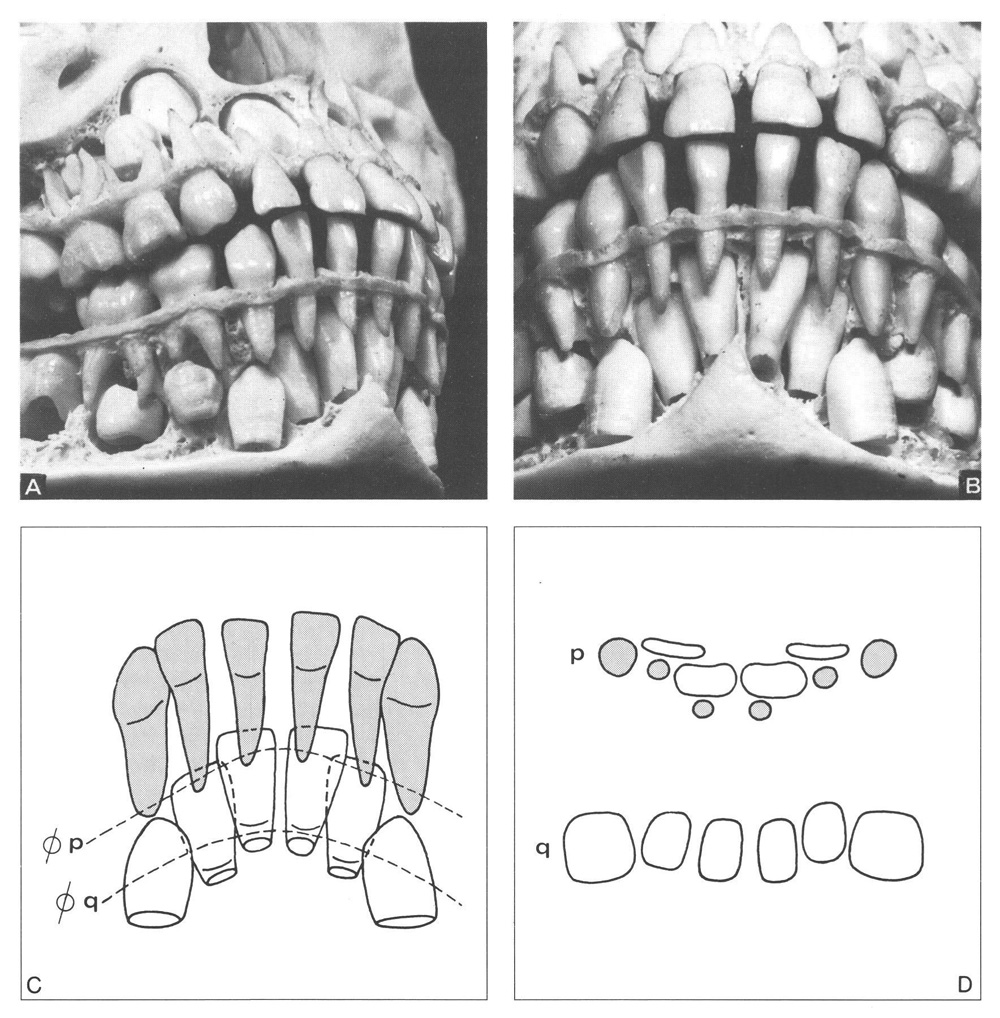
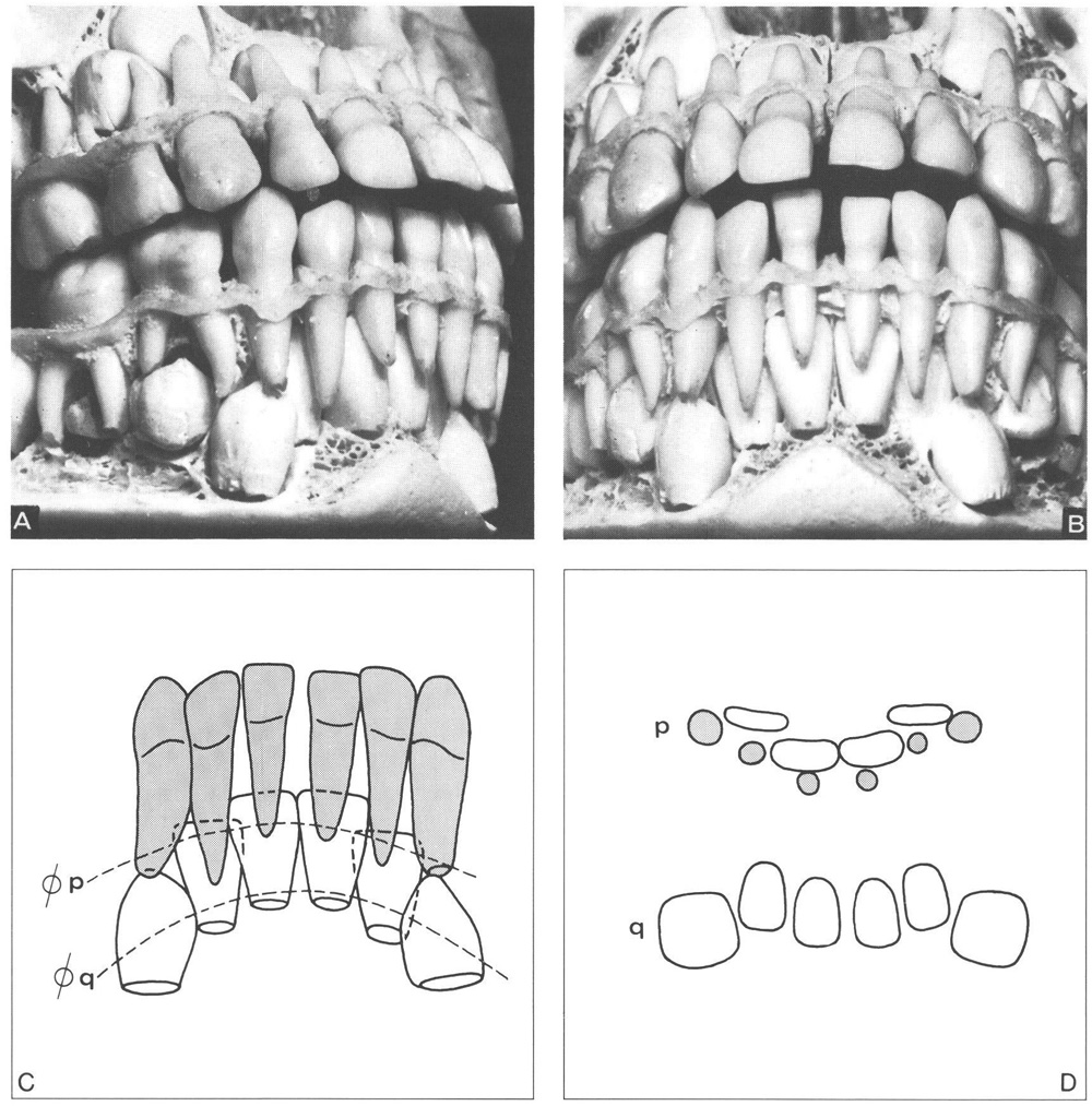
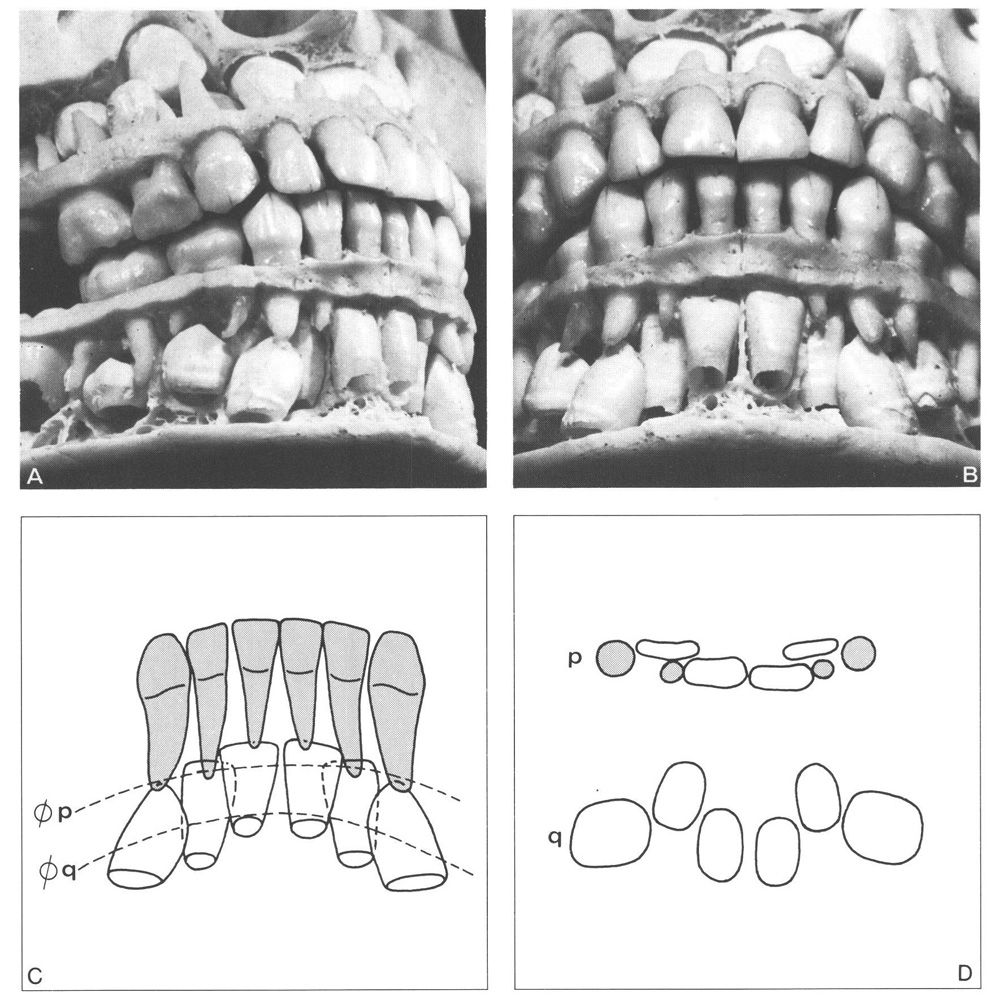
The arrangement of the tooth material within the anterior region of the mandible, before the emergence of the permanent incisors, differs, depending on the relation between the space available and the space needed (Figs. 2-1 to 2-3).

Fig. 2-1 Large anterior section of the apical area in the mandible.
A, B Many and large diastemata between the anterior deciduous teeth usually indicate the presence of a large section of the apical area. The mandibular permanent canines are only slightly lingually inclined and mesially angulated. The central permanent incisors are separated and overlap the neighbouring lateral incisors for about one quarter of their mesiodistal crown width. The cusp tips of the mandibular canines are located slightly distal to the apices of their predecessors.
C Schematic drawing with dotted lines indicating the sections presented in D. The central permanent incisor crowns do not contact the adjacent lateral deciduous incisor roots. The same holds true for the crowns of the permanent lateral incisors and the roots of the deciduous canines. Some asymmetry exists in the arrangement of the permanent incisors. The left central incisor is more lingually inclined and more distally angulated than the right one.
D In the region of the incisal borders of the permanent incisors (pj the lateral incisors are located lingual to the central incisors. Note the relations indicated under C. No overlapping exists of the permanent incisors (q) in the region of the cementoenamel junctions, where the central incisors are only slightly labial to the lateral incisors.

Fig. 2-2 Medium anterior section of the apical area in the mandible.
A, B Limited spacing between the anterior deciduous teeth. The mandibular permanent canines are lingually inclined and mesially angulated. The crowns of the lateral permanent incisors are partly overlapped by those of the central incisors and also by those of the permanent canines. The cusp tips of the permanent canines are close to the apices of their predecessors.
C The distal corners of the crowns of the lateral permanent incisors are in close proximity to the roots of the adjacent deciduous canines. The left permanent canine is more mesially angulated than the right one; the left lateral incisor is overlapped more than the right one.
D In the region of the incisal borders of the permanent incisors (p), the crowns of the permanent teeth and the roots of the adjacent deciduous teeth are in close proximity. In the region of the cementoenamel junctions of the permanent incisors (q), the available space does not markedly exceed the space required. Note the asymmetry in the relationship between the lateral permanent incisors and canines.

Fig. 2-3 Small anterior section of the apical area in the mandible.
A, B Few and only small diastemata between the anterior deciduous teeth. The mandibular permanent canines are markedly lingually inclined and mesially angulated. Their crowns overlap those of the lateral incisors. The cusp tips of the permanent canines are in close proximity to the apices of their predecessors.
C The central permanent incisor crowns are close together and their distal corners are in contact with the roots of the adjacent lateral deciduous incisors. The distal corners of the crowns of the lateral permanent incisors are slightly lingual to the roots of the adjacent deciduous canines.
D In the region of the incisal borders of the permanent incisors (p), the crowns of the permanent teeth and the roots of the deciduous ones are close together. In the region of the cementoenamel junctions (q), crowding exists where the lateral incisors are lingual to the central incisors and canines and are overlapped by the latter.
The anterior section of the apical area is large when the distance between the crowns of the mandibular permanent canines is large in relation to the mesiodistal crown dimensions of the permanent incisors. Favourable conditions are then present for the transition of the incisors. Frequently, a large anterior section of the apical area is associated with many and wide diastemata between the mandibular anterior deciduous teeth and a small anterior section of the apical area with few and small diastemata or even no diastemata. However, the correlation between the sum of the mesiodistal crown dimensions of the mandibular incisors of the two dentitions is rather low.144 So, large mesiodistal crown dimensions of deciduous incisors are not always associated with corresponding large permanent teeth, as well as vice versa. This has consequences when it is necessary to estimate the spatial requirements of the permanent anterior teeth.
2.3 Eruption before emergence*
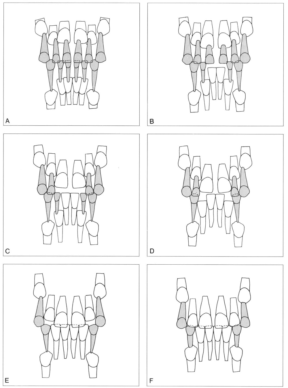
The mandibular central permanent incisors are the first anterior teeth to commence eruption. They move in the direction of the occlusal plane according to their orientation within the jaw. The overlapping of the crowns of the adjacent lateral incisors reduces. After about 1 year, when the central incisors have reached the level of the occlusal plane, the lateral incisors are free to start to erupt. Normally the overlapping is fully eliminated and the lateral incisors move in the direction of the occlusal plane. Before the onset of eruption and in the preemergence eruption phase, the lateral incisors are more lingually inclined than the central incisors, as indicated above. Correspondingly, the lateral incisors usually follow a more lingually inclined path of eruption than that of the central incisors, and their incisal edges emerge more lingually. Figure 2-4 shows the transition of the mandibular and maxillary incisors. Figure 2-5 shows six specimens of prepared skulls to illustrate the process of transition of the mandibular incisors and some variations in the size of the anterior section of the apical area.

Figure 2-4
Fig. 2-4 Schematic presentation of the transition of the mandibular and maxillary incisors. (From Van der Linden.223)
A Situation at approximately 5 years of age, prior to the start of eruption. All permanent crowns are placed lingually to the apices of their predecessors. The mandibular central permanent incisors are located slightly nearer to the occlusal plane than the adjacent lateral incisors. The crowns of the latter are lingual to those of the central incisors.
In the maxilla the incisal edges of the lateral permanent incisors are located nearer to the occlusal plane than those of the central incisors. The distoincisal corners of the central permanent incisors are in contact with the mesial surfaces of the roots of the adjacent lateral deciduous incisors.
B After loss of the mandibular central deciduous incisors the successors emerge, frequently in close proximity to each other. The overlapping of the mandibular lateral permanent incisors disappears before eruption of the central incisors is completed.
In the maxilla the erupting central permanent incisors pass the lateral incisors. The contacts between the distoincisal corners of the central permanent crowns and the mesial surface of the roots of the lateral deciduous incisor lead to a distal movement of the latter. The diastemata mesial to the deciduous canines reduce in width, and more space becomes available for the central permanent incisors to emerge.
C The mandibular central permanent incisors have reached the level of the occlusal plane before the lateral incisors start to erupt. In a medium anterior section of the apical area, the distoincisal corners of the mandibular central permanent incisors may contact the mesial surfaces of the roots of the lateral deciduous incisors, resulting in a distal displacement of the latter and a reduction of the diastemata mesial to the deciduous canines. In the maxilla, the central deciduous incisors are lost and their successors have erupted further. The concomitant distal displacement of the lateral deciduous incisors has continued./>
Stay updated, free dental videos. Join our Telegram channel

VIDEdental - Online dental courses


