CHAPTER 2 Osseointegration
Rationale for Two-Stage versus One-Stage Protocols
The word osseointegration consists of os, the Latin word for bone, and integration, derived from the Latin words meaning the state of being combined into a complete whole. Osseointegration is defined as direct bone anchorage to an implant body that can provide a foundation to support a freestanding fixed prosthesis; it has the ability to transmit occlusal forces directly to bone.1–3 To achieve osseointegration between the implant and the bone, the implant surface must be made of an inert material with affinity to establish a direct contact with the bony tissue, without a soft tissue interface. As Brånemark explains, “the bases for osseointegration, leading to permanent, functional and interactive coexistence between titanium and bone, is established when a topographically harmonious and precise recipient site—geometrically adjacent to the anatomy of the fixture—is gently prepared in bone.”4
The concept of osseointegration was initially discovered by accident as Brånemark was studying microcirculation in bone using titanium chambers inserted in the tibia of rabbits.5 Upon completion of the study, removal of the titanium chambers proved to be difficult. Further studies led to the understanding that titanium is the most favorable material for replacement of lost roots in the human alveolar bone.2,6,7 Once osseointegration was established, the junction of the implant and the bone—direct anchorage—was so strong that a 100-kg force did not separate and dislodge the implant,8 and further application of force would have led to fracture of the bone itself and not the osseointegrated junction between the bone and the implant. Therefore, a direct implant-to-bone contact without a soft tissue interface is paramount.
Considering a different point of view, Linkow and Chercheve9 and Weiss10 believed that a peri-implant membrane established between the implant surface and the bony osteotomy site acted like the periodontal membrane of teeth, cushioning the occlusal forces placed onto the prosthesis and transmitted to the alveolar bone. In later studies11 this fibrointegration could not withstand the forces applied under functional load and became larger, with an inflammatory reaction, gradual bone resorption, and loss of the fixtures. Brånemark further demonstrated that use of peri-implant membranes is consistent with failure of implants under load. In his studies, Brånemark12 described the biologic and the biomechanical differences between natural teeth, fibrointegrated implants, and osseointegrated implants. Three separate models were described as follows:
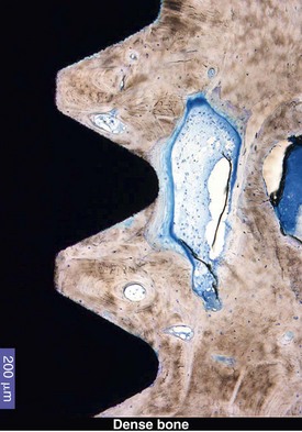
Teeth supported by differentiated, functionally oriented periodontal ligaments have a high threshold for tissue injury and demonstrate height mechanical resistance to applied occlusal loads. On the other hand, implants anchored in nonmineralized connective tissue demonstrate low resistance to injury with low mechanical resistance as the implants are surrounded by tissues that have low differentiation and functional fibers that are not generally functionally oriented. Osseointegrated implants, however, are surrounded by continuously remodeling differentiated and functionally oriented bone tissues with a high threshold for injury under functional load. From this observation, it was clear that implant-to-bone contact without a fibrous interface was needed for functional support under occlusal loading. To achieve predictable bone anchorage, a consistent composition and finish of the implant’s surface is essential,13 as is an intimate contact between the implant and the osteotomy site—the bone—which is achieved by threading the implant into the prepared site.14 The objective of a threaded endosteal implant is to establish stabilization of the fixture immediately after preparation of the osteotomy and during the initial healing period to achieve osseointegration. The dynamics of osseointegration and the relationship of an endosteal implant with its surrounding alveolar bone can be distinguished over time as three distinct and partially overlapping intervals or periods: healing, remodeling, and dynamic equilibrium (Figure 2-1).
During the first 18 months, referred to as the healing phase, primary and secondary bone healing occurs. The remodeling phase, which begins at 3 months and overlaps with the healing phase, ends at 18 months. After this period, the dynamic equilibrium phase begins, in which a balance is established between the forces acting on the fixture and the remodeling capacity of the anchorage bone.15
Key Factors Influencing Osseointegration
Bone healing begins within the first week after placement of the implant and reaches its peak around the fourth week. The interface between the implant and the osteotomy matures and gradually becomes bone tissue around 8 weeks.1 The dynamic condition of the bone continues after insertion of the prosthesis. Bony remodeling occurs for up to 18 months after insertion of the definitive prosthesis, with the resultant woven bone maturing into the denser laminar bone.5 Understanding the occlusal forces generated under functional loads, it was possible to develop a set of criteria for the evaluation of the continued maintenance of osseointegration over time. The occlusal forces generated by the dentate patient are approximately 100 psi. The occlusal forces generated by conventional complete denture patients are 15 to 20 psi.24–26 Patients with fully bone-anchored prostheses generate the same occlusal forces as dentate patients.27 In 1986, Albrektsson et al. proposed a standard for clinically defining osseointegrated implants under function as successful.28 The following criteria define successful implants:
Stay updated, free dental videos. Join our Telegram channel
VIDEdental - Online dental courses


