Neurophysiology
1. Describe the functional unit of a nerve system.
2. Explain the main function of the neuron.
3. Differentiate the role of the cell body in sensory and motor impulse transmission.
4. Describe the parts and functions of the neuron:
5. Compare the ions in nerve transmission in regard to their functional element relative concentrations and location during the resting stage, depolarization, and repolarization.
6. Explain the stages of nerve conduction.
7. Describe the Na+/K+ pump and differentiate between active and passive energy requirements for the stages of nerve transmission.
8. Describe action potential and describe saltatory conduction.
9. Explain the anatomy of a sensory neuron.
10. Differentiate between type A, B, and C fibers in terms of their function, size, and relative speed of impulse transmission.
11. Discuss speed of impulse propagation with myelinated versus non-myelinated nerves.
12. Describe and recite the stages of nerve transmission in detail.
13. Describe the refractory periods.
14. Explain the role of neurotransmitters in nerve conduction.
15. Discuss the mode of action of local anesthetic agents on nerves.
16. Correctly complete the review questions and activities for this chapter.
Parasympathetic nervous system
Coordinates the body’s normal resting activities and is known as the “rest or digest” response.
Introduction
A local anesthetic is a drug that causes reversible local anesthesia and a loss of nociception (also called a pain receptor) as a result of the depression of excitation in nerve endings or the inhibition of the conduction process in peripheral nerves. Local anesthetic agents used in dental practice prevent both the generation and conduction of a nerve impulse. When it is used on specific nerve pathways (nerve block), effects such as analgesia (loss of pain sensation) can be achieved. Basically, local anesthetics provide a chemical roadblock between the source of the impulse and the brain. The impulse therefore is unable to reach the brain.1,2
Local anesthetics are known as membrane-stabilizing drugs, which work by decreasing the rate of depolarization when a membrane potential is initiated. Local anesthetic drugs work essentially by inhibiting sodium influx through stimulus-gated sodium ion channels in the neuronal cell membrane. When the local anesthetic binds to the sodium channels, the influx of sodium is interrupted, and the action potential cannot rise and signal conduction is inhibited. The receptor site is located at the cytoplasmic (axoplasmic, inner) portion of the sodium channel. When neurons are firing quickly, causing sodium channels to be in their activated state, the local anesthetic drugs bind easier to the sodium channels, increasing the onset of neuronal blockade. This phenomenon is called state dependent blockade.2
Organization of the Nervous System
Central and Peripheral Nervous Systems
The central and peripheral nervous systems are divisions of the nervous system that are based upon location and direction of nerves (Figure 2-1).
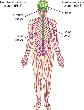
The central nervous system (CNS) is the structural and functional center of the entire nervous system that includes the brain, contained within the skull, and the spinal cord, contained within the vertebral canal. The CNS is responsible for receiving sensory information, processing the information, and initiating an outgoing response.3,4
The peripheral nervous system (PNS) are nerve tissues that lie in the periphery, or “outer regions,” of the nervous system, consisting of 31 pairs of spinal nerves arising from the spinal cord, and 12 pairs of cranial nerves arising from the brain.3,4
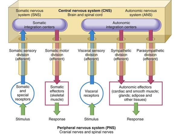
Nerve fibers that extend from the cell body toward the CNS are termed central fibers, and nerve fibers that extend from the cell body away from the CNS are termed peripheral fibers. Figure 2-2 illustrates the divisions and relationship of the CNS and PNS.
Afferent and Efferent Divisions
Afferent and efferent divisions of the CNS and PNS tissues are categorized according to the direction in which they carry information. The afferent division consists of all incoming information traveling along sensory or afferent pathways. The efferent division consists of all outgoing information along motor or efferent pathways. Figure 2-2 illustrates the afferent pathways in blue and the efferent pathways in red.
Somatic and Autonomic Nervous Systems
Another way to organize the nervous system is based on function. The somatic nervous system is a subdivision of the efferent division of the PNS and controls the body’s voluntary and reflex activities through somatic sensory and somatic motor components. External sense organs (including skin) are receptors. Somatic effectors are skeletal muscles and gland cells. The autonomic nervous system pathways carry information to the autonomic, or visceral, effectors that control involuntary (without conscious control) smooth muscle, cardiac muscle, or glandular tissue, and other involuntary tissue. The motor component controls smooth muscle contractions of viscera and blood vessels and the secretion of glands (eg: salivary glands). The autonomic nervous system is further divided into the sympathetic and the parasympathetic divisions. The sympathetic division prepares the body to deal with an emergency situation and is involved with the “fight or flight” response. The parasympathetic division coordinates the body’s normal resting activities and is known as the “rest or digest” response.3
Neuroanatomy
Nerve cells (neurons) are the basic functional unit of the nervous system which manipulate information and respond to either excitation or inhibition. Doing so involves changes in the bioelectrical or biochemical properties of the cell, which require a vast expenditure of energy for each cell. The nervous system, compared with other organs, is the greatest consumer of oxygen and glucose. These energy requirements arise directly from the metabolic demand placed on cells, which have a large surface area and concentrate biomolecules and ions against an energy gradient. Along with maintaining its metabolism, each neuron receives information from the environment or from other nerve cells, processes information, and sends information to other neurons.5–7
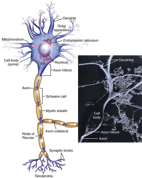
For neurons to carry out the three tasks of receiving, processing, and sending information, they must have specialized structures that are designed to carry out each of these tasks. Basically, they conduct electrical impulses and communicate with other neurons through long cellular extensions (called axons) and synapses. The basic parts of a neuron are illustrated in Figure 2-3. Additionally, specialized mechanisms and structures exist for neurons to maintain a difference in the concentration of ions across their membranes. First, the mix of ions inside neurons is different from the mix of ions outside the cell. Maintaining this difference requires huge amounts of energy, because ions must be pumped against electrical and diffusion gradients. The large surface area of neurons compounds this problem. Second, those neurons that send information over long distances must have a way to supply these distant sites with macromolecules and energy. To fully understand the cell biology of neurons, it is important to see the biochemical, anatomic, and physiologic properties of neurons as part of an integrated whole, the machinery that permits the neuron to do its specialized functions.5–7
The Structure of Neurons
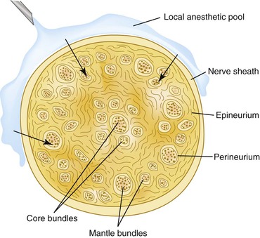
Each nerve is a cordlike structure that contains many axons. These axons are often referred to as nerve fibers. Within a nerve, each axon is surrounded by a layer of connective tissue called the endoneurium. The nerve fibers are bundled together into groups called fascicles, and each fascicle is wrapped in a layer of connective tissue called the perineurium. Finally, the entire nerve is wrapped in a layer of connective tissue called the epineurium (Figure 2-4).
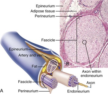
Fasciculi located in the mantle region are called mantle bundles, and fasciculi located in the core region are called core bundles. Mantle bundles are located near the outside of the nerve, and core bundles are located on the inside of the nerve. The location of the bundles in larger nerves has an impact on which bundles are affected by the local anesthetic first. The local anesthetic, when administered diffuses through the nerve to the mantle bundles (outer core) at a higher concentration first and then to the core bundles (inner core) at a more diluted concentration due to some anesthetic being absorbed by capillaries and lymphatics. Because the mantle bundles are affected by the local anesthetic first, these bundles also lose anesthesia before the core bundles (Figure 2-5).1,8
Classification of Neurons
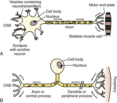
Nerves are categorized into three groups classified according to the direction that signals are conducted: Afferent nerves conduct signals from sensory neurons to the spinal cord or brain (carry toward). Efferent nerves conduct signals away from the brain or spinal cord along motor neurons to their target muscles and glands (carry away). Mixed nerves contain both afferent and efferent axons, and thus conduct both incoming sensory information and outgoing muscle commands in the same bundle. Although sensory and motor nerves are slightly different in structure, their main components of the neuron are the same, the cell body with a nucleus, axon, dendritic zone with free nerve endings (where stimulus is picked up) and terminal arborization (where the impulse is sent toward the CNS). The location of the cell body along the axon is the main difference between motor (efferent) neurons, and sensory (afferent) neurons. The cell body of the motor neuron participates in impulse conduction, and therefore is located at the terminal arborization. The cell bodies of sensory neurons do not participate in nerve conduction and therefore are located off the axon (Figure 2-6).
Functional Regions of a Sensory Neuron
There is a wide variation in the shape of sensory neurons, but they all have the same basic structures and functions (Figure 2-7).
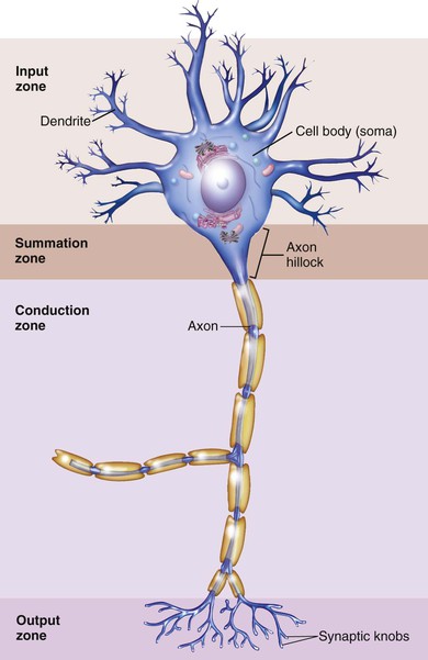
Dendritic (Input) Zone
The dentritic zone is the most distal section of the neuron and is an arborization of nerve endings. These free nerve endings respond to stimulation (e.g., bradykinin, a “pain mediator” produced during cellular injury) produced in the tissues in which they lie, initiating nerve conduction. The dendrites of a neuron are cellular extensions with many branches, and metaphorically this overall shape and structure is referred to as a dendritic tree. This is where the majority of input to the neuron occurs.3
Stay updated, free dental videos. Join our Telegram channel
VIDEdental - Online dental courses


