CHAPTER 2 Embryology and Histology
EMBRYOLOGY
Embryology is the study of prenatal development (gestation period), which begins with fertilization and continues until birth. Divided into three periods: preimplantation, embryonic, fetal.
 •
•  is recommended (see CD-ROM WebLinks).
DEVELOPMENT OF FACE AND ORAL CAVITY
Face and structures of the oral cavity begin development early in embryonic period. ALL three embryonic layers are involved in facial development (layers are discussed earlier). Depends on five facial processes (prominences) that form during fourth week and surround stomodeum (primitive mouth): (1) single frontonasal process; (2 and 3) paired maxillary processes; and (4 and 5) paired mandibular processes (Figure 2-1). In the future, the stomodeum will form the oral cavity.
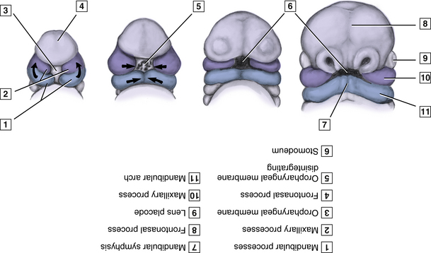
Figure 2-1 Embyro at third to fourth weeks of development.
(From Bath-Balogh M, Fehrenbach MJ: Illustrated dental embryology, histology, and anatomy, ed 2, St. Louis, 2006, Saunders/Elsevier.)
Stomodeum and Oral Cavity Formation
Before fourth week, stomodeum initially appears as a shallow depression in embryonic surface ectoderm at cephalic end, limited in depth by oropharyngeal membrane (Figure 2-1).
Mandibular Arch and Lower Face Formation
During fourth week, two bulges of tissue appear inferior to stomodeum; these paired mandibular processes are formed in part by neural crest cells that migrated to the facial region, covered externally by ectoderm and internally by endoderm (Figure 2-1).
Frontonasal Process and Upper Face Formation
During fourth week, frontonasal process is a bulge of tissue in upper facial area, at most cephalic end of embryo, which is also the cranial boundary of the stomodeum. In the future, frontonasal process will form upper face, which includes forehead, bridge of nose, primary palate, nasal septum, and all structures related to medial nasal processes (Figure 2-1).
Maxillary Process and Midface Formation
During fourth week, adjacent swellings, the maxillary processes, form from increased growth of mandibular arch. Maxillary processes grow superiorly and anteriorly on each side of stomodeum (Figure 2-1).
Branchial Apparatus Formation
Palatal Development
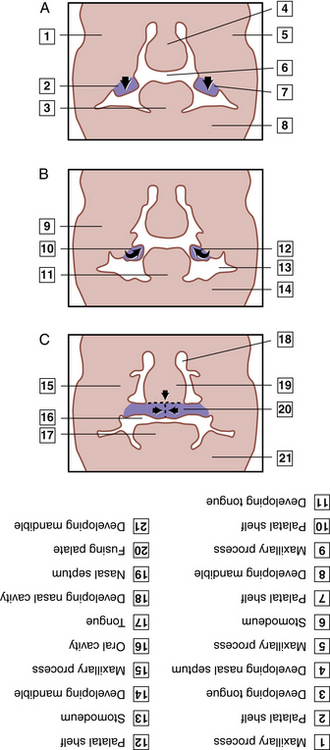
Formation of the palate starts in the embryo during fifth week, takes place during several weeks of prenatal development, and is completed in twelfth week in the fetus (Figure 2-2). Formed from two separate embryonic structures, primary palate and secondary palate. Palate is developed in three consecutive stages: formation of primary palate, formation of secondary palate, completion of final (definitive) palate.
Patient with Cleft Palate and/or Cleft Lip
CLINICAL STUDY
| Age | 21 YRS | SCENARIO |
| Sex |  Male Male  Female Female |
Upon extraoral examination during an initial appointment, the dental hygienist notes that there is a notch in the upper lip on the left side of the patient. When asked about it, the patient says it has been there since he was born. Nothing else is noted. |
| Height | 5’8” | |
| Weight | 150 LBS | |
| BP | 115/75 | |
| Chief Complaint | None | |
| Medical History | Hay fever | |
| Current Medications | OTC allergy medications prn | |
| Social History | College student in engineering |
TOOTH DEVELOPMENT
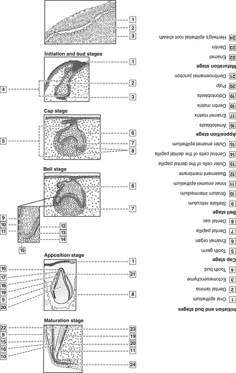
Child’s primary (deciduous) dentition develops during prenatal period and consists of 20 teeth, which erupt and are later shed (lost). As primary teeth are shed and jaws grow and mature, permanent dentition, consisting of as many as 32 teeth, gradually erupts and replaces primary dentition (Figure 2-3). Primary dentition begins on average with eruption of the mandibular central incisor at 6 to 10 months and is completed with eruption of the maxillary second molar at 25 to 33 months. Permanent dentition begins on average with the eruption of the mandibular first molar or central incisor at 6 to 7 years and is completed with the eruption of the third molars at 17 to 21 years. Discussion of tooth development will center first on the primary dentition and then include the permanent dentition.
GENERAL HISTOLOGY
 •
• Cell Structure
A cell is the smallest unit of organization in the body. Cell structure includes cell components, such as organelles, plasma, and cytoskeletal elements. All are IMPORTANT in cell function (Figure 2-4).
Stay updated, free dental videos. Join our Telegram channel
VIDEdental - Online dental courses


