Chapter 2
Diagnosis of Dental Caries
Aim
This chapter aims to update the principles of caries detection, diagnosis and record-keeping, and to give guidance on the appropriate use of adjuncts to caries diagnosis in the child and adolescent.
Outcome
On reading this chapter, the practitioner should feel more confident in caries diagnosis, especially in respect to the examination and caries risk assessment of children and be familiar with the relevant supporting theories in modern cariology.
Introduction
All children and adolescents are individuals and, as such, their dental care should be customised to their specific needs. The first stage in this process is to take a history (dental, social and medical) and complete a careful examination. The history and examination should be thorough and recorded in a systematic way, in order to gather all the available information and avoid omissions. The history must also be appropriate to the age of the child – for example, with older children it is important to take the child’s views into account, even at the expense of those of their parents or carers. The key to success is to get off on the right foot and gain the child’s trust and the parent’s confidence. Therefore, the initial greeting of the child and their carers is critical (Fig 2-1).
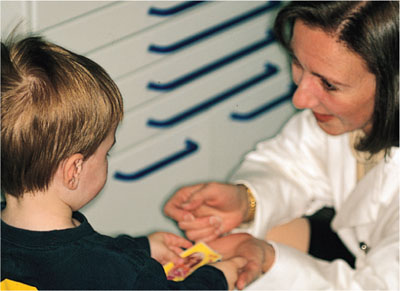
Fig 2-1 The initial greeting of the child is paramount.
The First Visit
A brief history of dental attendance should be recorded, as should the current complaint (if any). It is important that the family is made to feel welcome when they attend for care. Any implied criticism of past behaviour is likely to have a detrimental effect on future behaviour. In simple terms, child patients won’t wish to return for treatment or will be less compliant if they think they will be “told off”. History-taking helps with the assessment of the families’ attitudes to dental health and caries risk assessment (Fig 2-2).
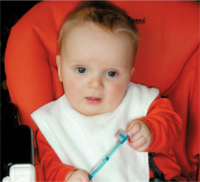
Fig 2-2 Dental care is needed as soon as the first teeth erupt.
A brief history of previous dental care is invaluable. For example, it is useful to know how well the child coped with the provision of previous restorations and local anaesthesia. The use of local anaesthesia is also an indication of the quality and prognosis of existing restorations. Such information can help with assessing the child’s ability to cope with any proposed treatment.
Examination
While talking to the child, it should quickly become apparent how agreeable to examination and treatment they are likely to be. The subsequent stages of the examination can then be tailored to the patient’s ability to cooperate. It may be that only a brief examination will be possible at the initial consultation.
Extra-oral Examination
-
Check general appearance
-
Demeanour
-
Swelling
-
Asymmetry
-
Lymphadenopathy
-
Skeletal pattern.
Intra-oral Examination
-
Soft tissues
-
Periodontal tissues
-
Plaque levels (oral hygiene)
-
Gingival bleeding (as well as being indicative of gingivitis, may indicate active approximal caries)
-
Teeth:
-
caries diagnosis
-
hypoplasia/opacities
-
-
Occlusion.
Caries Risk Assessment
A key element of the examination of a patient is the caries risk assessment. The results of this assessment should be recorded in the notes, as a patient-specific prompt, to encourage appropriate preventive management, such as the provision of fissure sealants. The recording of caries risk will also promote best practice by assisting with clinical audit.
Caries risk assessment is not a new concept to most dentists; it is something most dentists do implicitly for all patients. The step that is more novel is to make this an explicit action.
Previous Disease
Past behaviour and disease experience are one of the best predictors of future disease. Therefore, predictors of high caries activity are:
-
The presence of restorations
-
Previous extractions
-
New disease.
Dietary Factors
-
Non-intrinsic non-milk sugars in the diet cause caries.
-
Patients whose diet is high in such sugars, and particularly when these products are consumed frequently, are likely to be of high caries risk.
-
Time of consumption is also important:
-
access to any sugar at night increases caries risk,
-
milk can cause caries if a child is given free access at night, when the anti-caries benefits of saliva are reduced,
-
access to a sugary snack within an hour of bedtime in a young child is associated with increased caries risk.
-
Social Factors
Like heart disease and cancer, caries is a socio-economic disease. Therefore, in broad terms, the patient’s area post code helps predict their caries risk. For better management it is helpful to find out:
-
Who cares for the child?
-
How easy is it for them to attend?
-
Information about siblings, interests and pets helps with behaviour management by making the child feel special.
-
School performance might highlight a learning disability.
Fluoride Use and Plaque Control
-
The use of fluoride toothpaste is largely responsible for the reduction in caries prevalence seen in the Western World over the past 30 years.
-
Oral hygiene as a measure of the frequency and effectiveness of tooth-brushing (assuming a fluoride toothpaste is used) is therefore an important factor.
-
For those lucky enough to be living in fluoridated areas or with a history of use of fluoride supplements this must be included in an assessment.
Medical History
Medical history may influence caries risk in a number of ways:
-
A condition that predisposes to caries (e.g. xerostomia during chemotherapy).
-
A condition where the consequences of caries (either its treatment or sepsis) can be a threat to the patient (e.g. those at risk of infective endocarditis).
-
A condition that confers reduced ability to cooperate with dental care or to perform oral health procedures (e.g. cerebral palsy).
Saliva
Saliva plays a pivotal role in the prevention of caries, as it contains both specific and non-specific antibacterial agents such IgA, lysozyme and lactoferrin. It buffers acids and maintains the oral pH. A reduction in the production of saliva will therefore increase an individual’s caries risk. There is considerable individual variation in the quality of saliva, and although tests for the constituents of saliva and buffering capacity exist, these have a limited role at the chairside at present. However, if a dentist suspects a reduction in salivary flow this would influence a caries risk assessment.
Bacteria
Streptococcus mutans and Lactobacillus levels correlate with caries risk at a population level. Unfortunately, the levels of these bacteria are not predictive at an individual level.
Fissure Shape
Fissure shape is not a good predictor of caries risk. The identification of caries predilection sites, which includes the occlusal fissures of the permanent molars, does play an important role in caries diagnosis and prevention, but a clinician cannot reliably tell the depth or, more importantly, the shape of a fissure.
The Dentist’s Hunch
As stated earlier, most dentists make a risk assessment for their patients during the first meeting. It has been demonstrated that this assessment can be a valid predictor of risk.
Risk Categories
Generally the practitioner is advised to categorise caries risk into either high, moderate or low.
In the view of the authors, the moderate category does not add any value and it is better to use the dichotomy of “requires extra prevention” or “no intervention beyond what the patient is already doing”.
The Carious Process
In order to understand the problems associated with the diagnosis of dental caries, an understanding of the disease process is essential. This is also necessary if we are to adopt a modern biological and evidence-based approach to its prevention and management.
Coronal caries occurs on three sites:
-
Free smooth surfaces
-
Approximal surfaces
-
Pits and fissures.
Although not the only theory postulated for the aetiology of dental caries, the acidogenic theory has overwhelming evidence to support it. Miller, in 1883, concluded that caries results from decalcification caused by bacterial acid production followed by bacterial invasion and the destruction of any remaining tissues. The causes for the initiation and progression of caries are multifactorial. Over a period of time, caries can occur on a susceptible tooth surface in the presence of cariogenic bacterial plaque and bacterial substrate.
Pits and fissures are predilection sites for caries because they are stagnation areas, inaccessible to cleaning and thus the removal of bacterial plaque. Occlusal surfaces benefit less than smooth surfaces from the caries preventive action of fluoride.
The crown of a tooth consists of (from the surface inwards) enamel, dentine and the dental pulpal tissues (pulp). The dentine and pulp are often considered to be a single complex. Enamel is the most highly mineralised tissue in the human body (92% by volume; 97% by weight). The degree of mineralisation of dentine, although high, is less than that of enamel (48% by volume; 69% by weight).
Pits and fissures generally occur:
-
in the occlusal surface of the molars and premolars.
-
the palatal surface of incisors, canines and molars.
-
the buccal surface of lower molars.
The pathological features of caries in pits and fissures are similar to those seen in smooth surface caries but it is generally assumed that differences in surface form result in a later clinical (macroscopic) presentation.
Enamel Caries
In the mouth, the enamel surface is in a state of ionic flux. Below pH 5.5 mineral is eventually lost and this can lead to the formation of caries. At higher pH levels, and particularly in the presence of fluoride, this process is reversed.
White spot detection
The first visible sign of enamel caries, the white spot lesion, is relatively easily identified on free (exposed) smooth surfaces (Fig 2-3). However, it is often impossible to diagnose in pits and fissures because of surface morphology. White spot lesions are more obvious when teeth are dry. This is because of the different refractive indices of enamel, water and air. Sound enamel has a refractive index of 1.62. When demineralised, enamel becomes porous, if the teeth are wet the lesion has a refractive index approaching that of water (1.33) and will appear opaque compared to sound tissue. If dried, the water in the pores is replaced with air of refractive index 1.0 and the lesion becomes more obvious. To detect white spot lesions, teeth should be clean and dry.
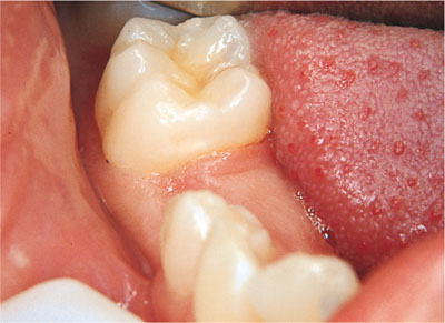
Fig 2-3 White spot.
Microscopy of a white spot
In ground section the established white spot lesion can be described as having four zones, the optical properties of which reflect differing degrees of mineralisation and lesion activity (Fig 2-4):
-
Zone 1 – The translucent zone is the first recognisable histological change at the advancing edge of the lesion.
-
Zone 2 – The dark zone is the second recognisable histological change. It is thought that the dark zone is na/>
Stay updated, free dental videos. Join our Telegram channel
VIDEdental - Online dental courses


