SECTION 2
DENTAL RADIOGRAPHY
Radiographs are important tools in the diagnosing of dental disease. There are many types of radiographs available, all of which are used for different purposes. There are two main types of dental radiographic films: intra-oral and extra-oral.
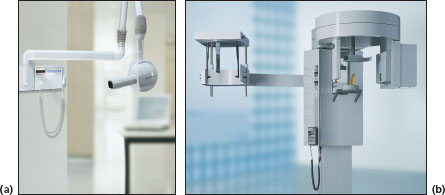
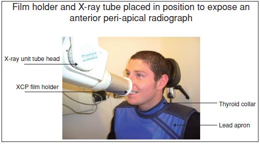
FIGURE 2.1a, b
Name
(a) Intra-oral X-ray machine (b) Extra-oral X-ray machine
Functions
- Intra-oral X-ray machines are used for exposing occlusal, peri-apical and bite-wing radiographs
- Extra-oral X-ray machines are used for exposing panoramic/OPG (orthopantomograph) radiographs and cephalometric radiographs
Varieties
Machines from different manufacturers may vary in design
FIGURE 2.2
Name
Lead apron and thyroid collar
Function and precautions
- A lead apron and thyroid collar must be used to protect the patient from radiation during X-ray procedures
- The lead apron is used with a thyroid collar that must cover the radio-sensitive tissues (from the thyroid downwards and including the lap area)
- The lead apron must be hung up, not folded because folding the apron will cause the lead inside to crack, causing radiation to pass through
Varieties
Different styles available from various manufacturers
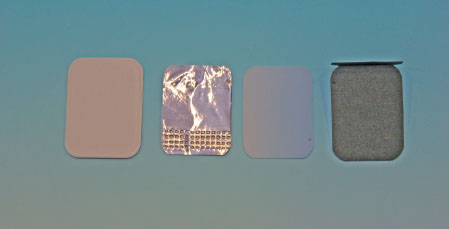
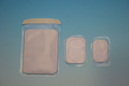
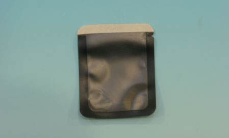
CONVENTIONAL INTRA-ORAL RADIOGRAPH FILM
FIGURES 2.3; 2.4
- A detailed radiograph film, which is exposed while in the patient’s mouth
- Used in conjunction with a film holder for accurate placement
- Some films may be covered in a clear plastic sleeve for infection control and prevention purposes (see Figure 2.4)
- Every film has a bump to assist in film orientation
- The bump always faces towards the X-ray tube
- Inside the film packet:
- Black paper on either side of film to protect film from light (see Figure 2.3)
- On the back of the film there is a thin sheet of lead foil to absorb stray/scatter radiation
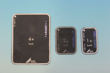
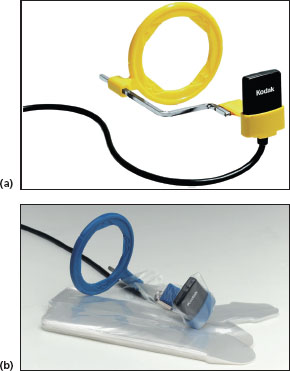
INDIRECT DIGITAL RADIOGRAPH FILM – PHOSPHOR PLATES
FIGURES 2.5; 2.6
- Phosphor plates are thinner than digital sensors and can be used with the extension cone paralleling (XCP) holders
- After exposure using a phosphor plate it is loaded into a scanner allows the operator to view the radiograph digitally
- Once the image is produced the phosphor plate is ‘wiped clean’ for reuse using a single use plastic sleeve for infection control and prevention
- A detailed radiograph film, which is exposed while in the patient’s mouth
- Used in conjunction with a film holder for accurate placement
- Phosphor plates are always covered in a plastic sleeve for infection prevention purposes (Figure 2.6)
- Every film has a bump to assist in film orientation
- The bump always faces towards the X-ray tube