2
Anatomy of the trigeminal nerve
The trigeminal nerve is the fifth cranial nerve (n. V), which plays an important role in the innervation of the head and neck area, together with other cranial and spinal nerves. Knowledge of the nerve’s anatomy is very important for the correct application of local anaesthetics.
2.1 Introduction
The trigeminal nerve contains a large number of sensory (afferent) and motor (efferent) neurons. The sensory fibres carry nerve impulses towards the central nervous system, while the motor fibres carry impulses away from the central nervous system. The trigeminal nerve has a wide innervation area (Figure 2.1). The nerve provides the sensitivity of the dentition, the mucosa of the mouth, nose and paranasal sinuses, and the facial skin. The nerve also contains motor fibres that innervate, among others, the masticatory muscles. Although the trigeminal nerve is the most important nerve for the sensory and motor innervation of the oral system, the facial (n. VII), glossopharyngeal (n. IX), vagus (n. X), accessory (n. XI) and hypoglossal (n. XII) nerves are also of significance. The n. VII, n. IX and n. X, for example, take care of the taste sense, the n. IX and n. X provide the general sensation (pain, touch and temperature) to the pharynx, soft palate and the back of the tongue, whilst the n. XII is responsible for the motor innervation of the tongue. Although these latter nerves do play an important role in innervating the oral cavity, they will only be mentioned marginally in this book.
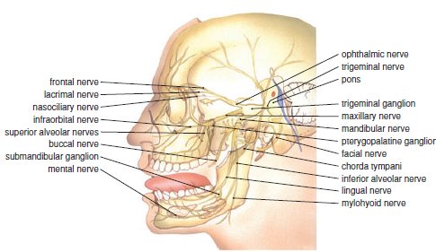
Figure 2.1 Overview of the trigeminal nerve (lateral view).
2.2 The central part of the trigeminal nerve
2.2.1 Origin
The trigeminal nerve emerges from the middle of the pons, at the lateral surface of the brainstem. The nerve consists of two parts here: the sensory fibres form a thick root and the motor fibres form the much thinner motor root. These two roots run to the front of the petrous part of the temporal bone where the large sensory trigeminal ganglion (semilunar or Gasserian ganglion) lies in a shallow groove surrounded by dura mater.
The trigeminal ganglion is formed by the aggregation of cell bodies of sensory neurons. Three main branches of the trigeminal nerve emerge from the ganglion: the ophthalmic nerve (n. V1), the maxillary nerve (n. V2) and the mandibular nerve (n. V3). The motor root joins the mandibular nerve only, once it has exited the skull via the foramen ovale. The sensory areas covered by the three main branches are generally as follows:
- The ophthalmic nerve carries sensory information from the skin of the forehead, the upper eyelids and the nose ridge, and part of the nasal mucosa.
- The maxillary nerve innervates the skin of the middle facial area, the side of the nose and the lower eyelids, the maxillary dentition, part of the nasal mucosa (including the maxillary sinus) and the palate.
- The mandibular nerve innervates the skin of the lower facial area, the mandibular dentition, the mucosa of the lower lip, cheeks and floor of the mouth, part of the tongue and part of the external ear.
Of all the areas that the trigeminal nerve innervates, the oral cavity is the most enriched with sensory neurons. The density of sensory neurons in the mouth is much larger than in any other area, e.g. the facial skin. This density of sensory neurons increases from the back to the frontal area of the mouth.
Most of the trigeminal ganglion neurons are pseudo-unipolar. This means that each neuron in the ganglion has a peripheral and a central process. The peripheral process (axon) is relatively long and carries the impulses coming from sensory receptors (Box 2.1). The central process (dendrite) is short, and enters the pons and synapses with the sensory trigeminal nucleus situated in the brainstem. The proprioceptive fibres in the trigeminal ganglion are an exception. Their cell bodies are not situated in this ganglion but in the mesencephalic nucleus of the trigeminal nerve. The proprioceptive fibres are found in the motor root of the trigeminal nerve and carry impulses from, among others, muscle spindles of the masticatory muscles.
The trigeminal ganglion has somatotopy. This means that the neurons in the ganglion are arranged in the same order as the areas that are innervated by the three main branches of the trigeminal nerve. The cell bodies of the ophthalmic nerve are grouped medially in the ganglion, while those of the mandibular nerve are grouped laterally. In the middle of these two groups the cell bodies of the maxillary nerve can be found.
2.2.2 Trigeminal nuclei
The trigeminal nerve has a sensory and motor nucleus within the brainstem (Figure 2.2). The sensory nucleus lies laterally and most of the sensory neurons of the trigeminal nerve contact (synapse) with the neurons in this nucleus. The nucleus forms a long column that extends from the midbrain to the spinal cord. It consists of (from cranial to caudal) the mesencephalic, the principal and the spinal trigeminal nuclei.
Proprioceptive information from, for example, the masticatory muscles is managed in the mesencephalic nucleus. The principal trigeminal nucleus mainly receives touch and pressure impulses from the entire oral area, whereas the spinal trigeminal nucleus receives information on pain, temperature and pressure from the entire trigeminal area. All information received via the sensory trigeminal nuclei is managed and integrated in, among others, the thalamus via ascending paths. After this the information is brought to various areas of the cerebral cortex, where perception occurs.
Figure 2.2 The motor and sensory nuclei of the third to twelfth cranial nerve in the brainstem (lateral view). The sensory nuclei are shown in blue, the motor nuclei in red.
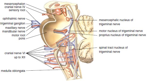
The motor neurons of the trigeminal nerve are grouped in a motor nucleus that lies medially to the sensory nucleus in the centre of the pons. The axons of these motor neurons run to (among others) the masticatory muscles. As previously described, these axons pass the trigeminal ganglion as an independent bundle (motor root), without synapsing within it. Similar to the motor neurons in the spinal column, the motor neurons in the motor trigeminal nucleus are directly stimulated via the corticobulbar tract, originating from the contralateral cerebral cortex. Within the motor nucleus there is a large amount of somatotopy, i.e. the motor neurons that innervate the different muscles are grouped together. Via fibres coming from the sensory mesencephalic nucleus, the motor trigeminal nucleus receives proprioceptive information from the masticatory muscles, temporomandibular joint and periodontium.
2.3 The peripheral part of the trigeminal nerve
2.3.1 Ophthalmic nerve
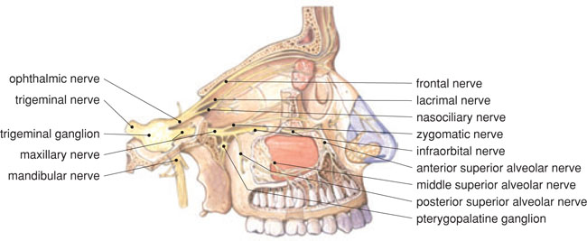
The ophthalmic nerve (n. V1) enters the orbit (Figure 2.3). This nerve carries only sensory fibres and, just before leaving the cranial cavity through the superior orbital fissure, it branches off into three: the nasociliary, frontal and lacrimal nerves. These nerves run in the roof of the orbit and are involved in the sensory innervation of a large number of structures, such as the mucosa of the nasal cavity and the sphenoid and ethmoid sinus, the skin of the nose ridge, the upper eyelid and forehead, and the mucosa that covers the eyeball and inside of the eyelids.
Figure 2.3 The branching of the ophthalmic and maxillary nerves (lateral view).
2.3.2 Maxillary nerve
The maxillary nerve (n. V2), too, is solely sensory. It enters the pterygopalatine fossa (see Section 2.4.1) via the foramen rotundum (Figure 2.3). Through the inferior orbital fissure it reaches the floor of the orbit and proceeds there as the infraorbital nerve, first in the infraorbital sulcus and then in the infraorbital canal. It then reaches the face via the infraorbital foramen.
Within the pterygopalatine fossa the maxillary nerve is connected via a number of branches to the upper side of the parasympatheti/>
Stay updated, free dental videos. Join our Telegram channel
VIDEdental - Online dental courses


