Temporomandibular Joint
Learning Objectives
• Define and pronounce the key terms in this chapter.
• Outline the movements of the temporomandibular joint as well as demonstrating them.
New Key Terms
Articular eminence (ar-tik-you-ler), fossa
Depression of the mandible (de-presh-in)
Disc of the joint (el-eh-vay-shun)
Elevation of the mandible (el-eh-vay-shun)
Joint capsule (de-vee-ay-shun)
Lateral deviation of the mandible (de-vee-ay-shun)
Muscles of mastication (mass-ti-kay-shin)
Postglenoid process (post-glen-oid)
Protrusion of the mandible (pro-troo-zhin)
Retraction of the mandible (re-trak- shun)
Subluxation (sub-luk-say-shun)
Synovial (sy-no-vee-al) fluid, cavities, membrane
Temporomandibular disorder (tem-poh-ro- man-dib-you-lar)
Temporomandibular Joint
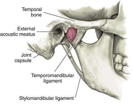
The temporomandibular joint (TMJ) is a joint on each side of the head that allows for movement of the mandible for mastication, speech, and respiratory movements; it is the most complex set of joints in the body. The TMJ can be palpated just anterior to each ear (see Figure 1-3).
Patients may have a disorder associated with the TMJ (discussed later). Thus, dental professionals must understand the anatomy, histology, and normal movements of the TMJ before being able to understand any possible disorders associated with the joint.
The TMJ develops in the eleventh to twelfth week of prenatal development, during the proliferation of the associated ligaments, muscles, and bones of the joint, as well as the joint spaces and articular disc.
Bones of the Joint
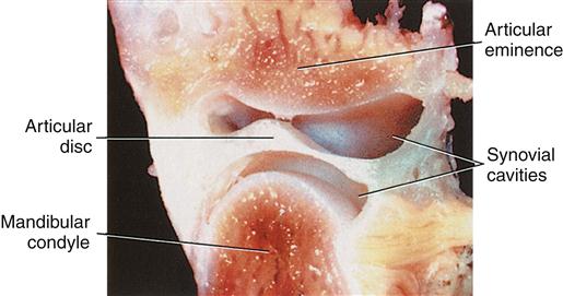
The TMJ is the articulation of the temporal bone and the mandible on each side of the head (Figure 19-1). Knowing the basic anatomy of the bones is necessary, as well as the histology and actions of the TMJ.
Temporal Bone
The articulating area on the temporal bone of the TMJ is located on the bone’s inferior aspect (Figure 19-2). This articulating area includes the bone’s articular eminence and the articular fossa. The articular eminence is positioned anterior to the articular fossa, and consists of a smooth, rounded protuberance on the inferior aspect of the zygomatic process.
The articular fossa, or mandibular fossa, is posterior to the articular eminence, and consists of a depression on the inferior aspect of the temporal bone, posterior and medial to the zygomatic arch (see Figure 1-3). Posterior to the articular fossa is a sharper ridge, the postglenoid process.
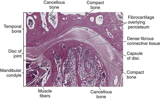
The temporal bone consists of compact bone overlying cancellous bone (Figure 19-3; see Chapter 8). The outermost surface of compact bone is covered by periosteum. Like all bones, the innermost part of the bone consists of endosteum and the medullary cavity with its bone marrow. The articulating bony surface of the joint is covered by fibrocartilage immediately overlying the periosteum.
Mandible
The mandible articulates with each temporal bone at the heads of the mandibular condyle, with their articulating surface of the condyle, which has histology similar to the articulating surface of the temporal bone. In a mature adult, each condyle consists of compact bone overlying cancellous bone (see Figure 19-2). Periosteum overlies the compact bone of the condyle, and the endosteum and bone marrow are located on the innermost part of the bone. Fibrocartilage then overlies the periosteum.
However, in contrast to the articulating surface of the temporal bone, a growth center is located in the head of each mandibular condyle before an individual reaches maturity (Figure 19-4). This growth center consists of hyaline cartilage underneath the periosteum on the articulating surface of the condyle. This is the last growth center of bone in the body and is multidirectional in its growth capacity, unlike a typical long bone.
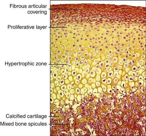
This area of cartilage within the bone grows in length by appositional growth as the individual grows to maturity/>
Stay updated, free dental videos. Join our Telegram channel
VIDEdental - Online dental courses


