19: Single-Tooth All-Ceramic Restorations

Chapter 19 Single-Tooth All-Ceramic Restorations

Brief History of the Clinical Development and Evolution of All-Ceramic Single-Tooth Restorations

Before the development of the all-ceramic restoration, crowns made of gold or gold alloys with an acrylic or porcelain facing were considered state of the art. Esthetics were lacking, as the resulting restorations were a far cry from the look of the natural dentition.

The porcelain jacket crown (PJC) was the first tooth-colored full-coverage restoration. Introduced by Dr. Charles H. Land in 1903, the porcelain jacket was made with feldspathic porcelain clay layers successively fired over platinum foil. The finished PJC was luted to the tooth using zinc phosphate cement. Although it could fulfill some of the esthetic drawbacks of its predecessor, it was not without its shortcomings. First, the removal of the platinum foil after the crown was fired meant that there was always a substantial gap left at the margin from which the zinc phosphate cement could leach out, leaving the tooth prone to caries. Second, the porcelain tended to be too opaque to match the surrounding teeth. Finally, this old-fashioned porcelain crown inherently lacked robust physical properties and strength. PJCs were definitely contraindicated for posterior teeth, as they were prone to failure even on anterior teeth.

Early PJCs tended to fail owing to the microfractures that occurred on the internal surface. The answer to this problem first appeared in the early 1950s with renewing interest in the PFM crown. Because the internal surface of the porcelain is bonded to a metal coping in PFM restorations, the metal-porcelain bond prevents the stress cracks from developing. In the 1960s the aluminous core PJC was developed. This crown was made on a refractory die rather than on platinum foil.

PFM restorations gained popularity exponentially and are to this day the most widely used type of single-tooth full-coverage restoration. The main disadvantage of the metal ceramic restoration still appears to be the ability of the overlying porcelain to mask the underlying metal, especially in porcelain-to–metal collar and porcelain-to-margin (combination) types. This esthetic barrier resulted in a burst of research and development in ceramic technology for anterior restorations.

In the 1970s this research culminated in the development of the collarless ceramometal crown and porcelain shoulder (butt) margins. The 1980s brought developments in the posterior dentition for single-unit crowns. These included early glass ceramics such as Dicor (DENTSPLY Prosthetics, York, Pennsylvania) and Cerestore (Johnson & Johnson Professionals, Inc., East Windsor, New Jersey). Although these were fairly good restorations, they were all conventionally cemented because resin bonding and resin cements were in early development. One of the first resin cements was Resiment (JL Blosser, Inc., Liberty, Missouri), an auto-curing, filled, multipurpose cement.

An early development was VITA In-Ceram ALUMINA (glass-infiltrated alumina crowns) (VITA Zahnfabrik, Bad Säckingen, Germany). These had a very hard aluminous core, creating challenges for the dentist if one had to be removed. These crowns tended to be a bit opaque, as the layering porcelain had the dubious task of hiding the opacious coping.

This paved the way for VITA In-Ceram SPINELL (glass-infiltrated magnesium alumina crowns), which sacrificed some physical properties in flexural strength and hardness in order to produce a coping with greater translucency.

These were followed by the high-strength VITA In-Ceram ZIRCONIA (glass-infiltrated alumina with partially stabilized zirconia) crowns, which failed esthetically but were useful as high-strength posterior crowns and bridge abutments.

From the early 1990s, various leucite-reinforced glass ceramic (LRGC) materials offered anterior single-unit esthetics using either the staining or the cut-back and layering technique. Empress (Ivoclar Vivadent, Amherst, New York) pressed-leucite porcelain was the most esthetic material, but it was typically a weak material when used in the posterior region, especially in the case of a full-coverage restoration. If this material was used, the restorations were typically stained and not layered.

Among the indirect resins tried in the posterior area were materials such as Concept (Ivoclar Vivadent), Artglass (Heraeus Kulzer, Hanau, Germany), Targis and Vectris (Ivoclar Vivadent), belleGlass (Kerr Corporation, Orange, California), Sinfony (3M ESPE, St Paul, Minnesota), and Cristobal (DENTSPLY Prosthetics) that were never really meant to be used for full-crown restorations. Although attempts were made, the results were quite disastrous, with severe wear being the main instigator of functional failure. These crowns typically lasted 5 years or less at best.

Better esthetics with greater strength was achieved with the development of Empress 2 (Ivoclar Vivadent), although this initially met with disastrous results because of the difficulty in getting the two coefficients to match the thermal expansion coefficient of the layering porcelain made of fluorapatite. In these restorations ceramists stacked layering powder-liquid ceramic, but cracking was a problem. This led to reworking the lithium disilicate–fluorapatite formula and renaming it Eris.

The Procera materials (Nobel Biocare, Yorba Linda, California), which are polycrystalline ceramics, were the next thing developed. With the very hard Procera cores, the color is opacious white, almost too white. Trying to hide the white core with the layering ceramic became a challenge.

Zirconia core restorations such as Lava (3M ESPE) were developed in response to the demand for an all-ceramic framework for fixed partial dentures (FPDs) in addition to the single-unit crowns. These zirconia restorations have an infrastructure that is designed using conventional waxing techniques or computer-aided design (CAD) technology. The infrastructure is milled from yttria-stabilized tetragonal zirconia polycrystal (Y-TZP) blanks using computer-aided manufacturing (CAM), after which the layering ceramic is stacked on to build the crown.

Lithium disilicate, Empress 2, has been re-introduced as e.max (Ivoclar Vivadent) and is available either in a pressable or a CAD-CAM form. This seems to be the ideal material because instead of cutting back the core and laying powder or liquid ceramic on top, the preparation is milled to anatomic form and stained, making this an extremely hard restoration. The whole crown is made up of lithium disilicate, which makes it monolithic rather than a bilayer ceramic, and accounts for its improved strength and esthetics. Although it can be cemented conventionally, the lithium disilicate restoration can also be bonded to the tooth with resin cement.

Material Options

All-purpose feldspathic porcelains are mainly used with PFM restorations and ceramic core–based all-ceramic restorations as the layering ceramic in a bi-layer restoration. They may be used to create porcelain veneers, inlays, and onlays as well.

The three main groups of materials used in the construction of an all-ceramic core are pressable glass ceramic, glass-infiltrated ceramic, and polycrystalline ceramic.

Pressable Glass Ceramics

Pressable glass ceramics are composed of two main groups: the LRGCs and the lithium disilicate glass ceramics (LDGCs). They both contain a fluid glassy phase and crystalline components.

LRGC restorations such as IPS Empress (Ivoclar Vivadent), OPC (Pentron Ceramics Inc., Somerset, New Jersey), Finesse All-Ceramic (DENTSPLY Prosthetics), and Authentic porcelain (Jensen Dental, North Haven, Connecticut) have been in use for over 20 years. LRGCs are highly translucent so they are good for esthetic restorations. Another advantage is the ability to wax the restoration to full contour, check the shape and occlusion, and invest and press it to form, resulting in a restoration that covers the site and can be bonded. The restoration is natural looking with accurate margins. Copings can be fabricated by using either a lost wax–heat pressing technique or CAD-CAM technology.

The disadvantage of this material is its inability to hide a dark discolored core. Because of the material’s high translucency, a discolored tooth, metal core buildup, or implant abutment will alter the final shade of the restoration. Anterior crowns made from a core material of this type have shown exceptional success rates. The flexural strength has been measured at 105 to 120 MPa, and the fracture toughness is 1.5 to 1.7 MPa m0.5.

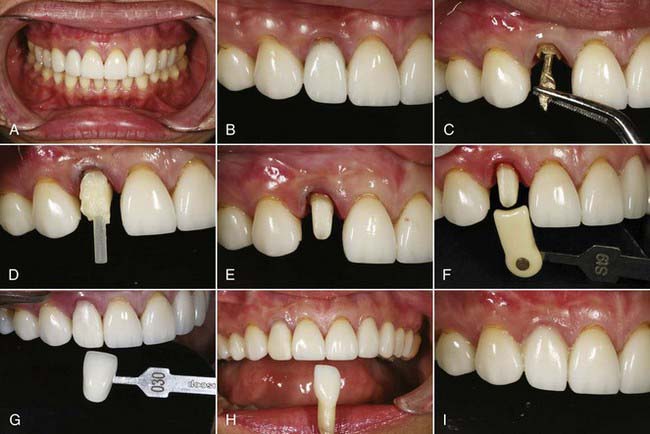
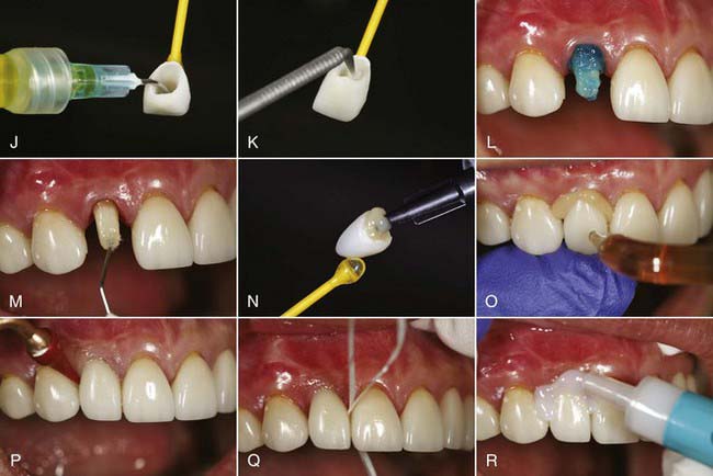
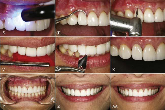
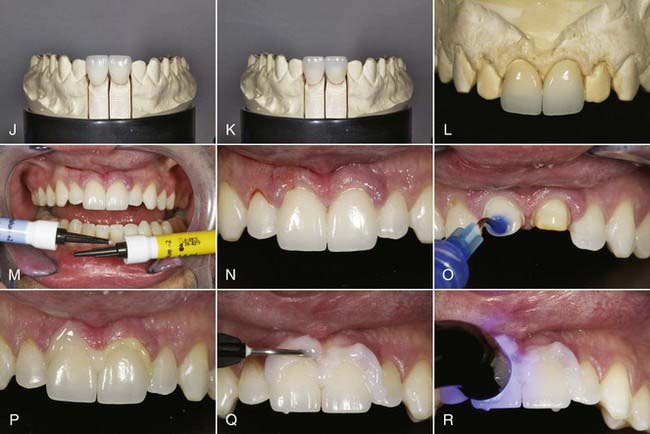
Materials such as these, with low strength, are best used in the anterior region. The strength of these restorations is supplemented by etching the internal surfaces with hydrofluoric acid, silanating, and bonding to treated tooth structure with resin cements (Figure 19-1).

image image image

FIGURE 19-1 Fiber post, composite core, and Empress pressed-leucite porcelain crown used to match existing veneers. A, Retracted pre-operative view of discolored tooth No. 7. Anterior restoration is 10 years old. B, Close-up pre-operative view showing shine-through of metal post and microleakage. C, Removal of gold cast post and core after crown removal. D, Composite core buildup around cemented fiber post. E, Finished post and buildup and tooth preparation. F, Preparation shade selected after the acrylic provisional has been placed. G, The final shade of the crown is selected. H, The shade of the new crown is checked on the preparation die against the anterior teeth. I, Try-in gel used to check the shade of the new restoration. J, Hydrofluoric acid porcelain etch is used on the intaglio surface of the crown for 15 seconds and then rinsed off. K, Silane coupling agent is applied to etched porcelain for 1 minute and then dried. L, The tooth is etched for 15 seconds with 35% phosphoric acid. M, Bonding agent is applied to the moist tooth surface. N, Light-cured resin cement is loaded into the crown. O, The crown is seated and spot tacked in the center of the facial surface with a 2-mm tacking tip for 1 second. P, Excess cement removed from the margins with a rubber tip. Q, The contacts are flossed and the floss pulled through to the lingual. R, Glycerin is placed around the margins to ensure curing of the oxygen inhibition layer. S, The crown is light cured into place for 20 seconds per surface. T, A scaler is used to remove excess cured cement. U, A 32-bladed finishing bur is used to remove cement from the margins. V, The bite is checked with articulation paper. W, Contact must be light, and one should be able to drag shim stock through when in maximum intercuspation. X, Close-up postoperative view of the completed Empress crown. Y, Retracted postoperative view of the leucite crown restoration on tooth No. 7. Z, Pre-operative smile. Note the protected smile and asymmetrical upper lip dynamics. AA, Postoperative smile. Note the fuller smile and symmetrical upper lip dynamics.

LDGC restorations such as IPS Empress II, IPS Eris, and IPS e.max (Ivoclar Vivadent) have been in use since 1999. LDGCs are recommended for anterior and posterior crowns as well as for three-unit FPDs from the second premolar and forward. These materials are not recommended for posterior bridges with a molar abutment. Flexural strength is about 360 MPa for e.max CAD and closer to 400 MPa for e.max Press. Fracture toughness is about 2.25 MPa m0.5 for e.max CAD and closer to 2.75 MPa m0.5 for e.max Press.

Once again, the core is fabricated with lost wax and heat-pressure techniques. These restorations are etched with hydrofluoric acid and adhesively cemented, although they are strong enough to be conventionally cemented—another advantage.

In using LDGC to fabricate a bridge, large connectors are needed between the abutments and the pontic tooth in both facial-lingual and incisal-cervical orientations. This material is not to be used for anterior bridgework when a 4.0-mm × 4.0-mm connector is not possible.

The earlier core materials made from lithium disilicate were originally not as translucent as the LRGCs. Both materials can be fabricated to full anatomic contour and characterized with surface stains, or the cores can be cut back and layered with feldspathic “effect” porcelains.

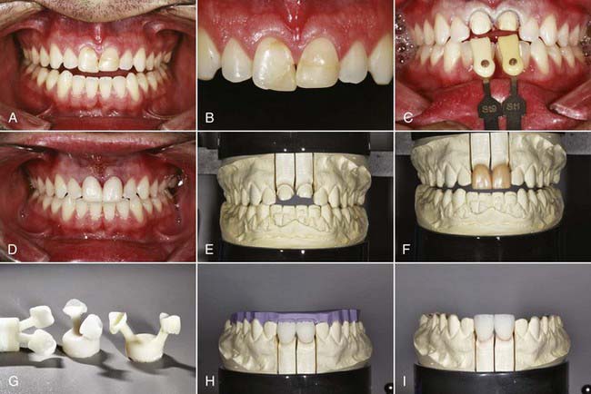
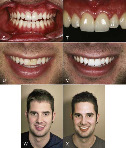
Like the LRGCs, the LDGCs have many indications and uses. The pressable lithium disilicate material (IPS e.max Press) is indicated for inlays, onlays, partial crowns, thin veneers, veneers, anterior and posterior crowns, three-unit anterior bridges, three-unit premolar bridges, telescope primary crowns, and implant restorations. In some cases in which minimal or no tooth preparation is desired (e.g., thin veneers), laboratories are able to press restorations as thin as 0.3 mm while still ensuring a strength of 400 MPa. Indications for the machinable lithium disilicate material (IPS e.max CAD) are inlays, onlays, veneers, partial crowns, anterior and posterior crowns, telescope primary crowns, and implant restorations. For a posterior crown fabricated to full contour using CAD methods, lithium disilicate offers 360 MPa of strength through the entire restoration. As a result, restorations demonstrate a “monolithic” strength unlike any other metal or metal-free restoration. With a flexural strength four times greater and a fracture toughness almost two times greater than those of LRGC, it is no wonder that use of LDGC core material in both its pressable and CAD-CAM versions (IPS e.max Press, IPS e.max CAD [Ivoclar Vivadent]) has grown in popularity (Figure 19-2).

image image image

FIGURE 19-2 e.max lithium disilicate crowns used for anterior restorations. A, Retracted pre-operative view. B, Close-up pre-operative view. C, View of teeth Nos. 8 and 9 prepared for crown restorations. D, The teeth provisionalized with acrylic crowns made from a pre-operative PVS impression and cemented. E, Model of the preparations. F, View of the full-contour wax-ups of the anticipated restorations. G, The individual wax patterns sprued and invested. H, Cut-backs are completed after the fit of the restorations was verified and the incisal matrix is placed on the model to verify the proper reduction. I, All contours are shaped (using a variety of diamond burs), and the surface anatomy and morphology added to make the restorations blend with the surrounding teeth. J, Small amounts of stain are applied where needed, and the restorations are fired. K, After the restorations have been fitted back on the solid model, contacts are checked, full embrasures are confirmed, and No. 2 pumice is used to finish the surface to the desired satin luster and reflectivity. L, View of the completed restorations on the master model at the dentist’s office. M, The appropriate shade of luting cement is chosen. N, The restorations are tried in with glycerin try-in gel. O, The preparations are etched with 35% phosphoric acid. P, The crowns are seated, and excess cement is allowed to escape from the margins before cleanup. Q, Glycerin is placed around the margins to ensure curing of the oxygen inhibition layer. R, The crown restorations are light cured into place. S, Retracted postoperative view of the lithium disilicate crown restorations on teeth Nos. 8 and 9. T, Close-up pre-operative view of the completed restorations. U, Pre-operative smile/>

Stay updated, free dental videos. Join our Telegram channel

Jan 3, 2015 | Posted by in Esthetic Dentristry | Comments Off on 19: Single-Tooth All-Ceramic Restorations

VIDEdental - Online dental courses

Get VIDEdental app for watching clinical videos