Use of Articulators in Occlusal Therapy
“THE ARTICULATOR: A TOOL, NOT THE ANSWER.”
—JPO
A DENTAL ARTICULATOR IS AN instrument that duplicates certain important diagnostic and border movements of the mandible. There are many different types of articulators and each is designed to serve the needs that the inventor believed were the most important for a particular use. With the enormous range of opinions and uses, dozens of articulators have been developed over the years. The instrument is certainly a valuable aid in occlusal therapy; however, it should always be considered as merely an aid and not, by any means, as a form of treatment. It can help accumulate information and, when properly used, will assist in some treatment methods. It cannot, however, give back proper information without proper handling by the operator. In other words, only when the operator has a thorough understanding of the capabilities, advantages, disadvantages, and uses of the articulator can the instrument become maximally beneficial in occlusal therapy.
Uses of the Articulator
The dental articulator can be helpful in many aspects of dentistry. In conjunction with accurate diagnostic casts that have been properly mounted, it may be used in diagnosis, treatment planning, and treatment.
In Diagnosis
Occlusal therapy involves two important phases: diagnosis and treatment. Because diagnosis always precedes and dictates the plan of treatment, it must be both thorough and accurate. Building a treatment plan on an inaccurate diagnosis will certainly lead to treatment failure.
Establishing an accurate diagnosis can be difficult because of the complex interrelationships between various structures of the masticatory system. To arrive at an accurate diagnosis, it is essential that all the needed information be collected and analyzed (Chapter 9). There are times during an occlusal examination when it may be necessary to evaluate the occlusal condition more closely. This is especially appropriate when a strong suspicion exists that the occlusal condition may be contributing significantly to the disorder or when the condition of the dentition strongly suggests the need for occlusal therapy. When these conditions are present, diagnostic casts should be accurately mounted on an articulator to assist in evaluating the occlusal condition. The casts are mounted in the musculoskeletally stable position (centric relation, or CR), so that the full range of border movements can be evaluated. If they are mounted in the intercuspation position and the patient has a CR-to-ICP slide, the more superoposterior position of the condyles cannot be located on the articulator and the occlusal conditions in this position cannot be properly evaluated.
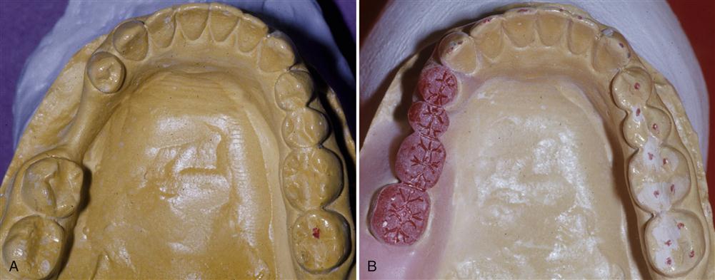
Mounted diagnostic casts offer two major advantages in diagnosis. First, they improve the visualization of both static and functional interrelationships of the teeth. This is especially helpful in the second molar region, where the soft tissues of the cheek and tongue often prevent good visibility. They also allow lingual examination of the patient’s occlusion, which cannot be viewed clinically (Figure 18-1). Often this is essential in examining the static and dynamic functional relationships of the teeth. The second advantage of mounted diagnostic casts involves the ease of mandibular movement. On the articulator a patient’s mandibular movements and resultant occlusal contacts can be observed without the influence of the neuromuscular system. Often when a patient is examined clinically, the protective reflexes of the neuromuscular system avoid damaging contacts. As a result interferences can go unnoticed and therefore undiagnosed. When the mounted diagnostic casts are occluded, these contacts become evident (Figure 18-2). Thus the casts can assist in a more thorough occlusal examination.
As has been emphasized in this text, however, the occlusal examination alone is not diagnostic of a disorder. The significance of the occlusal findings must be ascertained. Nevertheless, information received from properly mounted diagnostic casts can provide information useful for establishing an accurate diagnosis.
In Treatment Planning
The best method of providing treatment is to develop a plan that not only eliminates the etiologic factors that have been identified but does so in a logical and orderly manner. Sometimes it is difficult to examine a patient clinically and determine the outcome of a particular treatment. Yet it is essential that the final results of the treatment as well as each step needed to accomplish the treatment goals be visualized before treatment begins. When this is not possible, properly mounted diagnostic casts can become an important part of treatment planning. Diagnostic casts are used to assure that successful treatment will be achieved, and they can be employed in several ways depending on the treatment in question.
Selective grinding
Frequently it is difficult to examine a patient clinically and determine whether a selective grinding procedure can be accomplished without damage to the teeth. If a quick judgment is incorrect, the dentist may have ground through enamel and subjected the patient to an unplanned restorative procedure. In patients in whom the success of selective grinding is difficult to predict, the procedure is completed on properly mounted diagnostic casts and the end result visualized. When extensive tooth structure must be removed to meet the treatment’s goals, the patient can be informed in advance that additional treatment (e.g., crowns, orthodontics) will be needed and the additional expense discussed. This kind of planning builds the patient’s trust instead of eliciting doubt or disappointment.
Functional (diagnostic) prewax
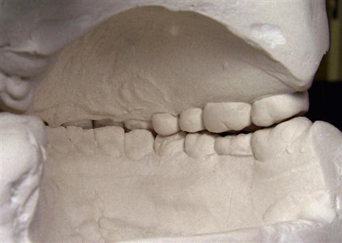
Often badly broken down or missing teeth require crowns or fixed prosthodontic procedures to restore normal function and occlusal stability. In some instances it is difficult to visualize exactly how the restorations should be designed to fulfill the treatment goals. Mounted diagnostic casts are useful in determining the feasibility of altering the functional relationship of the teeth as well as improving the selection of a method to accomplish the treatment goals. As with selective grinding, the suggested treatment is completed on the casts. A functional prewax is developed that fulfills the treatment goals (Figure 18-3). While the prewax is being fabricated, a proper design is developed that will be most appropriate for the specific situations encountered. The prewax will not only allow visualization of the expected final treatment but also give insight into any problems that may be encountered while reaching that goal. After it is completed, the treatment can begin with greater assurance of success.
Esthetic (diagnostic) prewax
It is very discouraging when time and money have been invested in the fabrication of anterior crowns or a fixed prosthesis and then the patient is not pleased with the esthetic results. Preexisting conditions must be examined carefully so the effects on the final esthetics of a restoration can be determined. Unusual interdental spacing, tissue morphology, or occlusion will often alter the final appearance of a crown or fixed prosthesis. If the final esthetics cannot be visualized because of unusual preexisting conditions, an esthetic prewax is indicated. This allows visualization of the most esthetically pleasing results achievable and gives the dentist and patient an idea of what can be expected when the final treatment is completed (Figure 18-4).
If, during the prewax, it becomes apparent that the esthetic results are undesirable, other types of treatments in conjunction with the fixed prosthesis may be needed. This may include dental implants, orthodontics, periodontics, endodontics, or a removable partial denture. After an aesthetic result has been achieved, both the dentist and the patient can visualize the expected appearance of the new restoration. The patient’s expectations now become realistic, which will minimize any disappointment. Treatment can begin with greater assurance of success.
Orthodontic setup
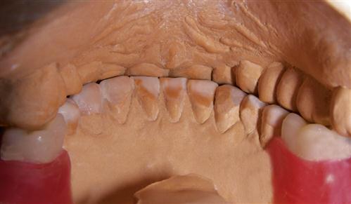
Malalignment of the dental arches is usually treated more appropriately by orthodontics. In simple, routine cases, orthodontic results can easily be predicted. However, on occasion, a particular alignment problem or crowding of the teeth may pose difficulty in visualizing the final results. When this condition exists, mounted diagnostic casts are very helpful. The teeth can be sectioned from the casts and repositioned in wax so that doctor and patient can visualize the expected final result (Figure 18-5). This also provides the doctor with insight as to any unexpected problems that may arise during the orthodontic therapy.
When extraction is being considered, the teeth to be removed are left out of the setup. The orthodontic results to be achieved by extraction can then be compared with the nonextraction results. The most appropriate treatment is selected by visualizing the final results of the different treatments available. An orthodontic setup therefore provides valuable information for treatment planning. It is especially helpful in developing a treatment plan for individual tooth movements (Figure 18-6). When complex orthodontic treatment is indicated, the orthodontic setup is helpful but cannot be the only indicator of treatment. A sound understanding of growth and development as well as the biomechanics of tooth movement is needed for a successful treatment plan.
Designing fixed restorative prostheses
The specific design of a fixed or removable prosthesis is generally dependent on the functional and esthetic considerations of the mouth. Mounted diagnostic casts are helpful in designing restorations that are best able to accommodate these considerations. The occlusal requirements from a single crown to a removable partial denture can be visualized and predicted on a mounted diagnostic cast.
When a tooth is weakened by caries or a preexisting restoration, a treatment must be selected to strengthen and preserve the clinical crown. If a single unit casting is the treatment of choice, properly mounted diagnostic casts are helpful in designing the type of restoration that will give the optimum form and function. Functional occlusal analysis of the casts can reveal areas needing additional strength for occlusal forces as well as areas where esthetics can be the prime consideration. In this manner a restoration is designed to meet the needs of both function and esthetics.
The same occlusal analysis of diagnostic casts is used to design a removable partial denture for the optimum occlusal condition. Mounted diagnostic casts provide information regarding the available interarch space for a removable partial denture base as well as which teeth are best positioned for occlusal and incisal rests. Even the prognosis of overlay dentures can be enhanced when mounted casts are used to help select the most desirable teeth to be maintained under the denture base or for the placement of dental implants when indicated.
Additional uses of the articulator and mounted diagnostic casts
Mounted diagnostic casts are often helpful for patient education. Usually patients more easily understand problems that exist in their mouth if these problems are identified on diagnostic casts. They can also understand a treatment plan more completely when it is demonstrated on their own diagnostic casts. This type of patient education enhances the establishment of a good working relationship with the patient. The foundation for successful treatment begins with the patient’s thorough understanding of the problems and the appropriate treatment.
In Treatment
Probably the most common use of the dental articulator is in treatment. The articulator cannot treat a patient, but it can be an indispensable aid in developing dental appliances that will help treat the patient. It can provide the appropriate information regarding mandibular movement that is needed to develop an appliance or restoration for occlusal harmony. Although this information could theoretically be acquired by working directly in the mouth, the articulator eliminates many factors that contribute to errors such as the tongue, cheeks, saliva, and neuromuscular control system. In some instances it is necessary to use materials that are not suitable for the oral cavity. Then the articulator becomes the only reliable method for developing an appropriate occlusal condition in the dental appliance. It is an intricate part of crown and fixed prosthodontic procedures. It is also a necessary part of the fabrication of removable partial dentures and complete dentures. Many orthodontic appliances also require the use of an articulator.
General Types of Articulators
Dental articulators come in many sizes and shapes. The designs are as individual as the purposes for which they are used. To discuss and understand articulators, it is helpful to separate the various types into three general categories—nonadjustable, semiadjustable, and fully adjustable—according to their ability to adjust to and duplicate the patient’s specific condylar movements. Generally the more adjustable the articulator is, the more accurate it can be in duplicating condylar movement.
In the following section each of these types of articulators is described along with the general procedures necessary for its use. The advantages and disadvantages of each are also discussed.
Nonadjustable Articulator
Description
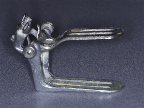
The nonadjustable articulator (Figure 18-7) is the simplest type available. No adjustments are possible to adapt it more closely to the specific condylar movements of the patient. Many of these articulators allow for eccentric movements but only average values. Accurate duplication of an eccentric movement for a specific patient is impossible.

The only accurate and reproducible position that can be achieved on a nonadjustable articulator is one specific occlusal contact position (e.g., the intercuspal position, or ICP). When the casts are mounted in this position on the nonadjustable articulator, they can be repeatedly separated and closed only to this position, which becomes the only repeatable and accurate position that can be utilized. Even the opening and closing pathways of the teeth do not accurately duplicate the pathways of the patient’s teeth, since the distance from the condyles to the specific cusps is not accurately transferred to the articulator. The ICP is reproducible only when the casts are mounted on the articulator in that position. All other positions or movements (e.g., opening, protrusive, laterotrusive) do not accurately duplicate the conditions found in the patient.
Associated procedures required for the nonadjustable articulator
Since only the occlusal position in which the teeth are mounted is accurately duplicated, arbitrary mounting procedures are used to locate and fix the casts. Generally the casts are held together with the teeth in maximum intercuspation and located equidistant between the maxillary and mandibular components of the articulator. Mounting stone is then placed between the mandibular cast and the mandibular component of the articulator, firmly joining these together. The maxillary cast is likewise attached to the maxillary component of the articulator. When the mounting stone is set, the casts can be separated and the simple hinge of the articulator will accurately return the casts to the position maintained during mounting.
Adva/>
Stay updated, free dental videos. Join our Telegram channel

VIDEdental - Online dental courses


