17: Surgery of the temporomandibular joint

17 Surgery of the temporomandibular joint

MINIMALLY INVASIVE TECHNIQUES

Injection into the joint

Entry into the joint with a needle, from the skin surface, may be necessary for the instillation of steroid or local anaesthetic solution and is a prerequisite for insertion of an arthroscope as the joint needs to be distended with fluid from within.

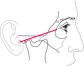
The upper joint space is most easily approached from below and behind, starting from a point 10 mm in front of the point of the tragus, just below a line that joins that point to the outer canthus (Fig. 17.1). With the mouth open, or the mandible protruded, the needle is inserted upwards, forwards and medially, until it penetrates the capsule just above and behind the condyle; this may be as deep as 2 cm from the surface. To check that the needle is in the joint, a small quantity of saline is injected then drawn back. It should be readily possible to flush fluid in and out of the joint. If no fluid can be withdrawn, the tip of the needle is unlikely to be in the joint and the position should be reassessed and adjusted. The average joint may be distended with about 2 mL of fluid.

Closed manipulation for adhesions: method and results

In cases of disc displacement without reduction, it is common that adhesions prevent the normal movement of the disc in the upper joint space. It is sometimes possible to release the lock and increase mobility by closed manipulation.

In general, pain during the procedure would be considerable if no form of anaesthesia were provided: muscle guarding in such circumstances is too powerful to permit the necessary manipulation. Local anaesthesia is often sufficient and is achieved by placing the local anaesthetic (lidocaine with epinephrine) posterior to the joint, close to the auriculotemporal nerve or in the joint itself. Patients should be warned in advance that occasionally local anaesthetic might diffuse from the site of injection to the facial nerve, giving rise to a temporary facial palsy. Should there be any weakness of the eyelids, the affected eye should be taped shut for the duration of the local anaesthetic.

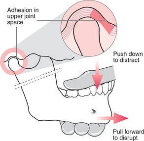
It is important, after the local anaesthetic has been placed, to measure the mouth opening, in order later to be able to determine the change that has taken place. The operator stands beside the patient, who is seated and leaning slightly backwards. The operator holds the mandible, with the thumb from the opposite hand inside the patient’s mouth, resting on the posterior teeth and the fingers placed beneath the body of the mandible (Fig. 17.2). The patient’s head is held fast against the operator’s body with the other hand, the fingers of which are placed over the TMJ to feel any movement within it. The thumb is used to push down on the posterior teeth and distract the joint, then slowly the mandible is drawn forwards with increasing force until increased mobility is achieved. This often happens as one or two sudden releases. When maximum movement is achieved, mouth opening should be measured again.

It is often possible to gain considerable mobility in closed lock by this technique. Unfortunately it can result in considerable pain over the following few days, leading to the patient moving their jaw little and the consequent reforming of adhesions. This effect can be reduced by giving NSAIDs before and after the treatment, but a considerable inflammatory exudate still remains within the joint. The pain and immobility can be eased further by irrigating the joint with saline and instilling a small quantity of steroid (see pp. 242, 245).

Arthroscopy

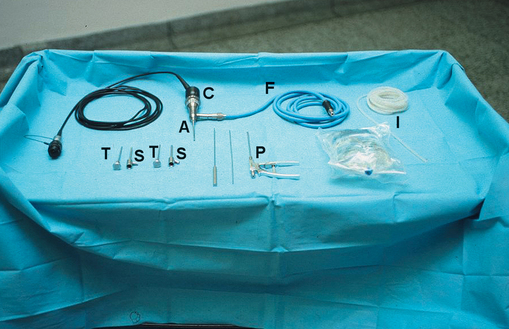
An arthroscope is an endoscope designed specifically for use within a joint (Fig. 17.3). TMJ arthroscopes are up to 2.8 mm in diameter and rigid. With these devices it is possible to inspect the whole of the upper joint space without formally dissecting the joint, all through a skin puncture a few millimetres in diameter.

The joint is distended by injection of about 2 mL saline (see above). Initially the sheath is inserted through a skin puncture with a pointed but round-ended trocar inside it. This is pushed upwards, inwards and forwards until it reaches the joint capsule. At this stage the trocar may be replaced with a more round-ended one to enter the upper joint space itself. Entry into the joint is far easier if the capsule has been distended with saline and is taut. Once into the joint, the trocar is removed, leaving the sheath in place, and is replaced with the arthroscope, which is attached to the fibreoptic light source and saline for irrigation. Because there tends to be some bleeding into the joint at the point of entry, vision would rapidly deteriorate if the joint were not washed through, but at this stage there is only one portal for both entry and exit. So, now a needle (or a second arthroscope port) is placed about a centimetre further forwards and parallel to the arthroscope itself; this can act as the exit (or egress) cannula. Saline is now slowly flushed through the joint as it is examined.

The joint can be examined by direct vision, but it is more common to attach a video camera to the scope and to display what is seen on a television monitor (Fig. 17.4).

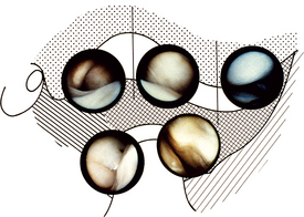
With the simplest of equipment the joint can be inspected, usually starting with the posterior recess, looking at the position of the disc, the condition of the posterior attachment tissues and the synovium on the medial aspect of the joint. The scope is then swept anteriorly over the top of the disc to look at the anterior parts of the joint. By inspection alone it is possible to detect disc displacement, adhesions, degenerative changes in the disc and cartilage over the glenoid fossa and articular eminence and synovial inflammation. It is possible, if adhesions are detected, to replace the blunt-ended trocar and sweep around within the joint to break them down. The joint must be thoroughly irrigated at the end of the procedure and many surgeons will finally instil a steroid before leaving the joint. It is often necessary to place one suture in the skin wound.

If two arthroscope ports are used it is possible to perform surgery within the joint under direct vision. Procedures including disrupting adhesions, biopsy and smoothing roughened areas may be relatively straightforward for the expert arthroscopist, but attempts are also being made to shorten the posterior attachment tissues and reposition the disc by laser surgery. In time such procedures may become more routine.

Selection of patients is very much a matter of personal judgement. Great reliance has been placed on a failure to respond to conservative measures, with persistent pain being a central criterion. Nonetheless, if arthroscopy is intended to be therapeutic and not just a diagnostic procedure there must be some real expectation of a mechanical problem within the joint. Therefore disc displacement disorders and degenerative joint disease resistant to conservative treatments may be indications for arthroscopy, whereas painful muscular conditions are not.

Studies of the effectiveness of arthroscopy have been very encouraging, with a success rate of approximately 90%, but there have been few randomized controlled trials against conservative or more aggressive surgical treatments to allow thorough scientific evaluation. Success rates must depend upon which patients are entered into the studies, and details of patient selection in reported trials have been scanty.

Arthroscopy is a procedure associated with few serious complications. The most common problem is failure to enter the joint cleanly, or at all, although the incidence of this decreases with experience. For this reason, considerable training is recommended before independent use of arthroscopes. Poor entry into the joint results not only in physical injury to the joint capsule and/or the joint surfaces but also in greater leakage of irrigant into the surrounding tissues leading to massive swelling. Bleeding along the entry tract or into the joint is an occasional problem, but is usually readily controlled with pressure. The scope may be misdirected towards the external ear canal or upward towards the middle cranial fossa (the bone of the glenoid fossa is paper thin) or may be placed deep to the joint in the pharyngeal wall. These are serious complications and great effort must be taken with technique to avoid them.

Only gold members can continue reading. Log In or Register to continue

Stay updated, free dental videos. Join our Telegram channel

Jan 14, 2015 | Posted by in Oral and Maxillofacial Surgery | Comments Off on 17: Surgery of the temporomandibular joint

VIDEdental - Online dental courses

Get VIDEdental app for watching clinical videos