15: The maxillary antrum

15 The maxillary antrum

THE IMPORTANCE OF THE MAXILLARY ANTRUM TO DENTAL SURGERY

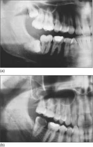
The maxillary sinus is often referred to as the maxillary antrum (or more simply the ‘antrum’). It is important to the dental surgeon because of its close relationship to the posterior maxillary teeth (generally from first premolar to third molar). The proximity varies between individuals (Fig. 15.1) but tends to increase with age as the antrum enlarges. The antrum can encroach into alveolar bone after tooth extraction, and periapical bone loss resulting from dental disease may further decrease the amount of bone separating it from the teeth. This close anatomical relationship can lead to diagnostic difficulties because both the antrum and the maxillary teeth are innervated by branches of the maxillary division of the trigeminal nerve. In addition, infection may spread from the periapical region of the posterior maxillary teeth to the antrum (and in the opposite direction) and the antrum is at risk of iatrogenic damage during certain dental procedures.

The ability to distinguish between dental and antral symptoms enables the dental surgeon to decide when dental treatment is indicated and when referral (e.g. to a general medical practitioner, an oral and maxillofacial surgeon or an ear, nose and throat (ENT) surgeon) is appropriate.

The antrum is visualized on oral and facial radiographs (notably periapical views of the posterior maxillary teeth, orthopantomogram (OPT) and occip-itomental views) and a knowledge of its normal radiographic appearance is essential in the diagnosis of both dental and antral disease. Evidence of antral pathology may be incidentally discovered by the general dental practitioner (GDP) on these radiographs.

Although the GDP is unlikely routinely to perform elective surgery within the antrum, there will be occasions when dentoalveolar surgery with a degree of antral involvement is necessary—for example, to repair a newly created oroantral communication. It is necessary for the GDP to be able to recognize and deal with problems such as oroantral communication and foreign body in the antrum and he or she should be capable of explaining the commonly performed antral investigations and surgical procedures to a patient who requests information. The GDP may also recommend other procedures that involve antral surgery (e.g. ‘sinus lift’/alveolar ridge augmentation procedures used prior to the placement of osseointegrated implants).

DISTINGUISHING SIGNS AND SYMPTOMS OF DENTAL PATHOLOGY FROM THOSE OF SINUSITIS

Maxillary sinusitis is common and the dentist needs to be able to distinguish it from dental disease. It is usually an acute condition, but chronic sinusitis may also develop following an acute episode and may persist or recur if drainage from the antrum to the nasal cavity is poor or when a foreign body is retained.

Acute sinusitis may affect any of the paranasal sinuses but is usually confined at any one time to a single sinus. It is a bacterial infection and typically occurs after a viral upper respiratory tract infection (URTI) (Haemophilus, Pneumococcus, Streptococcus, Staphylococcus species and anaerobes are commonly found).

Maxillary sinusitis is usually related to impaired antral drainage, which may be caused by:

Accumulated secretions become infected, allowing a collection of pus to develop in the antrum.

Dental diseases (including periapical pathology, infected odontogenic cysts and periodontal disease) may also cause sinusitis. Other possible causes include the presence of contaminated foreign bodies, trauma or infections of non-odontogenic cysts or malignant tumours.

Signs and symptoms of sinusitis

Symptoms of acute sinusitis usually appear a few days after the acute cold symptoms have resolved. Pain, headache, nasal obstruction, a purulent nasal secretion and ‘postnasal drip’ (a discharge of ‘mucopus’ into the pharynx) are commonly found and there may also be fever and malaise. The pain is dull, heavy, throbbing and located over the cheek and in the upper teeth. It may resemble toothache but is not related to hot, cold or sweet stimuli, although it may be increased by biting. Typically all of the posterior teeth on the affected side are painful and tender to percussion, although often no obvious dental cause can be found. Leaning the head forwards increases the pain (as it leads to an increase in venous congestion which, together with collected secretions, occludes the ostium and increases pressure within the antrum). However, leaning the head backwards reduces congestion, allowing the ostium to become patent, reducing internal antral pressure and consequently relieving pain. Although the pain usually overlies the affected sinus it may be referred from the antrum to the frontal and retro-orbital regions.

THE INVESTIGATION OF ANTRAL DISEASE

A diagnosis of sinusitis can usually be made from the findings of the history and examination. The teeth and surrounding tissues should be examined for disease (although remember that sinusitis may itself have a dental cause). Transillumination, using a torch shone from inside the mouth, may demonstrate antral congestion but this is a rather crude test and may fail to detect tumours. Radiographs are not usually required to confirm the diagnosis of sinusitis as the diagnosis can be made on clinical grounds alone. Radiographs are only required to exclude the possibility of dental disease, trauma or pathology within the antrum. If pathology or trauma is suspected, occipitomental radiographic views are often taken in specialist centres. A ‘fluid level’ may be visible which represents a collection of pus, although following trauma it may be due to the presence of blood and suggests that there is a fracture of the wall of the antrum. Opacities due to polyps, cysts, thickened antral lining, tumours and radio-opaque foreign bodies may also be demonstrated.

Other useful radiographs include:

Much useful information can be gathered by endoscopy of the antrum. This is better performed by someone with experience of endoscopy in the region (such as an ENT surgeon).

DENTAL EXTRACTIONS AND THE ANTRUM

The following complications may occur during the extraction of posterior maxillary teeth because of their close proximity to the maxillary antrum:

Maxillary premolars and permanent molars are most frequently implicated, although permanent canines may occasionally be very close to the antrum. Complications may also occur during the surgical removal of impacted partially erupted or unerupted teeth (e.g. canines, premolars and third molars). The extraction of deciduous teeth does not present a risk due to the relatively small size of the antrum in children and the presence of the developing permanent teeth.

An assessment of the size and proximity of the antrum before extraction or periapical surgery on a posterior maxillary tooth may help to anticipate and avoid these complications. When a complication does occur, it should be recognized and dealt with promptly.

The following factors should be taken into account during clinical and radiographic examination in order to anticipate and avoid complications:

If the dental history reveals the creation of an oroantral communication during a previous extraction there is likely to be an increased risk of this complication occurring during the subsequent extraction of adjacent or contralateral teeth. Similarly, if there is a history of difficult extractions, due perhaps to hypercementosis or dense bone, there may be an increased risk of tuberosity fracture.

Stay updated, free dental videos. Join our Telegram channel

Jan 14, 2015 | Posted by in Oral and Maxillofacial Surgery | Comments Off on 15: The maxillary antrum

VIDEdental - Online dental courses

Get VIDEdental app for watching clinical videos