16
Intra-oral examination
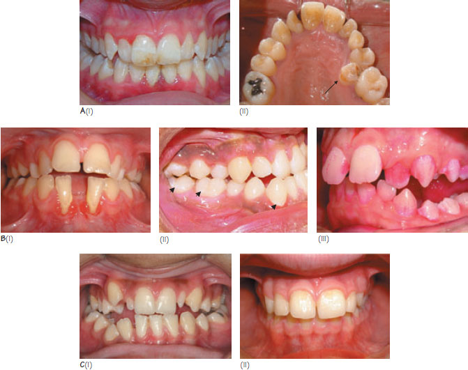
Figure 16.1 (A) (i) Enamel hypomineralisation should be documented before commencing treatment. It can affect dental aesthetics and bond strength of orthodontic attachments, (ii) Often severely hypoplastic premolars make good candidates for extraction if space is required for dental alignment. (B) Indicators of poor oral hygiene: (i) gingival inflammation, recession and plaque deposits are clearly visible, (ii) lines of decalcification are seen following the gingival margin of the first molars and (iii) plaque demonstrated using disclosing tablets. (C) Developmental disturbances in tooth size: (i) microdontia of the maxillary lateral incisors and (ii) megadontia of the upper left central incisor.
The aims of intra-oral examination are to:
- assess the mucosal/dental surfaces for pathology;
- determine the level of oral hygiene;
- establish whether dental development is normal;
- assess tooth position within and between the arches.
Assessment for pathology
Every patient should have a full examination of the mucosal surfaces during routine assessment. Dental pathology can have a significant influence on treatment planning. Of particular relevance are:
- dental caries;
- dental hypoplasia and hypomineralisation;
- toothwear;
- sequelae of traumatic injuries to the dentition;
- gingivitis, periodontitis and gingival recession.
All teeth that have previously suffered trauma or have advanced caries or large restorations should undergo thermal or electrical vitality testing. Areas of significant enamel hypoplasia/hypomineralisation should be documented and photographed (Figure 16.1A). Patients often become more aware of these defects during treatment as their primary focus of attention, the dental irregularity, is corrected. This may lead them to incorrectly attributing the enamel defects to orthodontic treatment. Periodontal charting is important if periodontitis is suspected. All dental disease must be controlled before contemplating orthodontic treatment.
Oral hygiene
The level of oral hygiene can be assessed by examining for gingivitis, probing to elicit gingival bleeding, and with visual aids such as disclosing tablets/solution (Figure 16.1B). The presence of a line of decalcification following the gingival margin is indicative of plaque accumulation and a cariogenic diet (Figure 16.1Bii). Poor hygiene during orthodontic treatment predisposes to decalcification, gingival hyperplasia, periodontal breakdown and removable-appliance-related stomatitis.
Assessment of dental development
It is important to note the teeth present and the mobility of the deciduous teeth that are present. A mobile deciduous tooth often indica/>
Stay updated, free dental videos. Join our Telegram channel
VIDEdental - Online dental courses


