16: Gingival and Periodontal Diseases

Gingival and Periodontal Diseases

Periodontal diseases range from simple and early inflammation of marginal gingiva to advanced gingivitis and subsequently periodontitis. A good classification system is crucial in identifying and differentiating the various kinds of periodontal diseases.

Periodontal disease classification is useful to help establish diagnosis, determine prognosis and facilitate treatment planning.

Different classification systems of periodontal diseases have been used for years and also replaced by newer classification systems as the knowledge of understanding microbiology, etiology, pathogenesis and host response have improved vastly over the past few decades.

Currently, the most accepted classification is the ‘1999 World Workshop Classification System’.

Classification System in Periodontal Disease

The classification is based on the 1999 International Workshop for Periodontal Disease.

Gingival diseases

Dental plaque-induced gingival diseases: These diseases may occur on a periodontium with no attachment loss or on one with attachment loss that is stable and not progressing.

Non-plaque-induced gingival lesions

1. Gingival diseases of specific bacterial origin

2. Gingival diseases of viral origin

3. Gingival diseases of fungal origin

4. Gingival lesions of genetic origin

5. Gingival manifestations of systemic conditions

6. Traumatic lesions (factitious, iatrogenic or accidental)

7. Foreign body reactions

8. Not otherwise specified

II Chronic periodontitis

III Aggressive periodontitis

IV Periodontitis as a manifestation of systemic diseases

Necrotizing periodontal diseases

VI Abscess of the periodontium

VII Periodontitis associated with endodontic lesions

VIII Developmental or acquired deformities and conditions

Localized tooth-related factors that modify or predispose to plaque-induced gingival diseases/ periodontitis

Mucogingival deformities and conditions around teeth

Mucogingival deformities and conditions on edentulous ridges

Occlusal trauma

The current classification has many changes seen which is drastically different from the previous classifications. The most noticeable change is that periodontitis is now classified based on the rate of progression of the disease and not on the age of onset.

Gingival Diseases

Increasing evidence indicates that gingivitis is not a single disease, but an assortment of diseases that are the end result of a variety of different processes. Inflammation of gingiva by bacteria is most common, but pathological changes in the gingiva can also result from systemic conditions (e.g. puberty), drugs (e.g. amlodipine) and neoplasms (e.g. leukemia). Hence, any disease that primarily affects gingival tissues should be primarily classified as a gingival disease.

The gingival diseases associated with children, adolescents and young adults have several common characteristics. Universal features include clinical signs of inflammation, signs and symptoms that are confined to the gingiva, reversibility of the disease on removal of the etiology, and the presence of microbial plaque to initiate or exacerbate the severity of the lesion.

Plaque Microbiology

Oral cavity can be regarded as a single microbial ecosystem or macroenvironment because the colonization of oral cavity starts at the time of birth. Within hours of birth the sterile oral cavity gets colonized by facultative and aerobic bacteria, which will be followed by anaerobic bacteria by 2nd day. By 2 years of age there will be around 400 different kinds of bacteria. After tooth eruption, a more complex oral flora is established. However, all these species are seen to be living in harmony with the host. But, however, there is an imbalance in this relationship between the host and microorganisms, disease prevails which is controlled by various other factors.

Oral environment is dominated with saliva, which is very complex in composition. Although saliva is not a good medium for supporting the growth of bacteria, it is likely that organisms shed from intraoral reservoirs find transient residence in saliva. Oral cavity provides two types of surfaces for colonization—soft and hard tissue (teeth)—main difference being soft tissue desquamation; hence the colonies are shed frequently whereas the hard surface provides a solid medium for the bacteria to adhere and develop complex layers.

Organisms in Various Periodontal Diseases

1. Periodontal health

2. Gingivitis

3. Chronic periodontitis

4. Localized aggressive

5. Generalized aggressive

6. NUG/NUP

7. Periodontal abscess

Host–Microbial Interaction

Health is not a static condition, it is a dynamic state in which the living and functioning individuals remain in balance with a constantly changing environment. These changes in environment also cause changes in tissue activity so that normal function can continue, a process known as homeo-stasis. If the environmental changes over-ride this homeo-stasis, the normal function cannot continue and this change is termed as disease.

Defense Mechanism

These protect the body from attack from microorganisms and can be classified as:

Specific mechanisms

image T-cell response (cell-mediated immune system)

image B-cell response (antibody-mediated immune system). Plaque bacteria produce a number of factors (virulence factor), which causes disease directly, or individually by stimulating the immune and inflammatory system. It is now known that individuals prone to periodontal disease have an aberrant immune inflammatory response to plaque which is genetically determined.

image Direct effects of bacteria

image Indirect effects of bacteria

Host modulation: Periodontal disease is multifactorial in nature as a result of interaction between plaque microorganism and host responses. Host modulation refers to the alteration/modification of host response to the microbial stimulus by the use of number of medications. The two major categories are NSAIDs and inhibition of MMPs.

Plaque-induced Gingivitis

It is the inflammation of gingiva which results from the bacteria located at the gingival margin. The association of plaque with gingival inflammation made it a frequently postulated cause of gingivitis. The initial histologic changes from health to plaque-induced gingivitis may not be evident clinically (Page and Shroeder, 1976; Bimstein et al, 1985) but as gingivitis progresses, clinical signs becomes more obvious.

Plaque-induced gingivitis begins at the gingival margin and can spread into the deeper gingival component. Clinical signs of gingival inflammation involve the change in color, contour, size, shape, consistency and surface texture are associated with a stable periodontium which exhibits no loss of periodontal attachment or alveolar bone. The classic clinical indicators of signs of inflammation are bleeding on probing and color change from pink/coral pink to reddish pink or erythematous gingiva. In children, gingivitis is not intense as that found in young adults with similar quantity of accumulation of dental plaque (Mattson and Goldberg, 1985).

In the initial and established stages of gingivitis, dental plaque is predominantly comprised of gram-positive aerobic microorganisms including Streptococcus mitis, S. sanguis, Actinomyces viscosus, A. naeslundii and Eubacterium spp. As age advances, development and severity of gingivitis is mainly dependent on the quality of the plaque rather than the quantity and the other contributing factors like the host immune response, environmental, genetic and the behavioral factors. The common clinical signs and symptoms of gingivitis include redness, edema, bleeding on probing, tenderness and enlargement (Loe et al, 1965; Suzuki, 1988) (Figure 1).
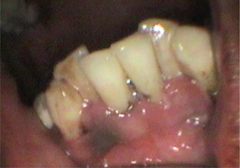
Radiographically no changes will be seen as the inflammation is confined only to the gingival sulcus. Histologic changes include proliferation of basal junctional epithelium leading to apical and lateral cell migration, vascular dilatation and vasculitis of blood vessels adjacent to the junctional epithelium, progressive destruction of collagen fibers, cytopathologic alteration of resident fibroblasts and progressive inflammatory cell infiltrate (Page and Shroeder, 1976).

Non-plaque-induced Gingivitis

Gingival diseases associated with endogenous sex steroid hormones

Since the 19th century, evidence suggests that the tissues of the periodontium are modulated by androgens, estrogens and progesterone. Much of this evidence has come from observing the changes in gingival tissues during distinct endocrino-logic events (puberty, menstrual cycle, pregnancy, etc.).

The principal explanation for sex steroid hormone-induced changes in the gingiva has pointed to changes of microbiota in dental plaque, immune function, vascular properties and cellular function in the gingiva. Sex steroid hormones will affect the host by influencing cellular function (in the blood vessels, the epithelium and the connective tissue) and immune function, and together with hormone-selected bacterial populations occupying the gingival sulcus, induce specific observable changes in gingival tissues (Mariotti, 1994).

Stay updated, free dental videos. Join our Telegram channel

Jan 12, 2015 | Posted by in Oral and Maxillofacial Radiology | Comments Off on 16: Gingival and Periodontal Diseases

VIDEdental - Online dental courses

Get VIDEdental app for watching clinical videos