Section 4: Osseous Surgery in Orthodontic Therapy
Chapter 15
Piezocision: Minimally Invasive Periodontally Accelerated Orthodontic Tooth Movement Procedure
INTRODUCTION
Surgical interventions on the alveolar ridges that are aimed at facilitating orthodontic treatment are not new. From the late 1800s to the late 1900s, we see the predominance of a mechanical concept that is prevalent in the surgical community: The orthodontic tooth movement is impaired by the physical presence of the alveolar cortex. Therefore, it becomes necessary to disrupt this cortical bone via surgery to allow for faster tooth movement. In 1892, Bryan (Guilford 1898) is credited with the first report in the literature in which alveolar corticotomy is used to correct a malocclusion. Several years later Kole (1959a, 1959b, 1959c), Generson (1979), and Suya (1991) use various types of corticotomies to achieve the same results. They cut through the alveolar cortex and created movable “blocks of bone” where the teeth are connected only by medullary bone. They believed that shorter treatment is due to removal of the cortical layer.
This concept was challenged by the Wilcko brothers in the late 20th century. They questioned the mechanical concept of bony block movement after reviewing radiographs and CT scans of their patients who had undergone corticotomy-facilitated orthodontic therapy. They noticed that following periodontally accelerated osteogenic orthodontics (PAOO), a demineralization of the jaw was taking place, followed by a remineralization. They hypothesized that rapid tooth movement resulted from marked but transient decalcification-recalcification process of the alveolus. This concept is known in the orthopedic literature as the regional acceleratory phenomenon (RAP), as described by Frost in 1983. He reported localized increased osteoclastic and osteogenic activity at the site of osseous surgery. There was a decrease in regional bone density accompanied by increased bone turnover. He noticed that the RAP begins within a few days of the surgery and usually peaks in 1–2 months. The Wilckos had understood and witnessed this occurrence in their own patients. The elevation of buccal and lingual full-thickness flaps, with extensive decortications of the buccal and lingual alveolar bone, resulted in a physical injury that was responsible for the initiation of a temporary demineralization process coupled with an increased regional bone turnover that characterizes the RAP. They surmised that this transient osteopenia (diminished bone density, same bone volume) is responsible for the rapid tooth movement, as the teeth move in a more “pliable” environment. Their pioneering work, combining alveolar decortication concomitant with bone grafting to expand alveolar volume and allow for rapid tooth movement into the newly expanded sites, stands out as seminal (Wilcko et al. 2001).
In 2007, Vercelotti and Podesta introduced the use of piezosurgery in conjunction with the conventional flap elevations to create an environment conducive to rapid tooth movement. Although quite effective, these techniques are also quite invasive in nature as they require extensive flap elevations and osseous surgery. They have the potential to generate postsurgical discomfort as well as postoperative complications. Because of these shortcomings, they have not been widely embraced by the patient or dental communities. Park et al. in 2006 and Kim et al. in 2009 introduced the corticision technique as a minimally invasive alternative to create surgical injury to the bone without flap reflection. In this technique, the authors use a reinforced scalpel and a mallet to go through the gingiva and cortical bone without raising a flap bucally and lingually. The surgical injury created is enough to induce the RAP effect and move the teeth rapidly during orthodontic treatment. This technique, although innovative, has two drawbacks: the inability to graft soft or hard tissues during the procedure to correct inadequacies and reinforce the periodontium and the repeated malleting, which may cause dizziness after surgery. We are describing here a new minimally invasive procedure that we called “piezocision.” This technique combines microincisions limited to the buccal side that will allow for the use of the piezoelectic knife and selective tunneling that allows for hard or soft tissue grafting.
INDICATIONS
Indications for using the piezocision technique include the following:
- Class I malocclusions with moderate to severe crowding (nonextraction)
- Correction of deep bite
- Selected class II malocclusions (end-on)
- Rapid adult orthodontic treatment
- Rapid intrusion and extrusion of teeth
- Simultaneous correction of osseous and mucogingival defects
- Prevention of mucogingival defects that may occur during or after orthodontic treatment.
ARMAMENTARIUM
The equipment needed to perform a piezocision include the following:
1. Topical and local anesthetic
2. Scalpel with blade #15C
3. Periosteal elevator (24G, Hu-Friedy, Chicago, IL)
4. Piezotome (Satelec, Acteon Group, Merignac, France), with insert BS1
5. Bone allograft or xenograft
6. 5-0 Chromic gut suture
7. Castroviejo needle holder
8. Surgical scissors
9. Peri-acryl, cyanoacrylate glue
10. Coe Pack if soft tissue grafting is needed
TECHNIQUE
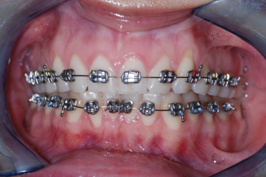
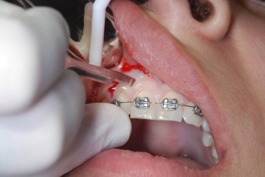
Piezocision is performed 1 week after the placement of orthodontic appliances (Figure 15.1). The patient is anesthetized using Xylocaine 2% with 1 : 100.000 epinephrine in infiltration. Once complete anesthesia is achieved, a small vertical incision is performed buccally and interproximally in the attached gingiva or mucosa. The incision into the attached gingiva is preferable as it will give less visible postoperative scarring. A midlevel incision is made between the roots of the teeth involved, keeping in mind that the soft tissues and the periosteum need to be cut to create an opening that will allow the insertion of the piezoelectric knife.
Figure 15.1 Class I malocclusion with moderate anterior crowding. Notice the mucogingival defect on tooth #11.
At this point, it is important to emphasize the following concept: Piezocision has a localized and selective effect on the bone. Only the teeth or arch(es) to be moved need to be operated on. The areas not undergoing surgery have a higher anchorage value because they are not affected by the demineralization process and thus can be used as such in the global treatment plan. Once the vertical interproximal incisions are completed on the maxillary and mandibular arches or in localized segments, the tip of the Piezotome (BS1) is inserted in the openings previously made, and a 3-mm piezoelectrical corticotomy is done (Figures 15.2–15.4).
Figure 15.2 Interproximal incisions done with blade #15.
Stay updated, free dental videos. Join our Telegram channel
VIDEdental - Online dental courses


