Chapter 15 Getting the appearance correct
Light
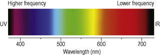
Visible light forms a small portion of the whole electromagnetic (EM) spectrum (Figure 15.1) and often takes the form of polychromatic light which is composed of electromagnetic radiation of more than one wavelength. The colour of an object that one observes is actually the reflection of the light that strikes it. For example, a red flower appears red because red light is reflected by the flower whilst the other colours of light are absorbed.
Description of colour
Munsell’s colour order system
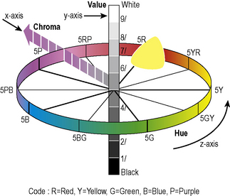
The Munsell colour system was devised by a painter, Albert Munsell, in 1905. The system’s attributes are hue, chroma and value (Figure 15.5).
Hue
Hue is the quality by which we distinguish one colour from another – for example, red from yellow, or green from blue. There are 100 Munsell hues: 10 major hues with each placed 10 steps apart in a horizontal plane (z axis, Figure 15.5) around a central axis. Teeth are found in the yellow and yellow–red region but the exact range of hues varies with the method of assessment and is different for extracted teeth. Dentine provides the main source of hue in a tooth but this is modified by the enamel.
Chroma
Chroma describes the intensity of the colour (hue) and distinguishes a strong colour from a weak one. The chroma scale starts at /0 and extends outwards; its maximum varies with each hue. The purest colours are found at the extremes of the colour cylinder (x axis, Figure 15.5). Reduced thickness or mineralization of dentine usually results in decreased chroma. The chroma of teeth is usually found within the range /1 to /5 but can range from /0 to /7.
Value
Value is the quality by which one distinguishes a light colour from a dark one and is therefore the brightness on a grey scale. The value symbol 0/ is used for absolute black and 10/ for absolute white (y axis, Figure 15.5). In a healthy young tooth there is less dentine thickness due to a reduced amount of secondary dentine and so the ratio of reflected to absorbed light radiation is increased compared to older teeth: as a tooth ages its value therefore decreases. Tooth value is usually found within the range 6/ to 8.5/ but can range upwards from 4/.
Tooth shade selection
‘Selecting the shade’ for a restoration belittles the complexity of the process of determining the shade and form for a restoration. Providing the technician with only the correct shade will not enable them to fabricate an aesthetic restoration, a substantial amount of additional information is required. Additionally, the clinician has to prepare teeth in such a way that the technician is able to recreate the desired shape and shade. (See chapter 10, 11 and 12). Methods of traditional and instrumental shade-matching will be described in this chapter, together with the supplemental information required for an aesthetic restoration.
Stay updated, free dental videos. Join our Telegram channel
VIDEdental - Online dental courses


