13: Inflammatory Jaw Lesions

Inflammatory Jaw Lesions

Osteomyelitis, by definition, is inflammation, not necessarily infection (by a microorganism), of bone and bone marrow. The term osteitis may be substituted for osteomyelitis to indicate inflammation of bone. In the mandible and maxilla, most cases are related to a microbial (usually bacterial) infection that reaches the bone through nonvital teeth, periodontal lesions, or traumatic injuries. This, coupled with the patient’s host resistance factors, determines the clinical presentation, the extent of the inflammatory process, and the speed with which the infection develops. Recognized subtypes of osteomyelitis are closely related and essentially represent differences in the causative agent and the host response. The primary justification for separation of osteomyelitis into various subtypes lies in the differences in treatment and prognosis for each. It is important to be aware of clinical and radiographic presentations when making differential diagnoses of bone lesions.

Pulpitis

All the principles of inflammation that apply to any other body organ apply to lesions of the dental pulp. In addition, dental pulp has some unique features that make it unusually fragile and sensitive. First, it is encased by hard tissue (dentin/enamel) that does not allow for the usual swelling associated with the exudate of the acute inflammatory process. Second, there is no collateral circulation to maintain vitality when the primary blood supply is compromised. Third, biopsies and direct applications of medication are impossible without causing necrosis of the entire pulp. Fourth, pain and increasing levels of sensitivity are the only signs that can be used to determine the severity of pulpal inflammation.

Because of referred pain and the lack of proprioceptors (position sensors) in the pulp, localizing the problem to the correct tooth can often be a considerable diagnostic challenge. Also there may be a poor correlation between clinical symptoms and pathologic changes occurring in the pulp. The level of pulpal inflammation is determined through a combination of clinical criteria. Results of electric, heat, cold, and percussion tests must be added to the patient history, clinical examination, and clinician experience to arrive at the most appropriate diagnosis for the correct tooth. Generally, the more intense the pain and the longer the duration of symptoms, the greater the damage to the pulp. Severe symptoms usually indicate irreversible damage.

Etiology

In the dental pulp, just as it is in any other tissue, inflammation is the response to injury. In addition, the pulpal response includes stimulation of odontoblasts to deposit reparative dentin at the site to help protect the pulp. If the injury is severe, the result is, instead, necrosis of these cells.

Caries is the most common form of injury that causes pulpitis. The degree of damage depends on the rapidity and extent of hard tissue destruction. Entry of bacteria into the pulpal tissue through a carious lesion is not necessary for pulpitis to occur, but this appears to be an important factor in the intensification of the inflammatory response. Pulpal microbiology adjacent to carious dentin demonstrates a diverse flora, including gram-positive anaerobes low numbers of lactobacilli. Operative dental procedures associated with cavity and crown preparations may also trigger an inflammatory response in the dental pulp. The heat, friction, chemicals, and filling materials associated with restoration of teeth are all potential irritants. It is well known that less damage occurs when a cooling water spray is used during tooth preparation than when no water is used. It is also well established that an insulating base (such as zinc oxide and eugenol under amalgam restorations or glass ionomer under a composite restoration) can provide significant protection of the pulp from irritating chemicals used in the preparation of nonmetallic restorative materials and from heat transferred through large metallic fillings.

Other types of injury that may trigger pulpitis are trauma, especially when it is severe enough to cause root or crown fracture, and periodontal disease that has extended to an apical or lateral root foramen.

Clinical and Histopathologic Features

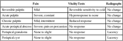
Several detailed classifications of pulpitis that are based on histopathologic changes have been proposed. Because of the difficulty in correlating clinical features with microscopy, these schemes have proved to be of little practical value. Instead, most practitioners prefer a simple classification that is helpful in the clinical setting relative to treatment and prognosis (Table 13-1).

TABLE 13-1

PULPITIS AND PERIAPICAL DISEASES

  Pain Vitality Tests Radiographs
Reversible pulpitis Mild Reversible sensitivity to cold No change
Acute pulpitis Severe, constant Hyperresponse to none No change
Chronic pulpitis Mild, intermittent Reduced response No change
Acute periapical abscess Severe; pain on percussion No response No change
Periapical granuloma None to slight No response Lucency
Periapical cyst None to slight No response Lucency

image

Acute Pulpitis

The inflammatory response of acute pulpitis may occur as progression of focal reversible pulpitis, or it may represent an acute exacerbation of an already established chronic pulpitis. Pulpal damage may range in severity from simple acute inflammation marked by vessel dilation, exudation, and neutrophil chemotaxis to focal liquefaction necrosis (pulp abscess) to total pulpal suppurative necrosis. Constant, severe, tooth-associated pain is the usual presenting complaint. Pain is intensified with the application of heat or cold, although in cases in which liquefaction of the pulp has occurred, cold may in fact alleviate the symptoms. If there is an opening from the pulp to the oral environment, symptoms may be lessened because of escape of the exudate that causes pressure on and chemical irritation of pulpal and periapical nerve tissues.

In the early phases of acute pulpitis, the tooth may be hyperreactive to electrical stimulation, but as pulp damage increases, sensitivity is reduced until there is no response. Because the exudate is confined primarily to the pulp rather than the periapical tissues, percussion tests generally elicit a response that differs little from normal.

Chronic Hyperplastic Pulpitis

This special form of chronic pulpitis occurs in the molar teeth (both primary and permanent) of children and young adults. Involved teeth exhibit large carious lesions that open into the coronal pulp chamber. Rather than undergoing necrosis, the pulp tissue reacts in a hyperplastic manner, producing a red mass of reparative granulation tissue that extrudes through the pulp exposure. This type of reaction is believed to be related to the open root foramen, through which a relatively rich blood supply flows.

Symptoms seldom occur because there is no exudate under pressure, and generally no nerve tissue is proliferating with the granulation tissue. Although the pulp tissue is viable, the process is not reversible, and endodontic therapy or tooth extraction may be necessary. The well-vascularized granulation tissue mass often becomes epithelialized, presumably by autotransplantation of epithelial cells from nearby mucosal surfaces.

Treatment and Prognosis

If the cause is identified and eliminated, focal reversible pulpitis should recede, returning the pulp to a normal state. If inflammation progresses into an acute pulpitis with neutrophil infiltrates and tissue necrosis, recovery is unlikely, regardless of attempts to remove the cause. Endodontic therapy or tooth extraction is the only available treatment at this stage.

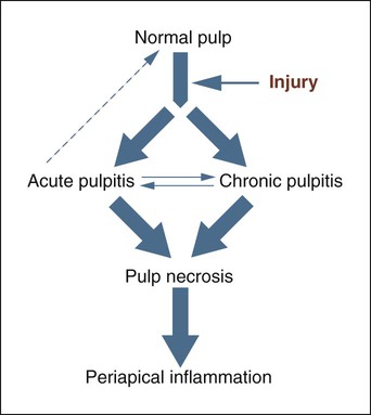
With chronic pulpitis, pulpal death is the characteristic end result (Figure 13-1). Removal of the cause may slow the process or occasionally may save the vitality of the pulp. Endodontic therapy or extraction is typically required. Chronic hyperplastic pulpitis is essentially an irreversible end stage that is treated with pulp extirpation and an endodontic filling or extraction.

image
FIGURE 13-1 Pulpitis pathways.

Periapical Abscess

Etiology

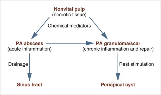
Numerous sequelae may follow untreated pulp necrosis and are dependent on the virulence of the microorganisms involved and the integrity of the patient’s overall defense mechanisms (Figure 13-2). From its origin in the pulp, the inflammatory process extends into the periapical tissues, where it may present as a granuloma or cyst (if chronic) or an abscess (if acute). Acute exacerbation of a chronic lesion may also be seen. Necrotic pulpal tissue debris, inflammatory cells, and bacteria, particularly anaerobes, all serve to stimulate and sustain the periapical inflammatory process.

Clinical Features

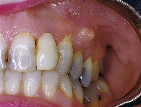
Patients with periapical abscesses typically have severe pain in the area of the nonvital tooth caused by pressure and the effects of inflammatory chemical mediators on nerve tissue. The exudate and neutrophilic infiltrate of an abscess put pressure on surrounding tissue, often resulting in slight extrusion of the tooth from its socket. Pus associated with a lesion, if not focally constrained, seeks the path of least resistance and spreads into contiguous structures (Figures 13-3 to 13-5). The affected area of the jaw may be tender to palpation, and the patient may be hypersensitive to tooth percussion. The involved tooth is unresponsive to electrical and thermal tests because of pulp necrosis.

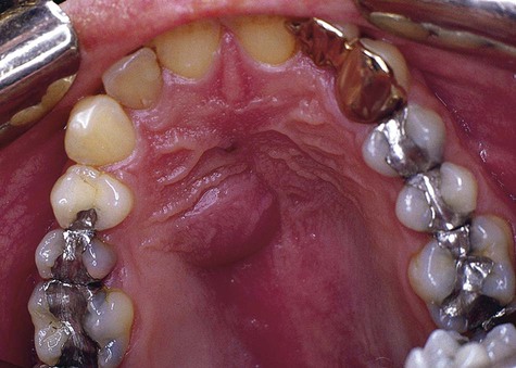
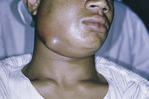
Because of the rapidity with which this lesion develops, time is generally insufficient for significant amounts of bone resorption to occur. Therefore, radiographic changes are slight and usually are limited to mild radiographic thickening of the apical periodontal membrane space. However, if a periapical abscess develops as a result of acute exacerbation of a chronic periapical granuloma, a radiolucent lesion is evident. The periapical granuloma represents the result of chronic inflammation at the apex of a nonvital tooth. This is a sequela of pulp necrosis, which may develop through acute or low-grade chronic inflammation. Notably, other, more serious conditions can occur in a periapical position (Box 13-1). Various clinical clues may alert the clinician that the periapical lesion may not be a simple dental granuloma (Box 13-2).

Histopathology

Microscopically, a periapical abscess appears as a zone of liquefaction composed of proteinaceous exudate, necrotic tissue, and viable and dead neutrophils (pus). Adjacent tissue containing dilated vessels and a neutrophilic infiltrate surrounds the area of liquefaction necrosis.

With chronicity, an abscess develops into a granuloma, which is composed of granulation tissue and fibrous tissue infiltrated by variable numbers of neutrophils, lymphocytes, plasma cells, and macrophages. (Note: Periapical granuloma is to be distinguished from granulomatous inflammation, which is a distinctive type of chronic inflammation that is characteristic of certain diseases [e.g., tuberculosis, sarcoidosis, histoplasmosis] and features a predominance of macrophages and often multinucleated giant cells.) Acute flare of a periapical granuloma would show an abundant neutrophilic infiltrate, in addition to granulation tissue and chronic inflammatory cells.

Treatment and Prognosis

Treatment of an acute periapical abscess requires observance of the standard principles of management of acute inflammation. Drainage should be established through an opening in the tooth itself or through the soft tissue surrounding the jaw, if cellulitis has developed. Antibiotics directed against the offending organism are required. Management must be thoughtful and skilled because the consequences of delayed or inappropriate treatment can be significant and occasionally life threatening.

Spread of an abscess may occur through one of several avenues. It may progress through the buccal cortical bone and gingival soft tissue, establishing a natural drain or sinus tract. The same type of situation may occur in the palate or skin; this depends on the original location of the abscess and the path of least resistance. If a drain is not established, the purulent exudate can cause an abscess or cellulitis in the soft tissues of the face, oral cavity, or neck. Cellulitis is an acute inflammatory process that is diffusely spread throughout the tissue rather than localized, as with an abscess. This variant is a result of infection by virulent organisms that produce enzymes that allow rapid spread through tissue. Bilateral cellulitis of the submandibular and sublingual spaces has been called Ludwig’s angina.

A dangerous situation occurs when acute infection involves major blood vessels, possibly resulting in bacteremia. Also, retrograde spread of the infection through facial emissary veins to the cavernous sinus may set up the necessary conditions for thrombus formation. Cavernous sinus thrombosis is an often fatal emergency situation.

Treatment of periapical granulomas and cysts is discussed in Chapter 10.

Stay updated, free dental videos. Join our Telegram channel

Jan 12, 2015 | Posted by in Oral and Maxillofacial Pathology | Comments Off on 13: Inflammatory Jaw Lesions

VIDEdental - Online dental courses

Get VIDEdental app for watching clinical videos