Tooth Morphology
Learning Outcomes
On completion of this chapter, the student will be able to achieve the following objectives:
• Pronounce, define, and spell the Key Terms.
• Identify the location of each permanent tooth.
• Use the correct terminology when discussing features of the permanent dentition.
• Describe the general and specific features of each tooth in the permanent dentition.
• Discuss clinical considerations of each tooth in the permanent dentition.
• Compare and contrast the features of the primary and permanent dentitions.
• Describe the general and specific features of the primary dentition.
Electronic Resources
 Additional information related to content in Chapter 12 can be found on the companion Evolve Web site.
• Labeling Exercise: Identify Each Tooth in the Permanent Dentition
• Labeling Exercise: Identify Each Tooth in the Primary Dentition
• WebLinks
Key Terms
Bicanineate A two-cusp type of mandibular second premolar.
Bifurcated Divided into two.
Bifurcation Area in which two roots divide.
Canine eminence (EM-i-nens) External vertical bony ridge on the labial surface of the canines.
Central groove Most prominent developmental groove on the posterior teeth.
Cingulum (SING-gyoo-lum) Raised, rounded area on the cervical third of the lingual surface.
Cusp Major elevation on the masticatory surfaces of canines and posterior teeth.
Cusp of Carabelli The fifth supplemental cusp found lingual to the mesiolingual cusp.
Diastema (dye-uh-STEE-muh) A space between two teeth.
Fossa (FOS-ah, FAW-seh) Wide, shallow depression on the lingual surfaces of anterior teeth.
Furcation (fur-KAY-shun) Area between two or more root branches.
Imbrication (im-bri-KAY-shun) lines Slight ridges that run mesiodistally in the cervical third of the teeth.
Incisal edge Ridge on permanent incisors that appears flattened on labial, lingual, or incisal view after tooth eruption.
Inclined cuspal planes Sloping areas between the cusp ridges.
Mamelon (MAM-uh-lon) Rounded enamel extension on the incisal ridges of incisors.
Marginal groove A developmental groove that crosses a marginal ridge and serves as a spillway, allowing food to escape during mastication.
Molars Teeth located in the posterior aspect of the upper and lower jaws.
Morphology (mor-FOL-uh-jee) Study of form and shape, as of the teeth.
Nonsuccedaneous (non-suk-se-DAY-nee-us) Pertaining to a permanent tooth that does not replace a primary tooth.
Pegged laterals Incisors with a pointed or tapered shape.
Succedaneous (suk-se-DAY-nee-us) teeth Permanent teeth that replace primary teeth.
Triangular groove A developmental groove that separates a marginal ridge from the triangular ridge of a cusp.
Tricanineate A three-cusp type of mandibular second premolar.
Trifurcated Divided into three.
Trifurcation Area in which three roots divide.
A thorough understanding of tooth morphology (the shape of teeth) is especially useful in the following clinical situations:
• Mounting dental radiographs (see Chapter 41)
• Assisting in charting a mouth with missing teeth and teeth that have “drifted”
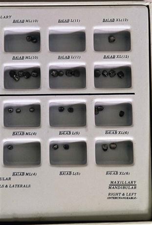
• Selecting temporary crowns from a box with a variety of shapes (Fig. 12-1)
• Forming matrix bands before application (see Chapter 48)
As you study tooth morphology, remember that there is always a certain amount of normal variation among individual teeth. Every tooth may not meet all the criteria for identification. When you understand the characteristics of each tooth, however, you will be able to differentiate among teeth, as well as between the left teeth and the right teeth in any particular group.
Anterior Permanent Dentition
The permanent anterior teeth include two central incisors, two lateral incisors, and two canines. The central incisors are closest to the midline, the lateral incisors are the second teeth from the midline, and the canines are the third teeth from the midline. All anterior teeth are succedaneous, which means they replace primary teeth of the same type.
The anterior teeth play an important role in a person’s appearance (Fig. 12-2). The size, shape, color, and position of the anterior teeth directly relate to how a person looks. Many people are extremely self-conscious about the appearance of their front (anterior) teeth. The anterior teeth also play an important role in speech and are necessary for formation of s and t sounds.

All anterior teeth have a cingulum, a rounded, raised area on the cervical third of the lingual surface. The lingual surface has rounded, raised borders on the mesial and distal surfaces called marginal ridges. Some anterior teeth have a fossa, a wide, shallow depression on the lingual surfaces (Fig. 12-3).
Rub your tongue up and down on the lingual side of your front teeth. The bump or raised area you feel near the gingiva is the cingulum, and the deeper area is the fossa.
Permanent Incisors
There are eight permanent incisors—four maxillary and four mandibular. The maxillary group comprises two central incisors and two lateral incisors, as does the mandibular group. These teeth complement each other in form and function. The central incisors erupt about a year or so before the lateral incisors do.
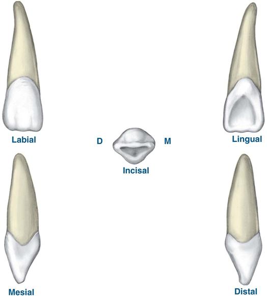
Maxillary Central Incisors
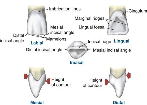
The maxillary central incisors (#8 and #9) have unique anatomic features (Fig. 12-4). They are larger than mandibular central incisors in all dimensions, especially in width (mesiodistally). The labial surfaces are more rounded from the incisal aspect. The root of the maxillary central is shorter than the roots of other permanent maxillary teeth. The marginal ridges, lingual fossa, and cingulum are more prominent on the maxillary central incisor than on the mandibular central incisor.
When an incisor is newly erupted, the incisal portion is rounded; this is called the incisal ridge. The term edge implies an angle formed by the merging of two flat surfaces. Therefore, an incisal edge does not exist on an incisor until occlusal wear has created a flattened surface on the incisal portion. The incisal edge is also known as the incisal surface or incisal plane. The incisal edges of maxillary incisors have a lingual inclination (slant). The incisal edges of mandibular incisors have a labial inclination. The incisal planes of mandibular and maxillary incisors are parallel to each other and work together to create a cutting action, similar to that produced by the blades of a pair of scissors.
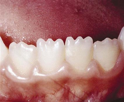
When newly erupted, the central and lateral incisors have three mamelons, or rounded enamel extensions, on the incisal ridge, or edge (Fig. 12-5). These mamelons usually undergo attrition (wearing away of a tooth surface) shortly after eruption. Then, the incisal ridge appears flattened and becomes the incisal edge.
Maxillary Lateral Incisors
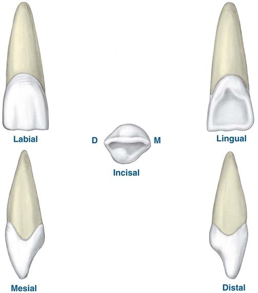
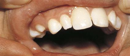
The maxillary lateral incisors (#7 and #10) are smaller than the central incisors in all dimensions except root length (Fig. 12-6). They usually erupt after the maxillary central incisors do. The crown of a maxillary lateral incisor has a single root that is relatively smooth and straight but may curve slightly distally. Remember this feature; it will be helpful when you are mounting radiographs.
The lateral incisors vary in form to a greater extent than any other tooth except the third molars, and frequently, they are congenitally missing. It is not uncommon to find maxillary lateral incisors with a pointed or tapered shape; such teeth are called pegged laterals (Fig. 12-7). Because of variations in form, the permanent maxillary lateral incisors can present challenges during preventive, restorative, and orthodontic procedures. Often, unusually large open contacts (spaces between teeth) called diastemas occur in this area because of variations in the size and shape of the lateral incisor and its position in the arch. Fortunately, dental materials and techniques are available today to correct these conditions.

Mandibular Incisors
The permanent mandibular incisors are the smallest teeth of the permanent dentition and the most symmetric. The central and lateral incisors of the mandibular arch closely resemble each other.
In contrast to the maxillary central and lateral incisors, the mandibular lateral incisor is larger than the mandibular central incisor. The lower, mandibular incisors generally erupt before the upper, maxillary incisors do. It is rare for developmental disturbances to occur with mandibular incisors.
Supragingival tooth deposits such as plaque, calculus, and stain tend to collect in the lingual concavity of the mandibular incisors. The buildup of these deposits is increased by the release of saliva, with its mineral contents, from the sublingual and submandibular salivary glands in the floor of the mouth.
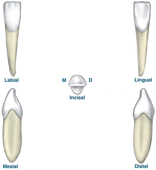
Mandibular Central Incisors
The mandibular central incisors (#24 and #25) are the smallest teeth in the dental arches. They have a small centered cingulum, a subtle lingual fossa, and equally subtle marginal ridges.
The crown of a mandibular central incisor is narrower on the lingual surface than on the labial surface (Fig. 12-8). Developmental horizontal lines on anterior teeth, or imbrication lines, and developmental depressions usually are not obvious.
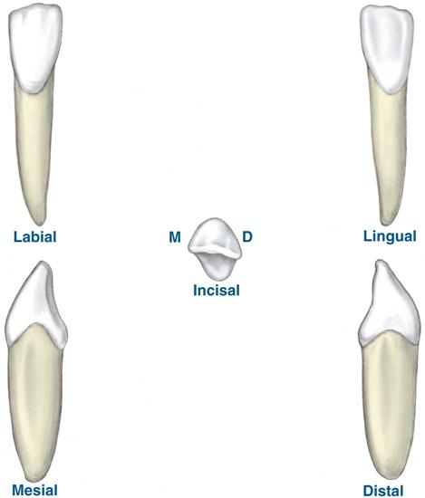
Mandibular Lateral Incisors
The mandibular lateral incisors (#23 and #26) are slightly larger than the mandibular central incisors but otherwise are similar. The lateral teeth usually erupt after the mandibular central incisors do. The mesial side of the crown is often longer than the distal side, causing the incisal ridge, which is straight, to slope downward in a distal direction. This helps in differentiating the right mandibular lateral incisor from the left incisor (Fig. 12-9).
Permanent Canines
The permanent canines are the four anterior teeth that are located at the corner of each quadrant for each dental arch (Fig. 12-10, A). They are commonly referred to as the “cornerstone” of the dental arches. These teeth are the most stable in the mouth. Their name is derived from the Latin word for dog (“canus”) because canines resemble dogs’ teeth. The permanent canines are the longest teeth in the dentition. The canine has a particularly long, thick root. The root is usually the same length as or twice the length of the crown. The crown of the canine is shaped in a manner that promotes cleanliness. Because of their self-cleansing shape and sturdy anchorage in the jaws, the canines are usually the last teeth to be lost.
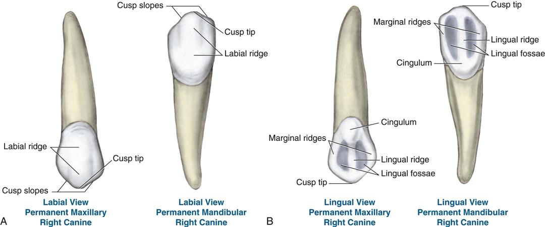
Another important characteristic of the canines is the cosmetic value of the canine eminence. This is the bony ridge over the labial portion of the roots of the canines that forms facial contours. Patients commonly call the canines their “eyeteeth” and often notice the normally slightly deeper yellow color of their canines compared with their incisors. You may hear the term cuspid used instead of canine. This is an older term that was used because these were the only teeth in the permanent dentition that had one cusp. A cusp is a major elevation on the masticatory surface of a canine or posterior tooth.
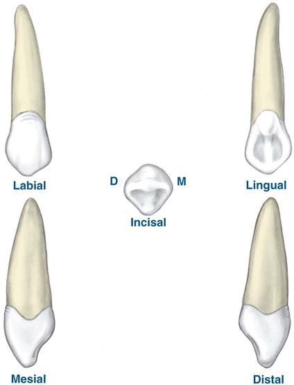
Maxillary Canines
The maxillary canines (#6 and #11) usually erupt after the mandibular canines, the maxillary incisors, and possibly the maxillary premolars do. The maxillary canines have a larger and more developed cusp compared with the mandibular canines (Figs. 12-10, B, and 12-11).
Stay updated, free dental videos. Join our Telegram channel
VIDEdental - Online dental courses


