PARANASAL SINUSES
Overview and Topographic Anatomy
Overview and Topographic Anatomy
GENERAL INFORMATION
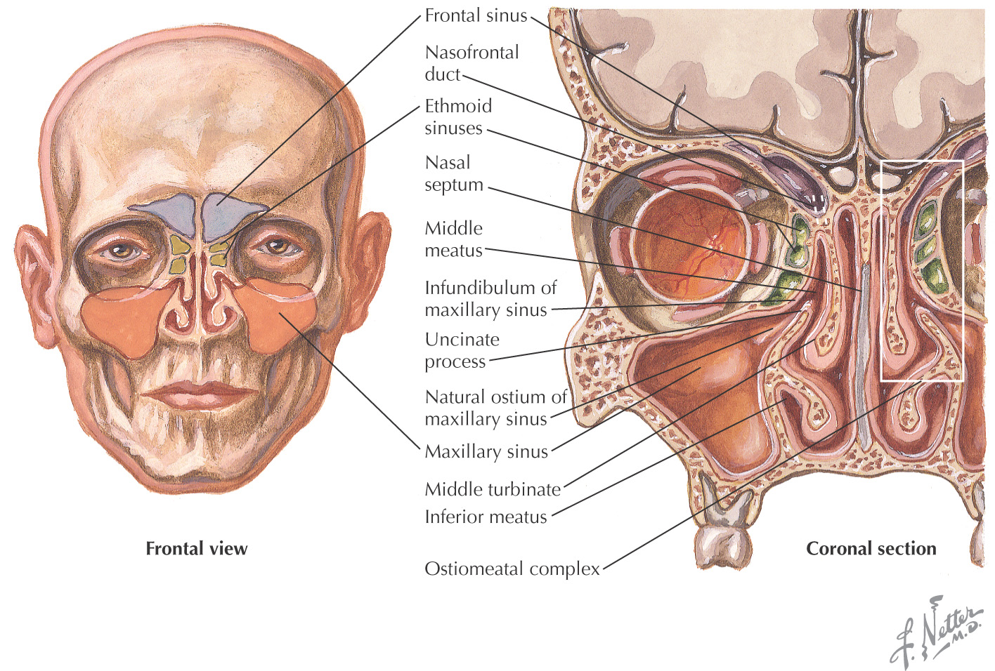
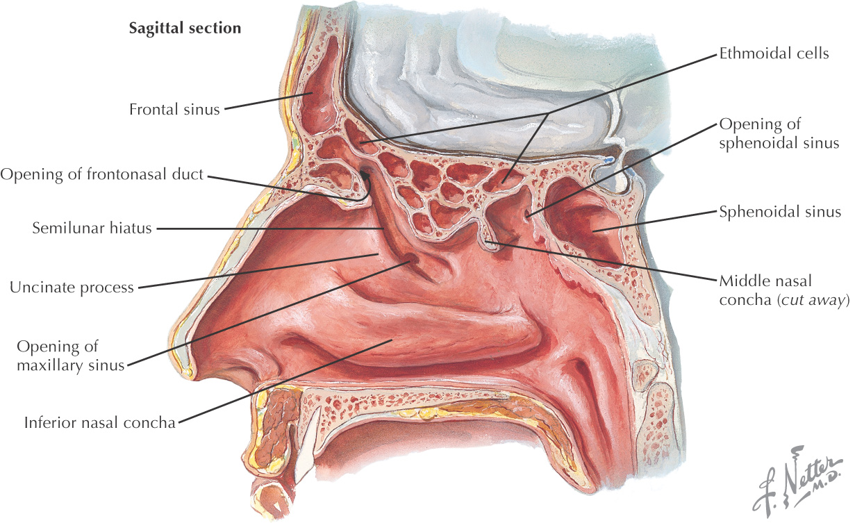
Paranasal sinuses: invaginations from the nasal cavity that drain into spaces associated with the lateral nasal wall
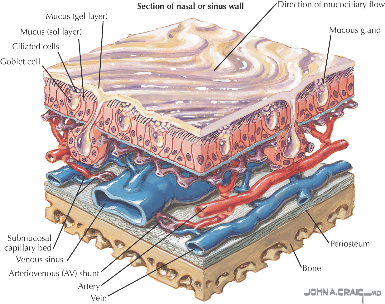
Each is lined by a respiratory epithelium
Morphology of the sinuses is highly variable
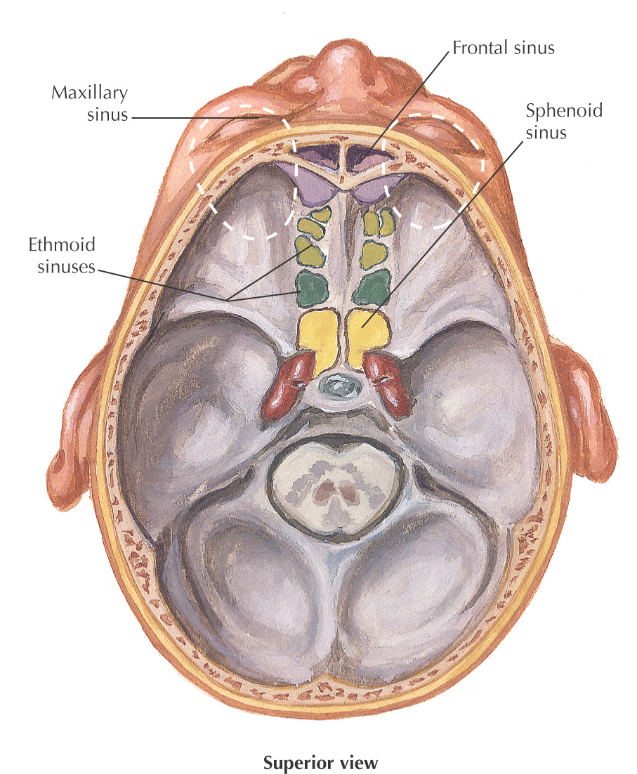
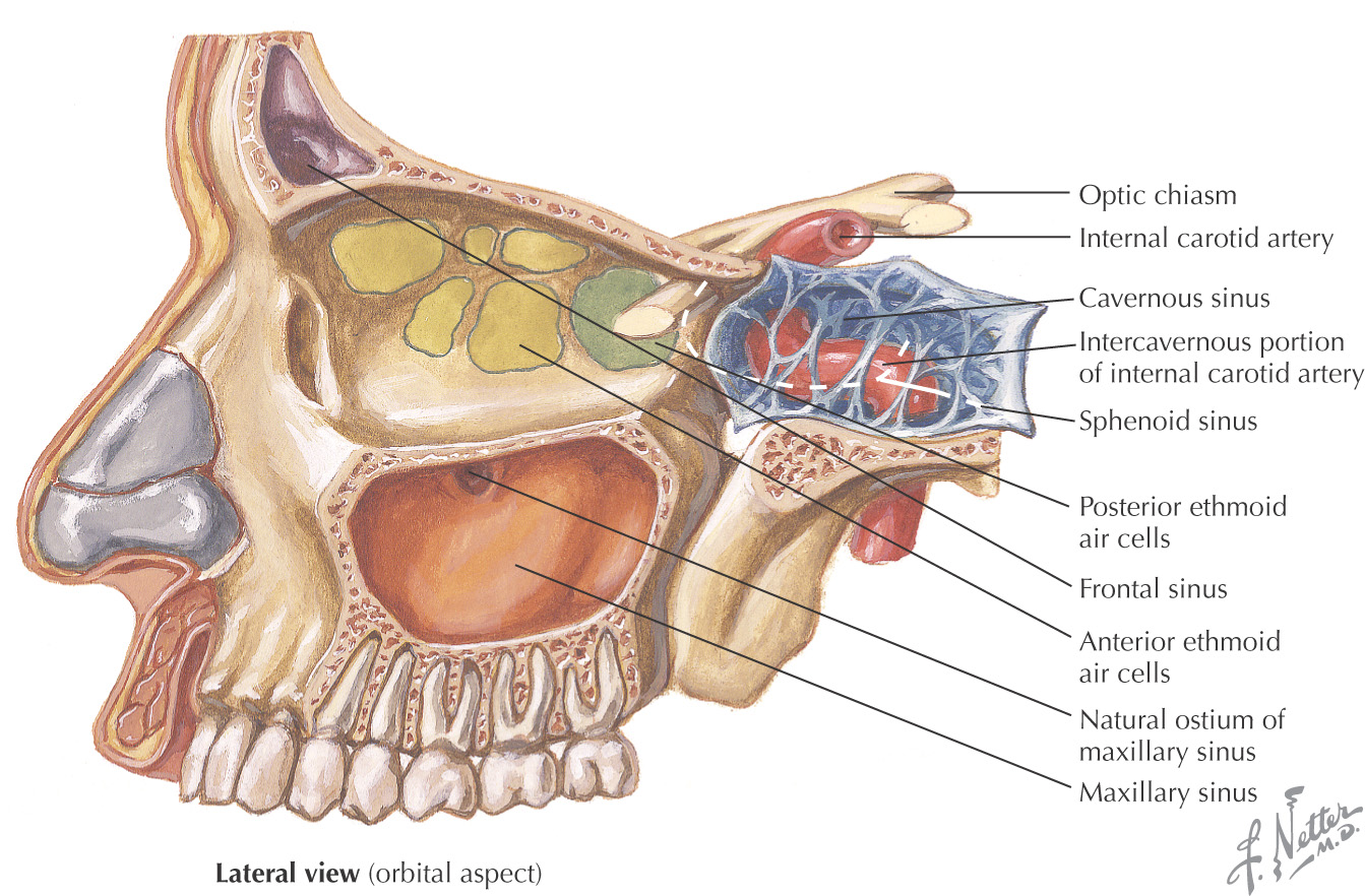
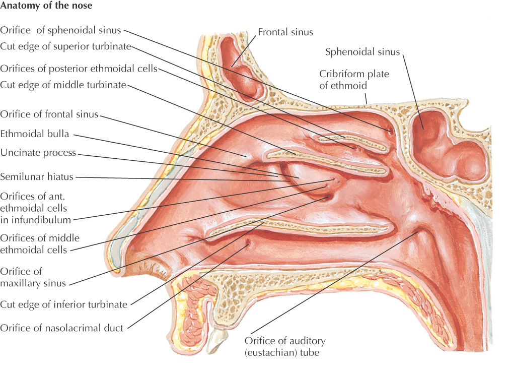
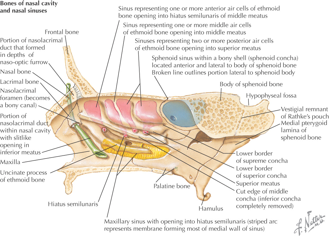
DRAINAGE OF THE PARANASAL SINUSES AND ASSOCIATED STRUCTURES
All paranasal sinuses drain into the nasal cavity
Different sinuses serve as drainage conduits for different regions

Frontal Sinus
GENERAL INFORMATION
The two frontal sinuses typically are asymmetrical
Rudimentary at birth and usually well-developed by the age of 7 or 8 years
Display a prime expansion when the 1st deciduous molars erupt and another when the permanent molars begin to appear at about age 6
Drainage varies; often drain in front of, above, or into the ethmoidal infundibulum
Primary lymphatic drainage is to the submandibular lymph nodes
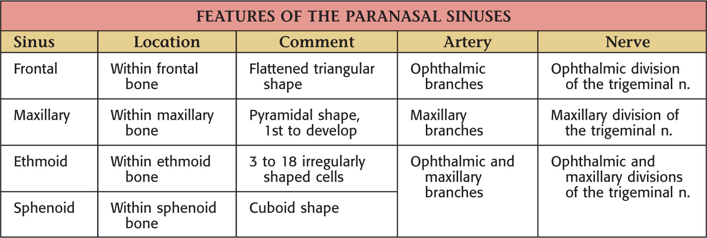
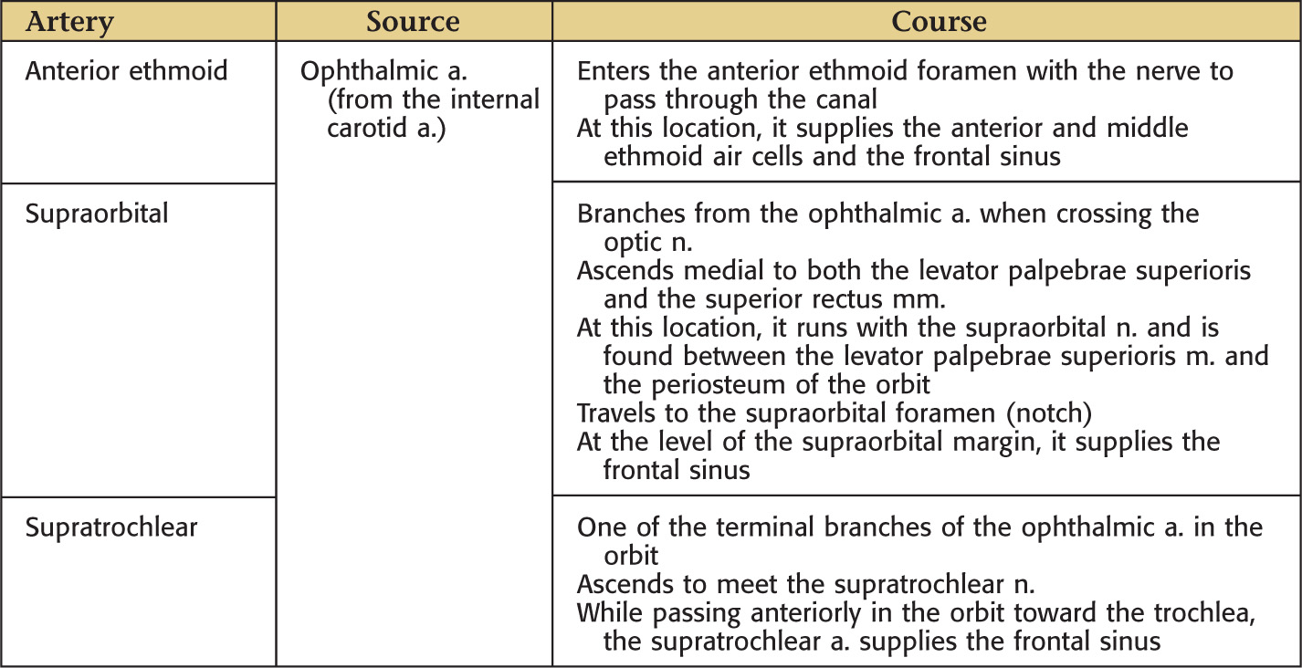
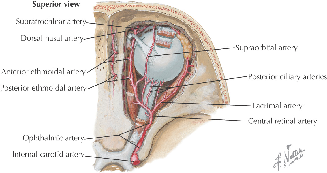
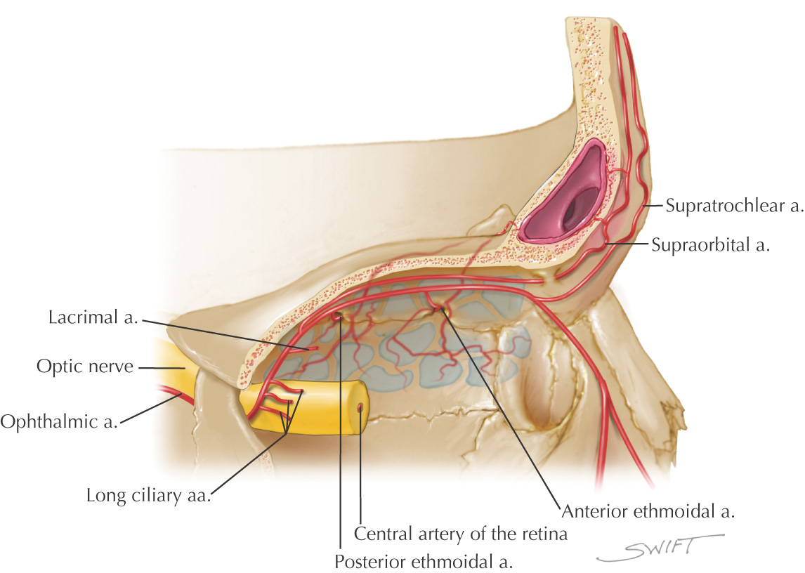
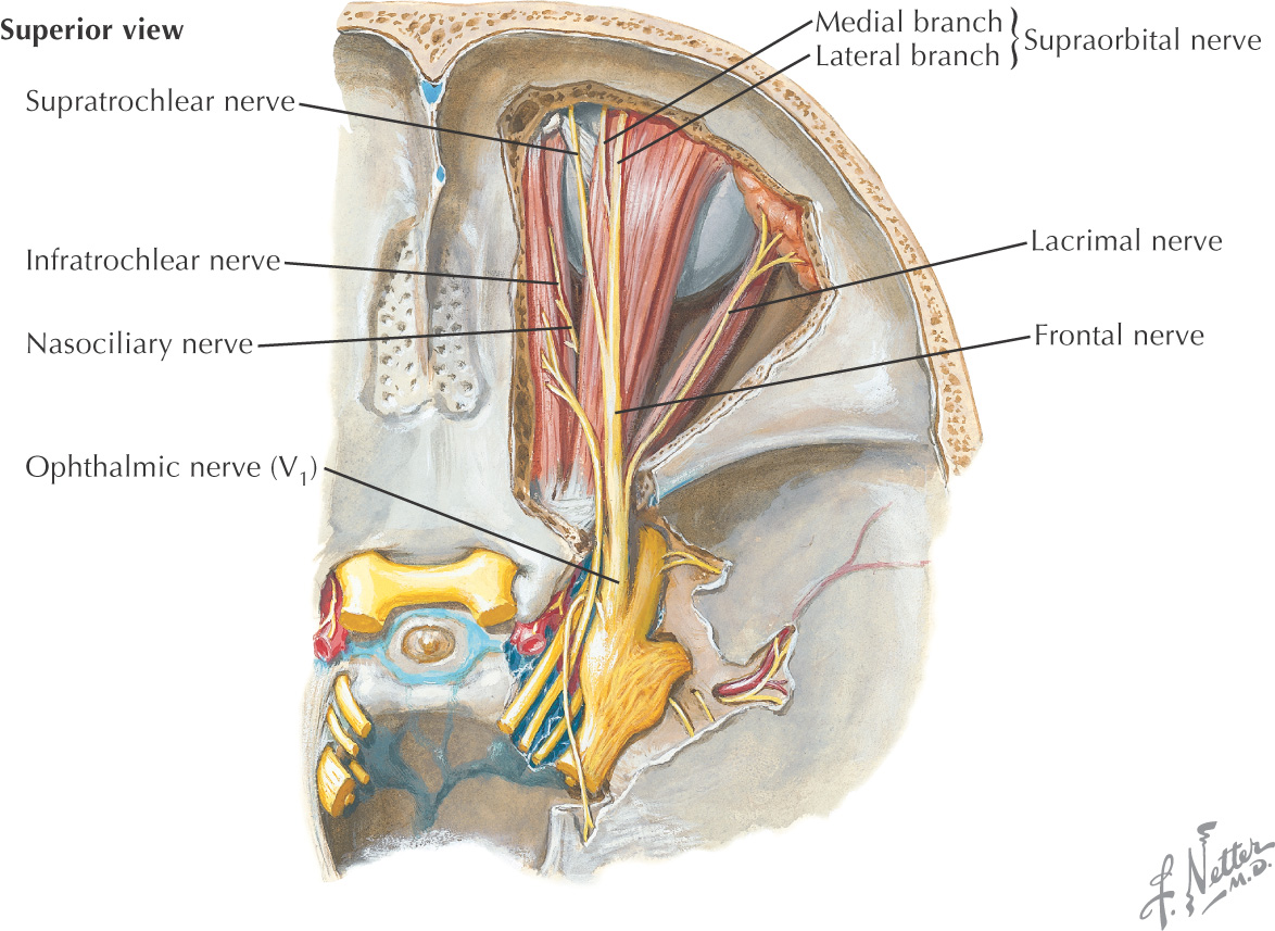
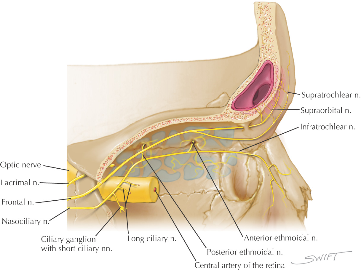
The frontal sinus receives its nerve supply from branches of the ophthalmic division of the trigeminal nerve
Relations of Sinus
• Superior: anterior cranial fossa and contents
• Inferior: orbit, anterior ethmoidal sinuses, nasal cavity
• Anterior: forehead, superciliary arches
• Posterior: anterior cranial fossa and contents
Location of Ostium
Middle meatus

ARTERIAL SUPPLY
NERVE SUPPLY

Ethmoid Sinus
GENERAL INFORMATION
May find 3 to 18 ethmoid air cells on each side
Ethmoid air cells may invade any of the other 3 sinuses
The middle ethmoid air cells produce the swelling on the lateral wall of the middle meatus called the ethmoid bulla
Primary lymphatic drainage is to the submandibular lymph nodes for the anterior and middle ethmoid sinuses; and the retropharyngeal lymph nodes for the posterior ethmoid sinus
Relations of Sinus
• Superior: anterior cranial fossa and contents, frontal bone with sinus
•/>
Stay updated, free dental videos. Join our Telegram channel

VIDEdental - Online dental courses


