11
Development of the Craniofacial Region
Embryonic Development of the Face
Figure 11.1 is a scanning electron micrograph of the face of a mouse embryo at around 10.5 days of development.
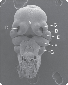
Name the Structures Labelled A–G
Briefly Describe How Structures A–E Contribute to Formation of the Developing Face
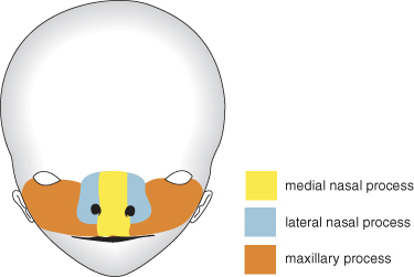
Structures A–E contribute to formation of the middle third of the face. In particular, the maxillary processes (E) grow medially towards the midline, fusing with the lateral nasal processes (C) to form the naso-lacrimal duct, cheek and alar base of the nose. Further growth results in fusion with the medial nasal processes (B) in the midline to form the middle part of the nose, lip philtrum and primary palate, including the incisor teeth (Figure 11.2).
What Contribution Does Structure F Make to the Face?
The mandibular process of the first pharyngeal arch will form the lower jaw or mandible, including the dentition. In addition, it will form the lower lip and muscles of mastication.
What Would Be the Equivalent Age of This Embryo in Relation to Human Development?
The equivalent is around 6 weeks.
What Contribution Does Structure G Make to the Developing Facial Region?
Structure G is the second pharyngeal arch and will form the upper part of the hyoid bone, the styloid process, stapes of the middle ear and stylohyoid ligament. In addition, it will form muscles innervated by the facial nerve, including the muscles of facial expression.
Tooth Development
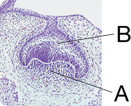
Figure 11.3 Shows a Developing Tooth Germ. Identify the Labelled Structures
What Parts of the Tooth Will These Structures Give Rise to?
The dental papilla is derived from neural crest cells, which form all the structures within the tooth (dentine of the crown and root, pulp, periodontal ligament) with the exception of enamel, which is derived from the epithelium of the early jaw.
What Stage of Development Are These Tooth Germs At?
They are at the cap stage.
What Is the Significance of This Stage?
The cap stage heralds the establishment of shape within the tooth germ in the human embryo, defining a tooth germ as incisor, canine, premolar or molar.
How Do the Tooth Germs of Different Tooth Classes Acquire Their Distinctive Shape?
Tooth development is controlled by molecular signalling interactions between the epithelium and underlying neural crest cells within the tooth germ. The molecular instructions for shape are established early in this process, by specific domains of homeobox gene expression specifying a tooth germ as ‘incisor’ or ‘molar’.
What Are the Main Features Identifiable on This Dental Panoramic Radiograph (Figure 11.4)?
- Congenital absence of the UR4, UR2, UL2, LL5; this has led to associated spacing of the remaining dentition
- Retained and infra-occluded LLE
- LL4 impacted against the LLE
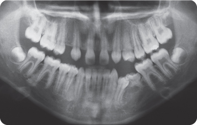
How Would You Describe the Condition?
Selective tooth agenesis or hypodontia.
What Is Hypodontia?
Hypodontia refers to agenesis of one to six teeth, excluding the third molars; whilst oligodontia refers to agenesis of more than six teeth, excluding the third molars.
In Caucasians, What Percentage of the Population Would Be Expected to Have a Congenitally Absent Maxillary Lateral Incisor?
Around 2%.
What Is the Cause of Selective Tooth Agenesis?
Selective tooth agenesis is primarily a genetic condition, caused by gene mutation. In the majority of cases it is non-syndromic and occurs in isolation, with no other anomalies being seen in the individual.
In a minority of cases, selective tooth agenesis can be seen in association with other anomalies in a syndromic condition.
How Can Tooth Agenesis Be Inherited Within Families?
All modes of inheritance have been described in association with tooth agenesis, including autosomal dominant and autosomal recessive Mendelian inheritance. Cases can also be sporadic, arising in families with no previous history.
Name Any Genes Known to Be Mutated in Cases of Non-Syndromic Tooth Agenesis
To date, relatively few human genes have been identified in cases of non-syndromic tooth agenesis. Examples include:
- MSX1: Encodes a transcription factor
- PAX9: Encodes a transcription factor
- AXIN2: Encodes a protein involved in stabilizing components of the Wnt signalling pathway
- WNT10A: Encodes a signalling protein
- LTBP3: Encodes a TGF-beta binding protein
- EDA: Encodes a signalling protein
- EDARADD: Encodes an intra-cellular adapter protein.
This Child Attended Complaining of Difficulty in Eating. What Are the Main Features of Their Dental Appearance (Figure 11.5)?
- Congenital tooth absence
- Microdont, poorly-formed conical teeth
- Dental spacing
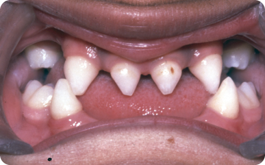
What Condition Is This Child Most Likely to Be Affected By?
Ectodermal dysplasia, a heterogenous group of conditions characterized primarily by:
- Hypodontia (or often oligodontia) affecting both primary and permanent dentitions, with those teeth that do develop often being poorly formed
- Sparse and poorly formed hair
- Absence of sweat glands
- Nail anomalies.
How Are These Conditions Inherited?
Sex-linked autosomal recessive inheritance is the most common form, with only males being affected. A less common autosomal recessive form also exists, affecting males and females.
Why Are the Teeth, Hair and Sweat Glands All Affected?
All of these organs develop in the embryo via very specific molecular interactions between their constituent epithelial and mesenchymal t/>
Stay updated, free dental videos. Join our Telegram channel
VIDEdental - Online dental courses


