1: Vesiculobullous Diseases

Vesiculobullous Diseases

Viral Disease

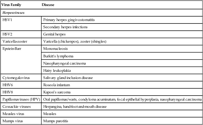
Oral mucous membranes may be infected by one of several different viruses, each producing a relatively distinct clinical pathologic picture (Table 1-1).

TABLE 1-1

VIRUSES RELEVANT TO DENTISTRY

Virus Family Disease
Herpesviruses
HSV1 Primary herpes gingivostomatitis
Secondary herpes infections
HSV2 Genital herpes
Varicella-zoster Varicella (chickenpox), zoster (shingles)
Epstein-Barr Mononucleosis
Burkitt’s lymphoma
Nasopharyngeal carcinoma
Hairy leukoplakia
Cytomegalovirus Salivary gland inclusion disease
HHV6 Roseola infantum
HHV8 Kaposi’s sarcoma
Papillomaviruses (HPV) Oral papillomas/warts, condyloma acuminatum, focal epithelial hyperplasia, nasopharyngeal carcinoma
Coxsackie viruses Herpangina, hand-foot-and-mouth disease
Measles virus Measles
Mumps virus Mumps parotitis

image

HHV, Human herpesvirus; HSV, herpes simplex virus.

The herpesviruses are a large family of viruses characterized by a DNA core surrounded by a capsid and an envelope. Seven types of herpesviruses are known to be pathogenic for humans, with six of these linked to diseases in the head and neck area.

Herpes Simplex Infection

Herpes simplex virus (HSVs) infections are common vesicular eruptions of the skin and mucosa. They occur in two forms—primary (systemic) and secondary (localized)—and may be localized or secondary in nature. Both forms are self-limited, but recurrences of the secondary form are common because the virus can be sequestered within ganglionic tissue in a latent state. Control of symptoms rather than cure is the usual goal of treatment.

Pathogenesis

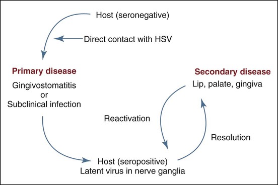
Physical contact with an infected individual or with body fluids is the typical route of HSV inoculation and transmission for a seronegative individual who has not been previously exposed to the virus, or possibly for someone with a low titer of protective antibody to HSV (Figure 1-1). The virus binds to the cell surface epithelium via heparan sulfate, which leads to transmembrane cytoplasmic insertion, followed by sequential activation of specific genes during the lytic phase of infection. These genes include immediate early (IE) and early (E) genes, coding for regulatory proteins and for DNA replication, and late (L) genes, coding for structural proteins. Documentation of the spread of infection through airborne droplets, contaminated water, or contact with inanimate objects is generally lacking. During the primary infection, only a small percentage of individuals show clinical signs and symptoms of infectious systemic disease, whereas a vast majority experience only subclinical disease. This latter group, now seropositive, can be identified through the laboratory detection of circulating antibodies to HSV.

The incubation period after exposure ranges from several days to 2 weeks. In overt primary disease, a vesiculoulcerative eruption (primary gingivostomatitis) typically occurs in the oral and perioral tissues. The focus of eruption is expected at the original site of contact.

After resolution of primary herpetic gingivostomatitis, the virus is believed to migrate, through some unknown mechanism, along the periaxon sheath of the trigeminal nerve to the trigeminal ganglion, where it is capable of remaining in a latent or sequestered state. During latency, no infectious virus is produced; early, but not late, genes are expressed; and no free virus is present. No major histocompatibility (MHC) antigens are expressed, no T-cell response occurs during latency.

Reactivation of virus may follow exposure to sunlight (“fever blisters”), exposure to cold (“cold sores”), trauma, stress, or immunosuppression causing a secondary or recurrent infection.

An immunocompromised host may develop severe secondary disease. HSV-seropositive patients being prepared for bone marrow transplants with chemotherapeutic drugs such as cyclophosphamide (with or without total-body radiation) are at risk for a secondary herpes infection that is particularly severe. Post-transplant chemotherapy also predisposes seropositive patients to severe recurrent oral infection. HSV-seropositive patients infected with human immunodeficiency virus (HIV) may also exhibit intense secondary disease. Uncommonly, HIV-positive patients may have lesions that are coinfected by both HSV and cytomegalovirus. The pathogenesis of dually infected ulcers is unclear. Seronegative patients may rarely be affected with herpetic disease during immunosuppressive transplant states.

The reactivated virus travels by way of the trigeminal nerve to the originally infected epithelial surface, where replication occurs, resulting in a focal vesiculoulcerative eruption. Presumably because the humoral and cell-mediated arms of the immune system have been sensitized to HSV antigens, the lesion is limited in extent, and systemic symptoms usually do not occur. As the secondary lesion resolves, the virus returns to and remains in latent form within the trigeminal ganglion with no evidence of viral particles remaining within the previously involved epithelium. It is believed that nearly all secondary lesions develop from reactivated latent virus, although reinfection by different strains of the same subtype is considered a remote possibility.

Most oral-facial herpetic lesions are due to HSV type 1 (HSV1), although a small percentage may be caused by HSV type 2 (HSV2) as a result of oral-genital contact. Lesions caused by either virus are clinically indistinguishable. HSV2 has a predilection for genital mucosa, with infection having a pathogenesis similar to that of HSV1 infection of the head and neck. Latent virus, however, is sequestered in the lumbosacral ganglion. Previous HSV1 infection may provide some protection against HSV2 infection because of antibody cross-reactivity.

Viral shedding, a phenomenon in which previously infected individuals may be capable of transmitting the virus, has been reported, although the relationship between frequency of shedding, viral titer, and actual transmission is unknown. Asymptomatic shedding of intact HSV particles in saliva can be identified in approximately 2% to 10% of healthy adults in the absence of clinical disease. The level of risk of infection from “shedders” to others has not been measured, although it is probably low and dependent on the quantity of shed viral particles and the susceptibility of the new host.

Based on clinical observations, it was long believed that an association exists between HSV2 and carcinoma of the cervix. Although this virus can transform cervical epithelial cells in culture, it is not generally found in cervical carcinoma and is no longer thought to play a central role in the disease. Similarly, some sort of association between HSV1 and oral cancer was long suspected. For example, in experimental studies of oral tissues, evidence suggests that HSV1 is oncogenic in vitro, provided that cytolysis is inhibited by ultraviolet (UV) light or chemicals. In the hamster cheek pouch model, HSV can induce genetic changes, including chromosome translocations, mutations, and gene amplifications; in other animal models, HSV acts as a cocarcinogen with tobacco and other chemical carcinogens. A high prevalence of HSV antibodies and antibodies to the immediate early proteins has been noted among patients with oral cancer, but the significance of this is unclear. Despite this, it is generally believed that little evidence suggests that HSV1 is etiologically linked to oral cancer.

Clinical Features

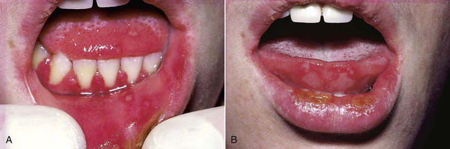
Primary Herpetic Gingivostomatitis

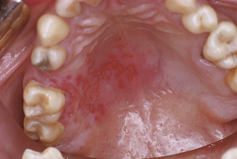
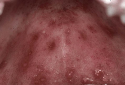
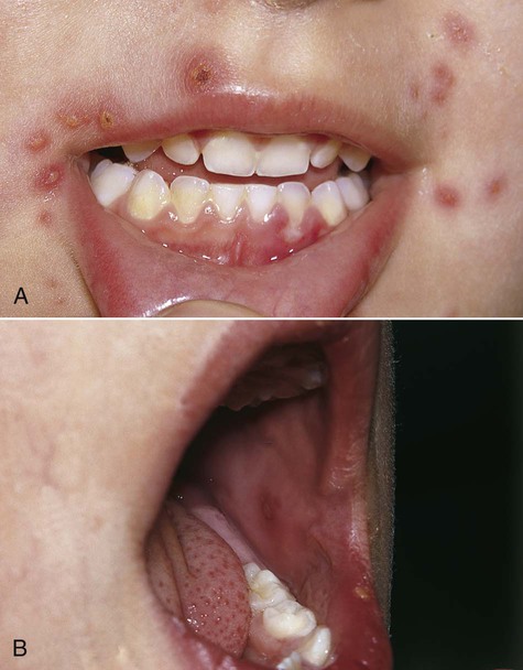
Primary disease is usually seen in children, although adults who have not been previously exposed to HSV or who fail to mount an appropriate response to a previous infection may be affected. By age 15, about half the population is infected. The vesicular eruption may appear on the skin, vermilion, and oral mucous membranes (Box 1-1 and Figure 1-2). Intraorally, lesions may appear on any mucosal surface. This is in contradistinction to the recurrent form of the disease, in which lesions are confined to the lips, hard palate, and gingiva. The primary lesions are accompanied by fever, arthralgia, malaise, anorexia, headache, and cervical lymphadenopathy.

After the systemic primary infection runs its course of about 7 to 10 days, lesions heal without scar formation. By this time, the virus may have migrated to the trigeminal ganglion to reside in a latent form. The number of individuals with primary clinical or subclinical infection in which virus assumes dormancy in nerve tissue is unknown.

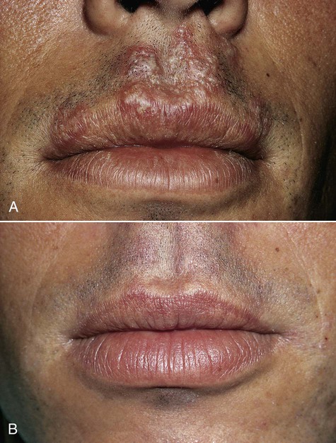
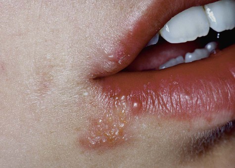
Secondary, or Recurrent, Herpes Simplex Infection

Secondary herpes represents the reactivation of latent virus. It is believed that only rarely does reinfection from an exogenous source occur in seropositive individuals. Antibodies to HSV are present in a large majority of the population (up to 90%), and up to 40% of this group may develop secondary herpes. The pathophysiology of recurrence has been related to a breakdown in local immunosurveillance or an alteration in local inflammatory mediators that allows the virus to replicate.

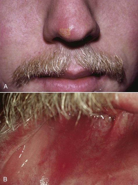
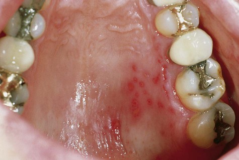
Patients usually have prodromal symptoms of tingling, burning, or pain in the site at which lesions will appear. Within a matter of hours, multiple fragile and short-lived vesicles appear. These become unroofed and coalesce to form maplike superficial ulcers. The lesions heal without scarring in 1 to 2 weeks and rarely become secondarily infected (Box 1-2; Figures 1-3 to 1-6). The number of recurrences is variable and ranges from one per year to as many as one per month. The recurrence rate appears to decline with age. Secondary lesions typically occur at or near the same site with each recurrence. Regionally, most secondary lesions appear on the vermilion and surrounding skin. This type of disease is usually referred to as herpes labialis. Intraoral recurrences are almost always restricted to the hard palate or gingiva.
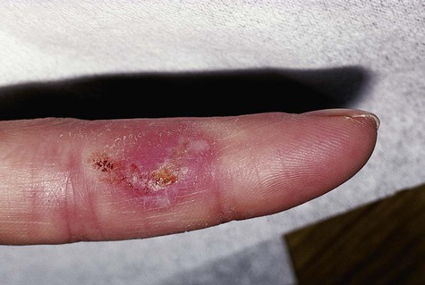
Herpetic Whitlow

Herpetic whitlow is a primary or a secondary HSV infection involving the finger(s) (Figure 1-7). Before the universal use of examination gloves, this type of infection typically occurred in dental practitioners who had been in physical contact with infected individuals. In the case of a seronegative clinician, contact could result in a vesiculoulcerative eruption on the digit (rather than in the oral region), along with signs and symptoms of primary systemic disease. Recurrent lesions, if they occur, would be expected on the finger(s). Herpetic whitlow in a seropositive clinician (e.g., one with a history of HSV infection) is believed to be possible, although less likely because of previous immune stimulation by herpes simplex antigens.
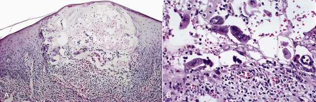
image
FIGURE 1-7 Herpetic whitlow.

Pain, redness, and swelling are prominent with herpetic whitlow and can be very pronounced. Vesicles or pustules eventually break and become ulcers. Axillary and/or epitrochlear lymphadenopathy may also be present. The duration of herpetic whitlow is protracted and may be as long as 4 to 6 weeks.

Histopathology

Microscopically, intraepithelial vesicles containing exudate, inflammatory cells, and characteristic virus-infected epithelial cells are seen (Figure 1-8). Virus-infected keratinocytes contain one or more homogeneous, glassy nuclear inclusions. These cells are also readily found on cytologic preparations. HSV1 cannot be differentiated from HSV2 histologically. After several days, herpes-infected keratinocytes cannot be demonstrated in biopsy or cytologic preparations. Herpes simplex lesions in HIV-positive patients may be coinfected with cytomegalovirus. The pathogenesis and significance of this phenomenon are undetermined.

Differential Diagnosis

Primary herpetic gingivostomatitis is usually apparent from clinical features. It can be confirmed by a virus culture (which requires 2 to 4 days for positive identification). Immunologic methods using monoclonal antibodies or DNA in situ hybridization techniques have become useful for specific virus identification in tissue sections.

Systemic signs and symptoms coupled with the oral ulcers may require differentiation from streptococcal pharyngitis, erythema multiforme, and acute necrotizing ulcerative gingivitis (ANUG, or Vincent’s infection). Clinically, streptococcal pharyngitis does not involve the lips or perioral tissues, and vesicles do not precede the ulcers. Oral ulcers of erythema multiforme are larger, usually without a vesicular stage, and are less likely to affect the gingiva. ANUG also commonly affects young adults; however, oral lesions are limited to the gingiva and are not preceded by vesicles. Moreover, considerable pain and oral malodor are often reported with ANUG.

Secondary herpes is often confused with aphthous stomatitis but can usually be distinguished from it on the basis of clinical features. Multiple lesions, vesicles preceding ulcers, and palatal and gingival location are indicative of herpesvirus infection. In contrast to herpetic lesions, aphthae are found almost exclusively on nonkeratinized mucosa, such as the floor of the mouth, the alveolar mucosa, and the buccal labial mucosa.

Treatment

One of the most important factors in the treatment of HSV infection is timing. For any drug to be effective, it must be used as soon as possible after recognition of early or prodromal symptoms. No later than 48 hours from the onset of symptoms is generally regarded as the ideal time to start therapeutic measures. A number of virus-specific drugs have been developed. Acyclovir and its analogs have shown the greatest efficacy in the treatment of mucocutaneous infection.

The rationale for the use of topical agents resides in their ability to interrupt viral replication through inhibition of DNA polymerization (acyclovir, penciclovir) or by interference with virus-epithelial interaction and prevention of intracellular access (docosanol). In herpes-infected cells, acyclovir is converted by a virus-induced enzyme (thymidine kinase) and other cellular enzymes to a form that inhibits primarily viral DNA polymerase rather than host cell DNA polymerase. The end result is interruption of viral DNA synthesis and relative sparing of cellular DNA synthesis.

Systemic agents including acyclovir (200- to 400-mg tablets 5 times per day) and valacyclovir are effective in the control of primary genital herpes and, to a lesser degree, primary oral herpetic gingivostomatitis. Supportive therapy (fluids, rest, oral lavage, analgesics, and antipyretics) is an essential component of any primary herpes simplex regimen.

Secondary herpes can be controlled to some degree with systemic acyclovir. Recurrences are not prevented, but the course and severity of the disease are favorably affected. Prophylactic systemic acyclovir is effective in problematic cases and in immunosuppressed patients. In HIV-positive patients with severe disease, aggressive therapy that may include intravenous acyclovir or ganciclovir may be necessary.

Topical acyclovir, although only somewhat effective, has been advocated by some for the treatment of secondary or recurrent herpes. A 5% acyclovir (or analog) ointment applied 5 times per day when symptoms first appear slightly reduces the duration of herpes lesions and may abort some lesions. Also, topical n-docosanol (10%) has been used effectively, although randomized clinical trials are lacking. Topical management does not prevent recurrence, however, and may be ineffective in some patients.

The question of chronic viral suppression in relation to multiple recurrent herpes simplex infections in some individuals has been addressed. Within this paradigm, treatment approaches include the use of intermittent episodic, intermittent suppressive, or chronic suppressive therapy, in conjunction with an evidence-based dosing regimen.

Varicella-Zoster Infection

The overall incidence of varicella infection the United States has been substantially reduced of late by 76% to 87% as a result of widespread vaccination programs. Primary varicella-zoster virus (VZV) infection in seronegative individuals is known as varicella or chickenpox; secondary or reactivated disease is known as herpes zoster or shingles (Box 1-3). Structurally, VZV is very similar to HSV, with a DNA core, a protein capsid, and a lipid envelope. Microscopically, striking similarities have been noted between herpes simplex conditions. A cutaneous or mucosal vesiculoulcerative eruption following reactivation of latent virus is typical of both VZV and HSV infections. Several signs and symptoms, however, appear to be unique to each infection.

Pathogenesis

Varicella

Varicella is believed to be transmitted predominantly through the inhalation of contaminated droplets. The condition is very contagious and is known to spread readily from person to person. Much less commonly, direct contact is an alternative way of acquiring the disease. During the 2-week incubation period, virus proliferates within macrophages, with subsequent viremia and dissemination to the skin and other organs. Host defense mechanisms of nonspecific interferon production and specific humoral and cell-mediated immune responses are also triggered. Overt clinical disease then appears in most individuals. As the viremia overwhelms body defenses, systemic signs and symptoms develop. Eventually, in a normal host, the immune response is able to limit and halt the replication of virus, allowing recovery in 2 to 3 weeks. During the disease process, VZV may progress along sensory nerves to the sensory ganglia, where it can reside in a latent, undetectable form.

Clinical Features

Varicella

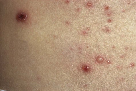
Because of widespread vaccination, varicella is uncommon today in developed countries. Historically, a large majority of the population experienced primary infection during childhood. Fever, chills, malaise, and headache may accompany a rash that involves primarily the trunk and head and neck. The rash quickly develops into a vesicular eruption that becomes pustular and eventually ulcerates. Successive crops of new lesions appear, owing to repeated waves of viremia. This causes the presence, at any one time, of lesions at all stages of development (Figure 1-9). The infection is self-limiting and lasts several weeks. Oral mucous membranes may be involved in primary disease and usually demonstrate multiple shallow ulcers that are preceded by evanescent vesicles (Figure 1-10). Because of the intense pruritic nature of the skin lesions, secondary bacterial infection is not uncommon and may result in healing with scar formation. Complications, including pneumonitis, encephalitis, and inflammation of other organs, may occur in a very small percentage of cases. If varicella is acquired during pregnancy, fetal abnormalities may occur. When older adults and immunocompromised patients are affected, varicella may be much more severe and protracted, and more likely to produce complications.

Herpes Zoster

Zoster is essentially a condition of the older adult population and of individuals who have compromised immune responses. The incidence of herpes zoster infection increases with age, reaching approximately 10 cases per 100,000 patient-years by age 80.The sensory nerves of the trunk and head and neck are commonly affected. Involvement of various branches of the trigeminal nerve may result in unilateral oral, facial, or ocular lesions (Figures 1-11 and 1-12). Involvement of facial and auditory nerves produces the Ramsay Hunt syndrome, in which facial paralysis is accompanied by vesicles of the ipsilateral external ear, tinnitus, deafness, and vertigo.

After several days of prodromal symptoms of pain and/or paresthesia in the area of the involved dermatome, a well-delineated unilateral maculopapular rash appears. This may occasionally be accompanied by systemic symptoms. The rash quickly becomes vesicular, pustular, and then ulcerative. Remission usually occurs in several weeks. Complications include secondary infection of ulcers, post-herpetic neuralgia (which may be refractory to analgesics), motor paralysis, and ocular inflammation when the ophthalmic division of the trigeminal nerve is involved.

Stay updated, free dental videos. Join our Telegram channel

Jan 12, 2015 | Posted by in Oral and Maxillofacial Pathology | Comments Off on 1: Vesiculobullous Diseases

VIDEdental - Online dental courses

Get VIDEdental app for watching clinical videos