1: Cariology and Caries Management

Chapter 1 Cariology and Caries Management

Section A Caries Risk Assessment

Brief History of Clinical Development and Evolution of the Procedure

Historically dentistry has treated dental decay with a surgical model, drilling the decayed tooth structure away and replacing it with a restorative material. Dental caries has been recognized for over 100 years as a disease that contributes to decay. Early pioneers—G.V. Black, Leon Williams, and others—recognized the relationship of dental plaque to decay. Over a period of decades, several bacteria have been identified and connected to the decay process. These bacteria include primarily Streptococcus mutans and Lactobacillus. Both of these types of bacteria are saccharolytic (metabolize carbohydrates), acidogenic (produce small molecular organic acids from the carbohydrate metabolism), aciduric (survive in acidic or low pH environments, pH ranges that dissolve the calcium and phosphate minerals from the teeth), and cariogenic (contribute to the decay process as a result of these characteristics). The prolonged periods of low pH on the teeth lead to a net mineral loss from the dental tissues and produce decay, cavitation, and loss of the teeth. Many studies over the last 30 years correlate high levels of mutans streptococci and lactobacilli with dental caries. However, this is more than a single- or double-pathogen disease process in the classic model of infection. Dental caries has multifactorial causation, with environmental risk factors, individual risk factors, and behavioral and dietary influences as well as the biofilm component. Literally any saccharolytic, acidogenic, and aciduric bacteria could contribute to the caries biofilm and lead to dental caries. In 1989 Philip Marsh demonstrated conclusively through a series of studies that it is not the sugar availability that leads to decay, but rather the acid production from the metabolism of the sugars. The resulting low pH environment provides the selection pressure to favor these bacteria in a patient’s mouth. Today up to 24 different bacterial species have been implicated in dental caries. Preza and co-workers demonstrated that additional species of bacteria need to be considered in the root surface caries biofilm, including Atopobium, Olsenella, Pseudoramibacter, and Selenomonas. In 2008, Takahashi and Nyvad demonstrated that during protracted periods of low pH in the oral biofilm, even the potential commensal oral streptococci become more acidogenic and aciduric and contribute to the disease process. They identified Streptococcus gordonii, Streptococcus mitis, Streptococcus oralis, and Streptococcus anginosus and termed these bacteria low-pH, non-MS streptococci. They described this phenomenon as an extension of Marsh’s earlier “ecological plaque hypothesis.” But it brings to light that it is important not only which bacteria are present in a patient’s biofilm, but what those bacteria are doing. In addition, other factors have been reported and studied with regard to their role in the disease process. Known risk factors now include previous history of decay, radiographic lesions, white spot lesions, visible plaque on the teeth, frequent snacking, low saliva and poor saliva buffering capability, xerostomia-producing medications, poor diet, suboptimal fluoride exposure, poor dental care habits, and low socioeconomic status. Today dental caries necessitates a caries risk assessment with a validated questionnaire to evaluate and correct the modifiable risk factors for an individual patient. It necessitates diagnosis of the bacterial infection using bacterial metric testing or culture. Finally, it necessitates specific targeted antimicrobial therapy of the biofilm infection to predictably and effectively treat the disease. Simply drilling and filling cavities, a surgical approach to treating a bacterial infection, does not diagnose or treat the disease and is no longer acceptable as a standard of care.

Material Options

Dental Caries Treatment Strategies

For all patients, any restorative and biomechanical needs must be addressed. Restoration of the defects may return the teeth to function but have little to do with correcting the dental caries biofilm disease. Many different options are available for treating the biofilm disease process. A comprehensive approach to treating the dental caries patient involves addressing every aspect of the disease. These strategies can be broken down into major groups and ideas. First in most treatment considerations are the reparative procedures required to correct the physical damage to the teeth. This includes remineralization of lesions that have not cavitated and still have an intact enamel surface with fluoride and calcium phosphate or hydroxyapatite, plus minimally invasive restorations using biomimetic materials for lesions that have cavitation and decay present. The next strategies are focused on the therapeutic approach to correcting the bacterial biofilm component of the disease. These procedures include antimicrobial agents, pH corrections, and metabolic agents (xylitol). Additional strategies include behavioral changes to improve the oral environment to favor a healthy biofilm. Typically this involves oral hygiene instructions for improved home care and plaque control plus dietary counseling. Some nonmodifiable factors may need to be addressed by adding more protective factors to the patient’s risk-and-caries balance equation. Special needs patients and those with xerostomia or medication-induced xerostomia fall into this category.

Remineralization Therapy

Remineralization has historically involved the use of topical fluoride. Stannous fluoride and acidulated fluorides were introduced first, but more recently neutral fluoride products have been used. The fluoride is applied in many different methods, such as 1 ppm public water fluoridation, 1100 ppm fluoride dentifrice, 5000 ppm fluoride gels and foams, 223 ppm fluoride rinse, and 23,000 ppm fluoride varnish. Fluoride’s basic mode of action enhances remineralization and inhibits demineralization. Fluoride ions incorporate into remineralizing enamel and dentin carbonated apatite to produce a more acid-tolerant fluorapatite-like form. Fluoride also makes hard tissues more acid resistant and inhibits bacterial intracellular enzymes.

More recently, nano-particle hydroxyapatite and CPP-ACP have made calcium and phosphate ions bioavailable to aid in the remineralization process. The benefits of additional sources of these ions is unclear. Some clinicians believe that the need to supplement sources of calcium and phosphate is limited to the xerostomic patient, in whom these molecules may be in short supply. Others believe there is added benefit to increasing the availability of calcium and phosphate in high-risk caries patients. Clearly, more studies are needed to answer this question. Products include Recaldent (Recaldent Pty Ltd, Australia), NovaMin (GlaxoSmithKline, United Kingdom), Trident (Warner-Lambert, Morris Plains, New Jersey), MI Paste (GC America, Alsip, Illinois), and pHluorigel HA and HA Nano Gel (Carifree, Albany, Oregon).

Restorative Strategies with Minimally Invasive Dentistry

Dental caries can be site, tooth, patient, and population specific. Ideally, successful caries prevention implies there will be no irreversible changes to any tooth site or surface (occlusal, approximal, smooth, or root surface). If prevention fails at any site, the greatest benefit for the patient begins with early lesion detection. Such detection should trigger protocols for chemical remineralization and interventions to arrest and reverse early damage caused by demineralization before surface cavitation occurs. Only if surface cavitation develops should surgical restoration be performed, and then it is done by using the most minimally invasive approach possible, maintaining the maximum amount of healthy tissue and structural integrity of the tooth. The restoration is completed with the most appropriate dental restorative material suited for that particular lesion and that particular patient.

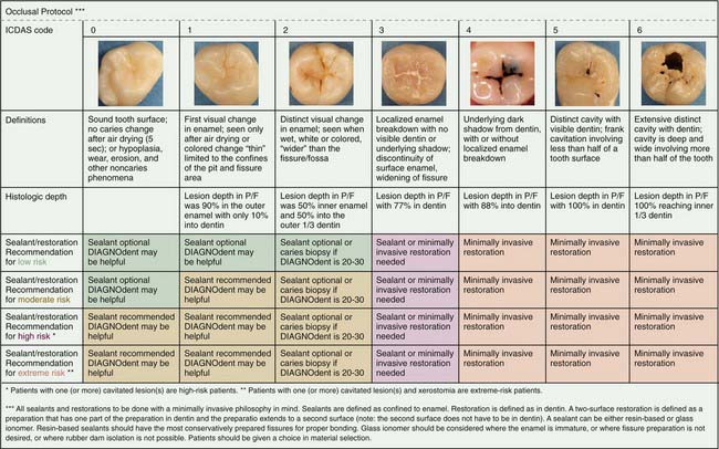
Traditionally dentists identified cavitated lesions using a sharp explorer tip, visual examination, and/or radiographs. The explorer in a given dental practice may not have been sharp, so defining lesions in specific states of cavitation varied from dentist to dentist. Numerous studies report that the use of a dental explorer is not adequate for detecting early occlusal lesions at all and not only may lead to a significant number of undetected lesions, including some false positives, but may, if it is sharp, cause traumatic surface defects in teeth. Radiographs are not useful for early occlusal lesions because of the masking effect of the facial and lingual enamel. New research has suggested the use of a visual ICDAS code system. ICDAS, an acronym for International Caries Detection and Assessment System, can be thought of as a coding system of 0 to 6 that correlates what is seen clinically with a definition and what research has reported histologically. The gradient starts with a code 0, which is a completely intact and healthy occlusal fissure system, and ends with a code 6, a fissure that is cavitated with a frank carious lesion. Recently Jenson and colleagues published various protocols based on ICDAS code and caries risk. Included is how one would use laser detection technology such as a DIAGNOdent (KaVo Dental, Charlotte, North Carolina) in the decision-making process (Figure 1-1).

image

FIGURE 1-1 The International Caries Detection and Assessment System (ICDAS).

(From Jenson L, Budenz AW, Featherstone JD, et al: Clinical protocols for caries management by risk assessment, J Calif Dent Assoc 35:714, 2007.)

With traditional methods of lesion detection, patients who saw different dentists could be given conflicting information as to whether they had cavities. Confusion led to issues of trust and situations in which needed care was not rendered (undertreatment) or restorations were placed when chemical remineralization would have been more appropriate (overtreatment). In the past the issue of cavitated and precavitated lesions was not relevant for many practitioners. Whether the enamel surface is cavitated or not is the determining factor in deciding to chemically remineralize a lesion. If the enamel surface is still intact, the bacteria are physically too big to diffuse through the enamel surface to infect the dentin and can successfully be managed with remineralization protocols. Interproximal or bitewing radiographs can be interpreted differently, so there is no definitive way to detect early lesions. Lesions penetrating minimally into the enamel may be surgically restored by some clinicians, whereas others wait for the dentin breech to be confirmed radiographically. Based on scientific evidence, current recommendations are to surgically intervene on the approximal smooth surface only if the bitewing radiograph shows both a solid enamel radiolucency going from the surface through the enamel and that the dentin has been penetrated. Digital radiographs have the benefit of exposing the patient to less radiation. The digital image can also be enhanced and enlarged easily, enabling better detection and monitoring of early lesions.

Lesion detection on the tooth root is best accomplished by visual inspection. The chemistry of remineralization is the same on the root (cementum) as it is for enamel. However, early lesion detection is difficult, because in theory no visible change is present in an early lesion. Many individuals have proposed that any exposed root is a root at risk because of its lower mineral content and vulnerability to acid and enzyme dissolution. Careful monitoring of remineralization of early lesions is needed because of the more porous nature of cementum and dentin (less mineral compared with enamel) and the close proximity to the dental pulp in deeper root lesions. If restoration is needed on a root surface, chemical adhesion and fluoride release (charging and recharging) of glass ionomer restorative materials should be considered, especially when use of a rubber dam is not feasible.

The current state of lesion detection leaves behind the dental explorer and involves a more scientific approach with the use of ICDAS, DIAGNOdent, digital radiographs, and some interesting new technologies. Quantitative light-induced fluorescence (QLF) measures the degree of demineralization by using the natural fluorescence of teeth. With this computer-assisted technology, white spot lesions can be monitored over time to determine if the lesion is progressing or remineralizing. Simply put, healthy enamel structure has different optical properties than decalcified enamel; it has a different optical signature. The fluorescent signal reflected from the decalcification (white spot lesion) of the enamel is captured by a fiberoptic sensor and interpreted through a computer-based algorithm to determine the amount of demineralization.

Therapeutic Caries Strategies

Antimicrobial therapy has traditionally involved chlorhexidine as a first defense in treating dental caries. It was thought to be effective at attacking the Mutans streptococci but has little effect on lactobacilli. Recent research has indicated that following chlorhexidine therapy, one phylotype of Mutans streptococci consistently remains and appears to be not only resistant but also strongly pathogenic. Ethyl alcohol and essential oils have also been used in the past. There have been new reports of efficacy with 10% povidone iodine and recommendations for 0.10% sodium hypochlorite as an antimicrobial rinse. A recent study raises the issue of a potential link between ethyl alcohol–containing oral mouthrinses and oral cancer in individuals otherwise not at risk for cancer. Products from the chlorhexidine category include Peridex and PeriGuard (DermaRite Industries, Paterson, New Jersey). In the sodium hypochlorite category, a product is CariFree Treatment Rinse (Oral BioTech, Albany, Oregon).

pH strategies include raising the pH or buffering the oral environment to promote remineralization and encourage repopulation of commensal bacteria. The demineralization-remineralization gradient for enamel peaks at roughly pH 5.5. Below this pH, demineralization occurs, whereas at above 5.5, remineralization occurs. Prolonged periods of low pH in the mouth favor acidogenic, aciduric, and cariogenic bacteria, so pH strategies should be a component of the overall plan for patients at high risk for caries. Products addressing pH include Arm and Hammer Baking Soda products and CariFree products (Oral BioTech). These are available as gums, dentifrices, oral neutralizing gels and sprays, and rinses.

Xylitol is a naturally occurring alcohol sugar not metabolized by mutans streptococci that has effective anticaries activity. In addition to inhibiting the attachment of the biofilm, it also interferes with intracellular metabolism. The mutans streptococci cannot use or break down xylitol and use up energy to expel it from the cell. Xylitol is available in many forms: gum, lozenges, mints, sprays, rinses, pastes, and a baking substitute for sugar or other sweeteners. Xylitol has the advantages of being low calorie and not stimulating insulin production in diabetics. Xylitol can create gastrointestinal distress at high levels of consumption and may be toxic for dogs. A multitude of xylitol products is available, including Omnii 3M (3M ESPE, St Paul, Minnesota), Epic (Epic Industries, Provo, Utah), Spry (Xlear, Orem, Utah), and CariFree (Oral BioTech).

Other Considerations

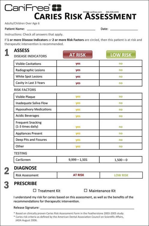
The most important consideration in performing a caries risk assessment and the medical management of dental caries is to use a standardized, validated caries risk assessment form. Several forms and sources are available (e.g., Journal of the California Dental Association, October-November 2007; www.cdafoundation.org; and other resources). In addition, in January 2009 the Scientific Council of the American Dental Association (ADA) endorsed caries risk assessment and provided a form available on the ADA website: www.ada.org/prof/resources/topics/topics_caries_over6.doc. Many forms include disease indicators, risk factors, and protective factors. Some forms stratify patients into high, moderate, or low risk categories; other forms also stratify them into extreme risk categories; and yet other forms include a determination of whether the caries is active or inactive. A simple caries risk assessment form that identifies the patient’s specific caries risk factors is all that is necessary. A simplified version based on a published form is presented in Figure 1-2.

image

FIGURE 1-2 Simplified caries risk assessment form.

(Courtesy Oral BioTech, Albany, Oregon.)

Performing caries risk assessment must be simple and straightforward. In private practice, most risk assessments are done in the hygiene operatory. The most important decision to be made in the operatory in real time is whether the patient is at risk for dental caries and continued or future lesion development. The decision must be made accurately and in relatively simple terms. Is the patient not presently at risk for disease, so he or she can be placed on an annual or biannual recare schedule with routine prevention measures and selection of restorative material? Or does the patient currently have dental caries and remain at continued risk for this disease, needing a more aggressive approach to treatment of the biofilm disease and specifically targeted prevention protocols in addition to any restorative needs? The assessment process should differentiate between these two groups of patients and identify the specific risk factors for each individual patient that potentially contribute to the biofilm disease process. This patient-specific information then becomes useful for treatment recommendations.

As already noted, risk factors that are not modifiable, such as age, amount and quality of saliva, or medication-induced xerostomia, must be counterbalanced in the treatment and prevention protocol with positive influences. With all the normal requirements and procedures already included in the hygiene operatory during a recare or new patient visit, this process must be precise and timely to be incorporated into an active dental practice. A simple risk assessment form that asks the right questions and produces the right patient profiles is the most desirable.

Special needs patients, patients with medication-induced xerostomia who cannot have their medication regimen altered, and children in low socioeconomic situations also have nonmodifiable risk factors. For these patients, it is important to increase the protective factors and help compensate for their risk factors to achieve a balanced state of health. This includes reinforcing home care instructions, modifying the toothbrush handle for a special needs patient if applicable, adding more fluoride to the daily regimen, providing dietary counseling, and prescribing pH-neutralizing products that can be used frequently during the day.

Innovative Elements

Technologic Advances

Traditionally, cariogenic bacteria levels were determined using a bacterial culture, typically by taking a saliva sample and culturing the saliva for Mutans streptococci and lactobacillus and extrapolating the data to determine the levels of these bacteria on the teeth. Disadvantages of the culturing technique as a screening test, diagnostic metric, or surrogate endpoint are that the cultures are not that sensitive or specific and do not correlate well with the patient’s actual caries risk. Cultures are helpful from an educational and motivational standpoint but are not accurate enough for more delineated decision making. They also are time-consuming; cultures typically require an incubation of 48 hours to yield a result, so in a busy general practice, adding this step is a management and systems issue. New applications of older technology have been developed, tested, and validated as screening tests for dental caries. All acidogenic and aciduric bacteria can survive in a low-pH environment through various adaptive mechanisms. For example, some bacterial intracellular enzymes can adjust activity to lower levels during prolonged periods of acidic pH (below 5.5). They can also pump the hydrogen (acidic) ions back out of the cell to retain intracellular neutrality while they are in an acidic environment. This hydrogen pump runs continuously and uses a tremendous amount of energy. All living cells derive energy from adenosine triphosphate (ATP). The ATP levels in a patient’s dental biofilm can be extrapolated to reflect energy use, which provides a high degree of correlation to the acidogenic and aciduric bacteria present. This can be accomplished in a simple chairside, real-time test with a light-sensitive meter and bioluminescence technology. The CariFree CariScreen system (Oral BioTech) is a newly developed test that has proved effective as a 15-second chairside screening test for dental caries.

Treatment Planning

Evidence-Based Principles

Several basic principles of caries risk assessment and the medical model of caries treatment have been validated by scientific studies. Caries risk assessment was validated in a large-scale study of two insured populations totaling 45,683 individuals. The results demonstrated that patients diagnosed as being at high caries risk were four times as likely as patients diagnosed as being at low caries risk to have a decay event during the test period. Patients identified as being at moderate risk were twice as likely as low-risk patients to have a carious lesion. This large-scale study validates the usefulness of caries risk assessment in predicting caries risk among adult patients. The study also demonstrates the lack of benefit from fluoride treatment in adults when stratified by risk category. Caries risk assessment forms and risk factors have been validated and weighted in two clinical studies. Caries risk assessment combined with the medical management of caries (caries management by risk assessment [CAMBRA]) has been validated in a clinical study. Patients demonstrated significantly reduced levels of cariogenic bacteria and carious events during the test period. Additional scientific studies provide evidence of the efficacy of fluoride regimens in children, the correlation of cariogenic bacterial loads to dental caries, and the valid use of ATP levels to the cariogenic bacterial loads. Xylitol has numerous scientific studies proving its efficacy in reducing both cariogenic bacterial load levels and caries rate. Biofilm studies are providing a clearer picture of the diverse nature of biofilms and the complexity of the dental caries biofilm disease.

Clinical Techniques

Case 2 Adult with High Caries Risk (Figure 1-4)

The patient is an ambulatory 47-year-old man. He has hypertension and is under the direct care of a physician. He is currently taking antihypertensive medication but has no allergies or other medical issues. He complains of a dry mouth and is concerned about losing his teeth. The patient has multiple missing teeth, visible plaque and carious lesions, multiple radiographic lesions, and a CariScreen score of 8756 (high risk). He is identified as at high risk on the risk assessment form. Among his multiple risk factors are dry mouth, poor oral hygiene, frequent snacking, medication-induced xerostomia, and suboptimal fluoride exposure. Treatment planning is broken into reparative processes and therapeutic processes. On the reparative side, remineralization is initiated with fluoride varnish and twice-daily use of fluoride rinses. Restoration consists of provisional restorations to repair all lesions with glass ionomer restorative material. The therapeutic regimen consists of antimicrobial rinses with sodium hypochlorite and fluoride twice a day for a month, followed by daily continued use of a pH-neutralizing fluoride and xylitol rinse and neutralizing xylitol gel twice a day until the 3-month recall interval. He is also given a pH-boosting oral spray to use between meals and immediately after any snacks. He is also given oral home care instructions and dietary counseling. He is placed on a 3-month recall schedule for review, reassessment, and retesting. This 3-month cycle of treatment and retesting is continued until health is achieved. Once the patient is determined to be caries free and at low risk for future disease, he is then scheduled for complete definitive restoration of his teeth, considering both function and esthetics.

Suggested Readings

Becker MR, Paster BJ, Leys EJ, et al. Molecular analysis of bacterial species associated with childhood caries. J Clin Microbiol. 2002;40(3):1001-1009.

Beighton D. The complex oral microflora of high-risk individuals and groups and its role in the caries process. Community Dent Oral Epidemiol. 2005;33(4):248-255.

Brailsford SR, Shah B, Simons D, et al. The predominant aciduric microflora of root-caries lesions. J Dent Res. 2001;80(9):1828-1833.

Ekstrand K, Qvist V, Thylstrup A. Light microscope study of the effect of probing in occlusal surfaces. Caries Res. 1987;21(4):368-374.

Featherstone JD. The science and practice of caries prevention. J Am Dent Assoc. 2000;131(7):887-899.

Featherstone JD, Domejean-Orliaguet S, Jenson L, et al. Caries risk assessment in practice for age 6 through adult. J Calif Dent Assoc. 2007;35(10):703-707. 10-3

Fontana M, Gonzalez-Cabezas C. Secondary caries and restoration replacement: an unresolved problem. Compend Contin Educ Dent. 2000;21(1):15-18. 21-4 passim; quiz 30

Fontana M, Zero DT. Assessing patients’ caries risk. J Am Dent Assoc. 2006;137(9):1231-1239.

Heinrich-Weltzien R, Kuhnisch J, van der Veen M, et al. Quantitative light-induced fluorescence (QLF)—a potential method for the dental practitioner. Quintessence Int. 2003;34(3):181-188.

Jenson L, Budenz AW, Featherstone JD, et al. Clinical protocols for caries management by risk assessment. J Calif Dent Assoc. 2007;35(10):714-723.

Katz RV. Assessing root caries in populations: the evolution of the root caries index. J Public Health Dent. 1980;40(1):7-16.

Lussi A. Comparison of different methods for the diagnosis of fissure caries without cavitation. Caries Res. 1993;27(5):409-416.

Lussi A. Validity of diagnostic and treatment decisions of fissure caries. Caries Res. 1991;25(4):296-303.

McCullough MJ, Farah CS. The role of alcohol in oral carcinogenesis with particular reference to alcohol-containing mouthwashes. Aust Dent J. 2008;53:302-305.

Mouradian WE, Wehr E, Crall JJ. Disparities in children’s oral health and access to dental care. JAMA. 2000;284(20):2625-2631.

Penning C, van Amerongen JP, Seef RE, ten Cate JM. Validity of probing for fissure caries diagnosis. Caries Res. 1992;26(6):445-449.

Pitts N. “ICDAS”—an international system for caries detection and assessment being developed to facilitate caries epidemiology, research and appropriate clinical management. Community Dent Health. 2004;21(3):193-198.

Pitts NB, Rimmer PA. An in vitro comparison of radiographic and directly assessed clinical caries status of posterior approximal surfaces in primary and permanent teeth. Caries Res. 1992;26(2):146-152.

Preza D, Olsen I, Aas JA, et al. Bacterial profiles of root caries in elderly patients. J Clin Microbiol. 2008;46(6):2015-2021.

Reynolds EC. Remineralization of enamel subsurface lesions by casein phosphopeptide–stabilized calcium phosphate solutions. J Dent Res. 1997;75(9):1587-1595.

Ricketts D, Kidd E, Weerheijm K, de Soet H. Hidden caries: what is it? Does it exist? Does it matter? Int Dent J. 1997;47(5):259-265.

Takahashi N, Nyvad B. Caries ecology revisited: microbial dynamics and the caries process. Caries Res. 2008;42(6):409-418.

van Houte J, Lopman J, Kent R. The predominant cultivable flora of sound and carious human root surfaces. J Dent Res. 1994;73(11):1727-1734.

Verdonschot EH, Bronkhorst EM, Burgersdijk RC, et al. Performance of some diagnostic systems in examinations for small occlusal carious lesions. Caries Res. 1992;26(1):59-64.

Young DA. New caries detection technologies and modern caries management: merging the strategies. Gen Dent. 2002;50(4):320-331.

Young DA, Featherstone JD, Roth JR. Curing the silent epidemic: caries management in the 21st century and beyond. J Calif Dent Assoc. 2007;35(10):681-685.

Stay updated, free dental videos. Join our Telegram channel

Jan 3, 2015 | Posted by in Esthetic Dentristry | Comments Off on 1: Cariology and Caries Management

VIDEdental - Online dental courses

Get VIDEdental app for watching clinical videos