Removable Partial Denture Considerations in Maxillofacial Prosthetics

CHAPTER 24 Removable Partial Denture Considerations in Maxillofacial Prosthetics

Maxillofacial Prosthetics

The preceding chapters have dealt with prosthesis considerations for partially edentulous individuals. In these patients, the extent of loss includes teeth and a varying degree of residual ridge bone, yet the remaining anatomy of the jaws and adjacent regions is functionally and physically intact. For these patients, the major distinguishing feature that affects removable partial denture design is whether the prosthesis will be tooth supported or tooth and tissue supported.

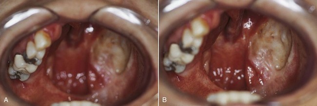
The maxillofacial patient can experience unique alterations in the normal oral/craniofacial environment; these result from surgical resections (Figure 24-1), maxillofacial trauma, congenital defects, developmental anomalies, or neuromuscular disease. In contrast to the above, when removable partial dentures are considered for these individuals, not only are tooth and tissue support considerations important, but the design must also take into account what impact the altered environment will have on prosthesis support, stability, and retention. In general, environmental changes reduce the capacity for residual teeth and tissue to provide optimum cross-arch support, stability, and retention.

As a subspecialty of prosthodontics, maxillofacial prosthetics is concerned with the restoration and/or replacement of the stomatognathic system and associated facial structures with prostheses that may or may not be removed on a regular or elective basis. This chapter discusses important background information related to maxillofacial prostheses and the principles involved in removable partial denture design for the maxillofacial patient.

Maxillofacial Classification

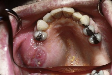
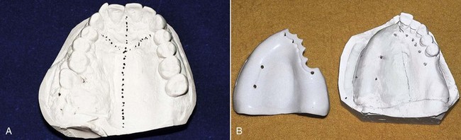
Patients can be categorized by maxillofacial defects that are acquired, congenital, or developmental. Acquired defects include those that are the result of trauma, or of disease and its treatment. These may include a soft and/or hard palate defect resulting from removal of a squamous cell carcinoma of the region (Figure 24-2). Congenital defects are typically craniofacial defects that are present from birth. The most common of these include cleft defects of the palate that may include the premaxillary alveolus. Developmental defects are those defects that occur because of some genetic predisposition that is expressed during growth and development (Figure 24-3). Such a classification order is helpful as patients within each category share similar characteristics (beyond those features specifically related to the prosthesis design), which become part of the total management plan. For example, prosthetic management of an adult who has experienced a maxillectomy procedure can be quite different from management of a patient with an unrepaired cleft palate.

Another helpful way to classify maxillofacial patients is by the type of prosthesis under consideration. Consequently, prostheses are said to be extraoral (cranial or facial replacement) or intraoral (involving the oral cavity); interim (for short periods of time, often perioperative) or definitive (more permanent); and treatment (used as a component of management, such as a splint or stent) prostheses.

Timing of Dental and Maxillofacial Prosthetic Care for Acquired Defects

Acquired defects are the most common maxillofacial defects managed by using removable prostheses. A conceptual framework for the timing of dental/oral care that best emphasizes the initial important surgical requirements, followed later by the important prosthetic requirements, is helpful to consider regarding the coordination of care for patients with acquired defects. Such a framework considers preoperative and intraoperative interim care and definitive care. Although it may seem unrelated initially, it is included in this discussion of removable partial dentures for maxillofacial applications because of the important impact that decisions made at all stages of management can have on prosthesis function and patient outcomes.

Preoperative and Intraoperative Care

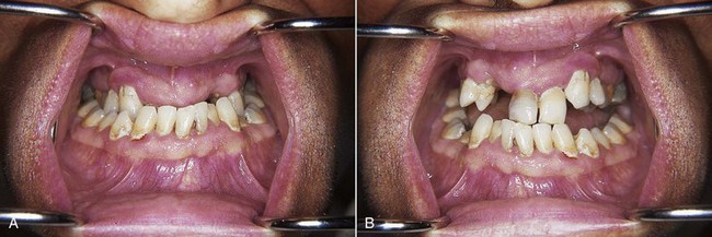
The planning of prosthetic treatment for acquired oral defects should begin before surgery. For the patient facing head and neck surgery, consideration should be given to dental needs that will improve the immediate postoperative course. Consequently, the prosthodontist who will help with management of the patient’s care should see the patient before surgery (Figure 24-4). The dental objectives of the preoperative and intraoperative care stages are to remove potential dental postoperative complications, to plan for the subsequent prosthetic treatment, and to make recommendations for surgical site preparation that improve structural integrity. Important patient benefits of such a preoperative consultation include the opportunity to develop the patient-clinician relationship, to discuss the functional deficits associated with the anticipated surgical procedure, and to describe how and to what extent the stages of prosthetic management will address them. The benefit from a prosthesis standpoint is that strategically important teeth, for definitive and/or interim prosthesis use, can be discussed with the surgical team and treatment planned for preservation.

The immediate postoperative period will be significantly challenging to the patient. If preexisting dental disease is severe enough to potentially create symptoms during the immediate postoperative period, treatment should be provided to remove such a complication. Large carious lesions, which could create pain, can be temporarily restored by endodontic therapy if they offer some advantage for postoperative prosthetic function. Teeth exhibiting acute periodontal disease (such as acute necrotizing ulcerative gingivitis) should be treated, as should any periodontal condition that could potentially cause postoperative pain because of excessive mobility or oral infection. Any tooth deemed nonrestorable because of advanced caries or periodontal disease, and not critical for temporary use during the interim care period following temporary treatment, should be removed before, or at the time of, surgical resection. Teeth that may appear to have a limited long-term prognosis may significantly enhance prosthetic service during the initial postsurgical period and should be maintained until the initiation of definitive care.

Impressions are made of the maxillary and mandibular arches to provide a record of existing conditions and occlusion to allow fabrication of immediate or interim prostheses (Figure 24-5) and to assess the need for immediate and delayed modification of the teeth or adjacent structures to optimize prosthetic care. It is important at this stage to begin planning for the definitive prosthesis because the greatest impact on the success of the maxillofacial prosthesis stems from the integrity of the remaining teeth and surrounding structures.

Interim Care

The major emphasis during this stage of care is the surgical (and adjunctive) management needs of the patient. In today’s environment of appropriately aggressive mandibular surgical reconstruction, mandibular discontinuity defects are seldom a surgical outcome. When discontinuity defects in the mandible result following surgery, interim prosthetic care is not indicated and the discussion will be directed to the maxillary defect.

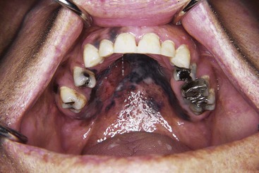
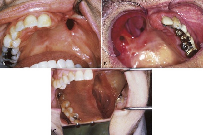
The typical maxillary acquired defect results in oral communication with the nose and/or maxillary sinus, although the composition of the surgical defect may vary widely (Figure 24-6). This creates physiologic and functional deficiencies in mastication, deglutition, and speech. Such defects have a negative impact on the psychological disposition of patients, especially if the defect also affects cosmetic appearance. The major deficiencies directly addressed by prosthetic management at this interim care time are deglutition and speech. This immediate postsurgical time is very challenging for patients, and it is important that they have been mentally prepared for it during the preoperative period. However, even with preliminary discussion, the impact of the surgery is often very distressing. An initial focus on improvement in swallowing and speech with the interim prosthesis can help boost the rehabilitation process significantly.

The patient is counseled that chewing on the defect side is not allowed because of its effect on prosthesis movement. The objective of this interim obturator prosthesis is to separate the oral and nasal cavities by obturating the communication. Such obturator prostheses most commonly refer to obturation of a hard palatal defect but conceptually can be considered the same for soft palatal defects at this stage of management, because both attempt to artificially block the free transfer of speech sounds and foods/liquids between the oral and nasal cavities. The advantages of having the ability to take nourishment by mouth without nasal reflux (allowing for nasogastric tube removal) and to communicate with family members are a significant component of early prosthetic management. How immediately such care should be provided depends on a number of factors.

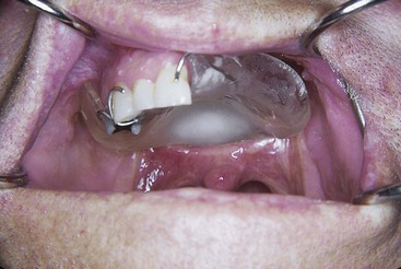
A prosthesis can be provided at surgery (see Figure 24-5B). Such a surgical obturator prosthesis is placed at the time of surgical access closure and serves to control the surgical dressing and split-thickness skin graft during the immediate postsurgical period. Such prostheses are best stabilized by appropriate wiring to remaining teeth or alveolar bone, or they may be suspended from superior skeletal structures. For some patients who have teeth remaining, such an immediate surgical prosthesis could be retained by wires in the prosthesis that engage undercuts on the teeth and would be removable; however, the ability to control the surgical dressing may be less predictable with such an approach. Immediate placement of a prosthesis has been suggested to improve patient acceptance of the surgical defect, although no measure of this psychological impact has been shown; this method offers greater assurance of adequate nourishment by mouth—potentially precluding the use of a nasogastric tube.

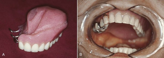
It may be preferable to stabilize the surgical dressing by suturing a sponge bolster to provide stabilization to the split-thickness skin graft. Following the primary healing stage, the sponge with packing (or the immediate prosthesis if used) is removed by the surgeon and an interim obturator prosthesis can be placed (Figure 24-7). For the patient who has been provided with bolster obturation, the presurgical prosthodontic evaluation is very important to ensure that the patient is prepared for the transition from bolster to prosthesis, and to ensure that plans for the prosthesis are made, especially if an interim prosthesis is to be fabricated. Interim prostheses are wire-retained resin prostheses that generally do not have teeth initially but may be modified with the addition of teeth after an initial period of accommodation (Figure 24-8).

When surgical defects become large, as in a near-total maxillectomy defect, prosthesis support, stability, and retention are not likely to be satisfactory unless extension into the defect can be accomplished. When teeth remain, the impact of the defect size is somewhat lessened. But when the remaining teeth are few or are located unilaterally in a straight line (see Figure 24-1), the mechanical advantage for prosthesis stability is less. The ability of the defect tissue to offer the needed mechanical characteristics to the interim prosthesis is unpredictable at best. It is this patient who benefits the most from a well-planned surgery that preserves oral and defective anatomy to the advantage of the prosthesis.

Potential Complications

The interim phase of prosthetic management can last for three or more months. The primary objective is to allow the patient to pass from an active surgical (and adjunctive treatment) phase to an observational phase of management with minimal complications. During the transition, the patient recovers from the systemic effects of the treatment, deals with the psychological impact of the defect using his or her own coping strategy, and becomes more aware of the functional deficits associated with the surgical defect(s). Minimizing potential complications during the transition, which includes preparing the patient for those anticipated to occur, facilitates the process for the patient and family. Common interim prosthetic complications are related to tissue trauma and the associated discomfort; inadequate retention (looseness) of the maxillary prosthesis; incomplete obturation with leakage of air, food, and liquid around the obturator portion of the prosthesis; and the tissue effects of chemotherapy and radiation therapy.

Discomfort related to the use of interim prostheses can be due to surgical wound healing dynamics, defect conditions, mucosal effects of adjunctive treatment, and/or prosthetic fit. Common areas of surgical wound pain include junctions of the oral and lip/cheek mucosa, especially at the anterior alveolar region for maxillectomy patients. The lateral scar band produced when the skin graft heals to the oral mucosa can be the site of discomfort in some patients. When a split-thickness skin graft is not placed, discomfort caused by the prosthesis fit within the defect can be a consistent and long-term problem. The hard palate surgical margin when not covered with surgically reflected oral mucosa most often will be covered by nasal epithelium, which is also very prone to discomfort. Alveolar bone cuts that have not been rounded will perforate the oral mucosa and will be painful whether or not a prosthesis is worn. This is most frequently a finding for mandibular resection superior alveolar margins when the reconstruction has restored the lower and labial contour to the mandible, but the intraoral mucosa at the superior surface is under tension because of a difference in height.

The prosthesis can create discomfort via excessive static pressure from the internal surfaces or from overextension into the vestibular tissue. The prosthesis can also create discomfort caused by functional movement associated with swallowing and speech. As was discussed previously, prosthesis movement is dependent on the quality of the supporting structures. Teeth offer the best support, followed by firm edentulous ridges, and lastly, surgical defect structures. The tongue, opposing dentition, and cheek/lips place force on the prosthesis that must be resisted over a large area to prevent movement. Because the defect is least likely to be able to resist movement, the relative size and structural integrity of the defect compared with the remaining teeth and/or edentulous ridge determine the potential prosthesis movement and most affect the discomfort related to such movement.

When teeth are available (and especially if located both close to and far away from the defect), retention is enhanced by engaging them with prosthetic clasps. Clasp retention is the most efficient means of effectively resisting dislodgment. The clasps will require periodic adjustment to maintain their effectiveness as the movement of the prosthesis flexes the clasps beyond their elastic recovery capacity. For edentulous patients, because the surgical defect allows communication between cavities, the fitting surface of the prosthesis can no longer create a closed environment to develop a seal for resisting dislodgment. Consequently, during the interim phase, when complete engagement of the defect is not possible because of tissue sensitivity, the careful use of denture adhesives is required to facilitate retention. The patient should be instructed that adhesives can alter the prosthesis fit and disrupt the close adaptation of the prosthesis to the remaining tissue. Used adhesive must be removed before new adhesive is reapplied, to maintain fit and hygiene. Also related to retention is the inability to completely place the prosthesis, which for maxillectomy patients can be due to contracture of the scar band. When the maxillary resection leaves the cheek unsupported by bone, the prosthesis provides the necessary support for wound maturation. If the patient removes the interim obturator prosthesis for a period sufficient to allow contraction, the prosthesis will be more difficult to place. Once placed, however, the scar band will relax and subsequent removal and placement will be more easily accomplished. The discomfort associated with this phenomenon is mostly due to patient anxiety and can be effectively addressed by reassuring the patient that this is an easily handled complication.

During the immediate postoperative healing stage, the surgical defect will undergo a change in dimension that affects the prosthesis fit and seal. If space is created with the change, speech will be altered (increase in nasality) and nasal reflux with swallowing will occur. The interim prosthesis is made of easily adjustable material to allow accommodation for such changes. The most common manner of adjustment is through the use of temporary resilient denture lining materials, which offer the ability to mold to the tissue directly and reduce the mechanical effects of movement by virtue of their viscoelastic nature. Leakage can occur quite easily when swallowing unless the patient follows certain instructions. Because the prosthesis cannot offer a watertight seal that matches the presurgical state, patients will be instructed not to swallow large quantities at one time, and to hold their heads horizontal when swallowing. When the head posture is forward, as when one is taking soup from a spoon, leakage easily occurs around the obturator component of a prosthesis. Another difficult condition that presents difficulty in controlling leakage on swallowing is the midline soft palatal resection. The functional movement of the remaining soft palate is often very difficult to retain with a prosthesis. It is also difficult to provide an adequate seal during the interim prosthesis stage.

When combination treatment is prescribed for the patient, it is commonly provided during the postsurgical phase, when the patient is using an interim prosthesis. The major intraoral complication associated with both radiation therapy and chemotherapy, which affects interim prosthetic service, is mucositis. A careful balance between comfort and adequate fit for speech and swallowing needs must be determined with input from the patient. If prosthesis adjustment can offer relief to ensure completion of treatment and the patient understands the impact adjustment may have on speech and swallowing, then it should be accomplished.

The long-term effects of radiation therapy, especially radiation-induced xerostomia and capillary bed changes (obliterative endarteritis) within the mandible, present a potentially significant threat to any remaining dentition and to the development of osteoradionecrosis. During the interim prosthesis stage, the patient will begin to notice the xerostomic effects, which include development of thick, ropy saliva that makes swallowing more difficult, and an increase in discomfort associated with removable prostheses.

Defect and Oral Hygiene

Following surgical pack removal, the defect site will mature with time and exposure to the external environment. Initial loss of incompletely consolidated skin graft, mucous secretions mixed with blood, and residual food debris within the cavity are common oral findings for the patient with a maxillary defect. These cause concern for patients who are unprepared and unfamiliar with these new oral findings. As they become more familiar with the surgical defect, patients should be encouraged to clean the defect of food debris and mucous secretions routinely. Defect hygiene will allow quicker healing and will improve the ability to adequately fit a prosthesis. Common defect hygiene practices include (1) lavage procedures, which include rinsing of the defect during normal showering, (2) rinsing of the defect using a bulb syringe or a modified oral irrigating device (modified to provide a multiple orifice “shower” effect), and (3) manual cleaning procedures, such as the use of a sponge-handled cleaning aid. Frequently, dried mucous secretions are difficult to remove and require adequate hydration before mechanical removal.

Following surgical pack removal, the patient may be reluctant to begin oral hygiene practices because of oral discomfort. As patients use the interim prosthesis, which requires daily removal and cleaning at a minimum, they will realize the need for and benefit of normal oral hygiene practices because of improved prosthesis fit and tolerance. When teeth are remaining, it is important to the success of long-term prosthesis care to maintain a high level of oral hygiene. This is more critical for patients who exhibit xerostomia and have increased risk of caries. For these patients, daily application of fluoride in custom-formed carriers is prescribed along with frequent professional cleanings. The successful use of maxillofacial prostheses is enhanced greatly by the support provided by natural teeth. Consequently, during the interim prosthetic period, periodontal management procedures are begun in anticipation of the definitive treatment to allow a smooth transition from the interim to definitive prosthetic stages.

Definitive Care

Only gold members can continue reading. Log In or Register to continue

Stay updated, free dental videos. Join our Telegram channel

Mar 13, 2015 | Posted by in Prosthodontics | Comments Off on Removable Partial Denture Considerations in Maxillofacial Prosthetics

VIDEdental - Online dental courses

Get VIDEdental app for watching clinical videos