Fig. 5.1
Access cavity in maxillary first molar. (a) Conservative access, allowing straight-line access to main canals. Note cavity extension mesial for MB2 canal. (b) Access cavity and canal orifices filled with sodium hypochlorite. Note change of canal visibility due to light refraction through liquid and the isolation with transparent rubber dam (Case by H. W.)
Based on differences in study methodology, operator expertise, degree of mineralization, and the classification used MB2 was reported to exist in 19–96% of the cases [28]. According to published studies, the possibility of finding a second mesiobuccal canal increases with the use of magnification and illumination, particularly a dental microscope, as well as the increasing experience of using it [29, 33–37].
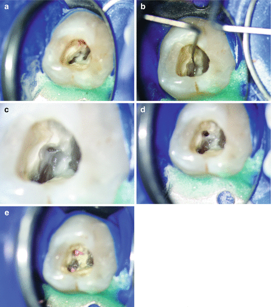
Typically, the canal entrance is hidden beneath dentin. This overhanging dentin shelf needs to be removed to detect the orifice with a mesial extension to allow appropriate access to the canal. In many clinical situations for about 1–2 mm the coronal portion of mb2 runs very shallow from distal to mesial before the canal curves towards distal, following the mesiobuccal root (Fig. 5.2).


Fig. 5.2
Identification and instrumentation of a calcified MB2 of a first maxillary molar. Access cavity overview with dentin obstructing the canal entrance to MB2 (10× magnification). (b) Canal identification after ultrasonic dentin removal (10×). (c) Initial preparation of MB2 for straight-line access (16×). (d) Completely instrumented MB2 canal prior to root filling (10×). (e) Root fillings in mesiobuccal canals (10×) (Images a, b, and d from Setzer and Kim [38]; with permission)
There are different approaches to gain access to the middle and apical third to avoid iatrogenic complications such as blockage or transportation. Using a long shank #2 round bur or LN bur, one can directly attempt to locate the main portion of the canal from the coronal aspect. Alternatively, the canal path can be carefully modified using a combination of stainless steel Hedström and K-files size #8 and #10 hand instruments or rotary instruments that allow lateral brushing, e.g., ProTaper S1and Sx or TRUShape Orifice modifiers. Micro-openers #10/0.04 or path or scout files may be of help to clear obstructions. Frequent irrigation is advised to clean debris to avoid blockage of the orifice and the coronal canal portion. The clinician is advised to proceed with caution with the instrumentation of mb2. Not only is it the smallest of all canals in the maxillary first molars, but it also curves, often sharply, towards the mb1 canal if these canals are joining. Abrupt curvatures may pose a great risk for instrument fractures. It is often advisable to complete the instrumentation of mb1 before approaching mb2 to gain a better understanding of the root morphology, leading to an optimal clinical result (Fig. 5.3).


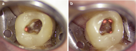
Fig. 5.3
Situation after root canal filling. Straight-line access allowed direct instrumentation of all canals; however, this must not mean complete visibility of all canals. Note color difference between pulp floor (dark) and pulp chamber walls (light), as well as developmental lines connecting the main canals. (a) Palatal canal. (b) Distobuccal and mesiobuccal canals. Note isolation with transparent rubber dam
Other counts for the total number of canals were reported, including 5, 6, 7, or 8 main canals, including variations such as the palatal canal splitting in three individual canals [39].
5.2.2 Maxillary Second Molars
Maxillary second molars are generally smaller than the first maxillary molar. Four canals are found less frequently than in first maxillary molars. Although access should follow the same guidelines as for first maxillary molars, it is expected that all root canal orifices are much closer to each other. If four orifices are present, they are frequently found within a diamond shape area or even almost a line connecting the mesiobuccal with the palatal canal. Fused roots are common in second maxillary molars, a fact that accounts for the close proximity of the canal entrances that is often encountered in these teeth.
5.2.3 Mandibular First Molars
Most mandibular first molars typically have two roots with either three or four root canals. Commonly, two root canals are found in the mesial root and one or two canals in the distal root. In order not to miss any canals in the distal root, the access cavity should not be triangular, but rather trapezoid to rectangular in shape, and in most cases be located in the mesial two-thirds of the occlusal surface (Fig. 5.4). Mesiobuccal and mesiolingual canals are found slightly centrally in relation to the mesiobuccal and mesiolingual cusp tips. The distal canal(s) will be found distally to the furcation, respectively distal to the buccal developmental groove. Due to the curvature of the distal root, it is mostly sufficient to limit the access cavity extension in distal direction. Straight-line access will still be possible. If only one distal canal is present, it is located centrally in buccolingual direction. It commonly has a long oval canal cross-section stretching buccolingually.


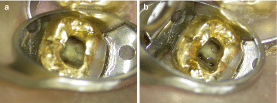
Fig. 5.4
Access cavity in mandibular first molar showing a conservative access through an existing coronal restoration. (a) Access cavity and canal orifices dried. (b) Access cavity and canal orifices filled with sodium hypochlorite. Note change of canal visibility due to light refraction through liquid
Careful examination of both lingual and buccal canal walls will sometimes allow to locate a second distal canal, which may separate off below the level of an oval orifice. If two canals are present in the distal root, they are most likely centered buccally and lingually of the mesiodistal axis. According to the literature, one canal in the distal root is found in about 60 % and two canals in about 40 % of mandibular first molars; however, one should always take the presence of two canals into consideration (see Chap. 1 in this book). Generally, the developmental grooves help to identify the root canal orifices. Mesiobuccal and mesiolingual are often connected by a developmental groove, which may extend into a partial or complete isthmus between the canals. In approximately 5 % of the clinical cases a third mesial canal or mid-mesial canal, between the mesiobuccal and mesiolingual canals, has been reported. Isthmus structures between the mesial canals should be carefully instrumented, to allow for the removal of vital or necrotic tissue remnants and to disinfect these ramifications. The opening and cleaning of the isthmus will also allow to easier identify any mid-mesial canals, which may only become accessible at a deeper level. Four canals are rarely present in the mesial root.
A three-rooted configuration of the mandibular first molar is found particularly in East or Southeast Asian populations; a second distal (distolingual) root is reported in 5–15 % of clinical cases (“radix entomolaris”) [30–37, 39–41]. In these situations, the main distal canal in the distobuccal root is found closer to the mesiodistal midline, whereas the distolingual canal entrance is found distinctively closer towards the distolingual cusp. For these situations the access cavity must be extended in the direction of the distolingual canal to allow for an almost vertical access to the canal. This is necessitated by the sharp buccal curvatures that are often found in combination with a distinctive apical kink in these separate distolingual roots. This curvature is often invisible on an ortho-radial projection. A third, distolingual root is often considerably shorter than the mesial or distobuccal roots.
5.2.4 Mandibular Second Molars
Similar to the mandibular first molars in anatomy, two roots with three or four root canals are common in the second mandibular molar, with about 80 % for three and 20 % for four root canals. However, there are greater variations in the root and root canal morphology than in first mandibular molars. Pulp chamber and canal entrances are smaller than in first mandibular molars. Roots are more often fused. Mesiobuccal and mesiolingual canal entrances may be in very close proximity, sometimes the root canal orifices appear as one large central canal entrance of oval shape. The clinician must carefully inspect these orifices. In a number of cases, both mesial canals split immediately below the pulp floor in their mesiolingual and mesiobuccal directions. Straight-line access must be created towards buccal/mesiobuccal for the mesiobuccal canal and towards lingual/mesiolingual for the mesiolingual canal, to avoid blockage of the canals by debris during access. The techniques are similar to the relocation and instrumentation of mesiobuccal and distobuccal canals that are in close proximity to each other in maxillary second molars.
A complicated morphology variation is the C-shape root and root canal system configuration [42, 43]. When cross-sectioned through the single C-shaped root in a horizontal plane, the root will appear to have the form of a letter “C”, with the open portion of the C mostly facing lingually.
Common radiographic misinterpretations are the anticipation of one root with one large central canal or two fused roots. However, what appears to be one “canal” is the radiographic appearance of the surface of the root folded into a C-shape. The canals in C-shape molars may have very small diameters and can be radiographically masked by hard tissue structure. While radiographically a C-shape tooth may appear as having one root with a large central canal, two root tips mesial and distal to the “canal” can often be seen. Second mandibular molars with one root and one real central canal should demonstrate the radiographic appearance of a single root tip. In C-shape molars the variations of anatomy are manifold. There can be one canal system connected as one single C-shape web, or as partial C-shape canal morphology with main canals that are completely or partially connected by isthmuses. These types have been referred to as semicolon-types, with the isolated canal located at the mesiolingual aspect.
Upon access of these teeth, the clinician must exert great caution not to perforate outside the C-shape configuration. Depending on the extent of the C-shape area and the existence of main canals, various techniques may be required to gain access and to instrument the root canal system. If main canals are present, their instrumentation is advised first. The cleaning of the connecting isthmuses should follow as a second step. This will allow an easier management and will reduce the risk of iatrogenic complications. If only a thin isthmus structure exists and the C-shape is complete, it will need to be carefully instrumented from coronal to apical. This can be done initially by using small long-shank round burs, then followed by LN-burs and ultrasonic instrumentation. It will require direct visibility of the area to be instrumented to prevent perforations or transportations.
5.2.5 Third Molars
Both maxillary and mandibular third molars may present with great variability in root and root canal system anatomy. No simple general guidelines can be given. Roots are often completely or partially fused and may number from one to four or even more. Root lengths can be very variable, even within the same tooth. Third molar root canal therapy may be limited by the access to the tooth, not only by tooth inclinations in mesiodistal or buccolingual directions, but also due to rotations, soft tissue impactions, or limited mouth opening by the patient. Canals may be difficult to negotiate due to size or abrupt curvatures. If limited opening is encountered, a children’s head handpiece and endodontic handpieces with reduced head size can provide better access and correct alignment of the bur. Pre-bendable rotary nickel-titanium files manufactured from controlled memory may be another aid for easier access to these root canals.
Stay updated, free dental videos. Join our Telegram channel

VIDEdental - Online dental courses


