In esthetic dentistry, expanding the evaluation beyond the teeth is necessary to achieve a truly desirable result. The lips, attached and unattached mucosa, free gingival margin, and osseous position and contours must be considered and changed if necessary. Although many treatment modalities are available to accomplish these modifications, the use of lasers of varying wavelengths provides advantages not possible by other means. Lasers are often thought of as generic instruments, but different laser wavelengths function differently, and each has its place in the esthetic continuum. Diode, neodymium:YAG, CO 2 and erbium lasers each have advantages that can be exploited to maximum effect and disadvantages that must be taken into consideration. A thorough understanding of their mechanism of action, their tissue effects, and laser safety is vital to obtaining excellent results.
In endeavoring to improve a patient’s smile, various means of altering tooth morphology, position, and color have been used. These include orthodontics, bleaching, bonding, full porcelain jackets, and porcelain veneers. These techniques involve only tooth structure and do not address the equally important relationship of the soft tissues to the teeth or the relationship of the teeth and soft tissues to the patient’s face. If disharmonies between these elements are not corrected, the resulting smile enhancement will not achieve the full desired effect.
Lasers for soft tissue and osseous recontouring
Of the means at our disposal for esthetic alteration of the soft tissues, the availability of lasers of different wavelengths provides us with the greatest range of options. Using lasers we can alter the mucosa and gingival tissues without causing bleeding, which provides better visualization, and recontour the osseous crest in a “flapless” procedure. Laser wounds exhibit histologic features that confer significant advantages over those created by scalpel or radiosurge. Most significantly, laser wounds have been found to contain significantly lower numbers of myofibroblasts, resulting in a minimal degree of wound contraction and scarring, and allowing for improved postoperative function . For esthetic dentistry, this is particularly important in dynamic soft tissues, such as those of the labial mucosa and frenum.
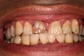
Frequently, the cause of a diastema and abnormal tissue architecture is a maxillary or mandibular frenum attached too close to the free gingival margin. This fibrous attachment of the lip to the alveolar mucosa may pull on the interdental papilla during lip movement, resulting in forces that separate the teeth, change the shape of the interdental papilla, and, in extreme cases, cause a malformation of the lip. In our esthetic evaluation, these tissues must be included in the overall planning to insure that the final result is not compromised. Fig. 1 shows an example of an extremely wide frenum that was attached so low it altered the contour of the lip to an unnatural, everted appearance. The frenum was excised with a CO 2 laser (Opus Duo; Lumenis, Santa Clara, CA) set on 6 W in continuous wave and attached to a focusing handpiece. The labial mucous membrane was then recontoured to eliminate the bulge of hyperplastic tissue by defocusing the laser beam and removing the tissue layer by layer in a vaporization procedure. The abnormal angle of the papilla between the central incisors was narrowed, and the gingival zenith was corrected using an Nd:YAG laser (Pulsemaster 6; American Dental Technologies, Corpus Christi, TX) set at 150 mj and 20 Hz with an initiated tip ( Fig. 2 ).