Initial Placement, Adjustment, and Servicing of the Removable Partial Denture

CHAPTER 20 Initial Placement, Adjustment, and Servicing of the Removable Partial Denture

Initial placement of the completed removable partial denture, the fifth of six essential phases of removable partial denture service mentioned in Chapter 2, should be a routinely scheduled appointment. All too often the prosthesis is quickly placed and the patient dismissed with instructions to return when soreness or discomfort develops. Patients should not be given possession of removable prostheses until denture bases have been initially adjusted as required, occlusal discrepancies have been eliminated, and patient education procedures have been continued.

Although it is true that some accommodation is a necessary part of adjusting to new dentures, many other factors are also pertinent. Among these are how well the patient has been informed of the mechanical and biological problems involved in the fabrication and wearing of a removable prosthetic restoration, and how much confidence the patient has acquired in the excellence of the finished product. Knowing in advance that every step has been carefully planned and executed with skill, and having acquired confidence in both the dentist and the excellence of the prosthesis, the patient is better able to accept the adjustment period as a necessary but transient step in learning to wear the prosthesis. This confidence could be lost if the dentist does not approach the insertion and postinsertion phases as equally important for the success of the treatment.

The term adjustment has two connotations, each of which must be considered separately. The first is adjustment of the denture bearing and occlusal surfaces of the denture made by the dentist at the time of initial placement and thereafter. The second is the adjustment or accommodation by the patient, both psychologically and biologically, to the new prosthesis.

After the resin bases have been processed and before dentures are separated from the casts, the occluding teeth must be altered to perfect the occlusal relationship between opposing artificial dentition or between artificial dentition and an opposing cast or template. Denture bases must be finished to eliminate excess and perfect the contours of polished surfaces for the best functional and esthetic results. This is made necessary by the inadequacies of casting procedures, because both the metal and resin parts of a prosthetic restoration are produced by casting methods. Unfortunately, such procedures in the laboratory rarely eliminate the need for final adjustment in the mouth to perfect the fit of the restoration to the oral tissue.

Included in this final step in a long sequence of finishing procedures necessary to produce a biologically acceptable prosthetic restoration are the following: (1) adjustment of the bearing surfaces of the denture bases to be in harmony with the supporting soft tissue; (2) adjustment of the occlusion to accommodate the occlusal rests and other metal parts of the denture; and (3) final adjustment of the occlusion on the artificial dentition to harmonize with natural occlusion in all mandibular positions.

Adjustments to Bearing Surfaces of Denture Bases

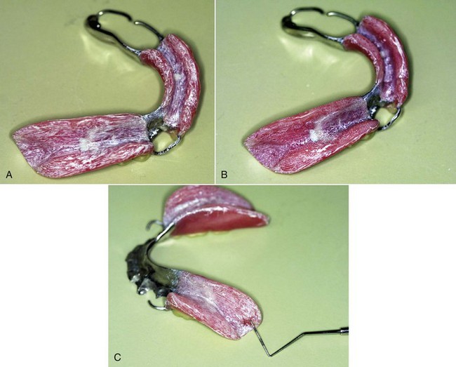
Altering bearing surfaces to perfect the fit of the denture to the supporting tissue should be accomplished with the use of some kind of indicator paste (Figure 20-1). The paste must be one that will be readily displaced by positive tissue contact and that will not adhere to the tissue of the mouth. Several pressure indicator pastes are commercially available. However, equal parts of a vegetable shortening and USP zinc oxide powder can be combined to make an acceptable paste. The components must be thoroughly spatulated to a homogeneous mixture. A quantity sufficient to fill several small ointment jars may be mixed at one time.

image

Figure 20-1 A, Tissue side of finished bases of a Kennedy Class I modification 1 removable partial denture, where pressure indicates that paste has been applied. Paste was applied following careful inspection of the tissue surface for irregularities or sharp projections, which must be eliminated before fitting in the mouth. The entire tissue surface of the bases was dried before it was coated with a thin coat of pressure indicator paste using a stiff-bristle brush. Brush marks are evident, and it is the change in the pattern of brush marks that guides adjustment. It is important to avoid thick application of indicator paste, which can hide the presence of significant pressure. B, The prosthesis can be dipped in cold water or sprayed with a provided release agent before placement in the patient’s mouth, to prevent paste from sticking to oral tissues. After careful seating of the denture, the patient can close firmly on cotton rolls for a few seconds, or the dentist can alternately apply a tissue-ward pressure over the bases to simulate functional movement. The presence of tissue contact is evident in the pattern of the paste, which is different from the brushed pattern. There is no suggestion of excessive pressure in this tissue contact pattern. However, it is not uncommon to relieve the area adjacent to the abutment sparingly. Several placements of the denture with indicator paste are usually necessary for evaluation of the accuracy of the bases. C, A different denture base recovered from the mouth after manipulation simulating function. The tissue contact reveals excessive pressure at the region lingual to the retromolar pad.

Rather than dismissing the patient with instructions to return when soreness develops and then overrelieving the denture for a traumatized area to restore patient comfort, use a pressure indicator paste with any tissue bearing prosthetic restoration. The paste should be applied by the dentist in a thin layer over the bearing surfaces. The material should be rinsed in water so it will not stick to the soft tissue, and then digital pressure should be applied to the denture in a tissue-ward direction. The patient cannot be expected to apply a heavy enough force to the new denture bases to register all of the pressure areas present. The dentist should apply both vertical and horizontal forces with the fingers in excess of what might be expected of the patient. The denture is then removed and inspected. Any areas where pressure has been heavy enough to displace a thin film of indicator paste should be relieved and the procedure repeated with a new film of indicator until excessive pressure areas have been eliminated. This is particularly difficult to interpret when patients exhibit xerostomia. An area of the denture base that shows through the film of indicator paste may be erroneously interpreted as a pressure spot, when actually the paste had adhered to the tissue in that area. Therefore only those areas that show through an intact film of indicator paste should be interpreted as pressure areas and relieved accordingly. The decision to relieve an area of pressure must consider whether the pressure is in a primary, secondary, or nonsupportive denture bearing area. The primary denture bearing areas should be expected to show greater contact than other areas.

Pressure areas most commonly encountered are as follows: in the mandibular arch—(1) the lingual slope of the mandibular ridge in the premolar area, (2) the mylohyoid ridge, (3) the border extension into the retromylohyoid space, and (4) the distobuccal border in the vicinity of the ascending ramus and the external oblique ridge; in the maxillary arch—(1) the inside of the buccal flange of the denture over the tuberosities, (2) the border of the denture lying at the malar prominence, and (3) the point at the pterygomaxillary notch where the denture may impinge on the pterygomandibular raphe or the pterygoid hamulus. In addition, bony spicules or irregularities in the denture base that will require specific relief may be found in either arch.

The amount of relief necessary will depend on the accuracy of the impression, the master cast, and the denture base. Despite the accuracy of modern impression and cast materials, many denture base materials leave much to be desired in this regard, and the element of technical error is always present. It is therefore essential that discrepancies in the denture base are detected and corrected before the tissues of the mouth are subjected to the stress of supporting a prosthetic restoration. One of our major responsibilities to the patient is that trauma should always be held to a minimum. Therefore the appointment time for initial placement of the denture must be adequate to permit such adjustment.

Adjustment of Occlusion in Harmony with Natural and Artificial Dentition

The final step in the adjustment of the removable partial denture at the time of initial placement is adjustment of the occlusion to harmonize with the natural occlusion in all mandibular excursions. When opposing removable partial dentures are placed concurrently, the adjustment of the occlusion will parallel to some extent the adjustment of occlusion on complete dentures. This is particularly true when the few remaining natural teeth are out of occlusion. But where one or more natural teeth may occlude in any mandibular position, those teeth will influence mandibular movement to some extent. It is necessary therefore that the artificial dentition on the removable partial denture be made to harmonize with whatever natural occlusion remains.

Occlusal adjustment of tooth-supported removable partial dentures may be performed accurately by any of several intraoral methods. Occlusal adjustment of distal extension removable partial dentures is accomplished more accurately with the use of an articulator than by any intraoral method. Because distal extension denture bases will exhibit some movement under a closing force, intraoral indications of occlusal discrepancies, whether produced by articulating paper or disclosing waxes, are difficult to interpret. Distal extension dentures positioned on remounting casts can conveniently be related in the articulator with new, nonpressure interocclusal records, and the occlusion can be adjusted accurately at the appointment for initial placement of the dentures (Figure 20-2).

Only gold members can continue reading. Log In or Register to continue

Stay updated, free dental videos. Join our Telegram channel

Mar 13, 2015 | Posted by in Prosthodontics | Comments Off on Initial Placement, Adjustment, and Servicing of the Removable Partial Denture

VIDEdental - Online dental courses

Get VIDEdental app for watching clinical videos