Implant Restoration of the Growing Patient
Clark M. Stanford
University of Illinois at Chicago College of Dentistry, Chicago, Illinois, USA
There is a number of reasons why teeth may be missing in the growing patient, including disease and its management, trauma, and/or congenital anomalies. Orofacial congenital anomalies occur in about 2–3% of newborns, with multiple environmental, genetic, and complex interactive etiologies being proposed.1 There are multiple complex forms of craniofacial anomalies but for the purposes of this discussion, we will use the ectodermal dysplasias as an illustrative example of the complex management issues that arise in treating any of these conditions. The ectodermal dysplasias are not one disorder but a complex polyphenotype with more than 210 different forms being recognized.2 The most common form is hypohidrotic ectodermal dysplasia (HED), which has an incidence rate of about 1 in 5000–10 000 live births.3 Many of these conditions involve a loss or lack of formation of dentition, supportive skeletal and facial structures, and the need for a comprehensive, age appropriate orofacial care for the developing child and young adult.
One of the most common abnormalities affecting the dentition is congenital anomalies, followed by trauma or iatrogenic conditions caused by cancer management. In regard to congenital anomalies, the ectodermal dysplasias are defined as a diverse set of syndromic conditions derived from mutations affecting communication between the overlying ectoderm and the underlying mesoderm in embryonic development. The condition is seen throughout the animal kingdom. Conventionally, a clinical definition of this phenotype has been two or more ectodermally derived structures (hair, teeth, nails, sweat glands, etc.) being affected. Under some of these conditions, the structures either do not form (aplasia) or form an altered phenotype (dysplasia), such as conical‐shaped or microdontic teeth.4 Originally described in its current classification by Frieire‐Maia and Pinheiro,5 the most common form of ectodermal dysplasia syndrome is referred to as X‐linked hypohidriotic ectodermal dysplasia or HED; note there is currently a significant effort to revise this system using molecular classification. The X‐linked HED is defined by a combination of features including hypodontia (missing fewer than six teeth), oligodontia (missing six or more teeth) (Figure 13.1a, b), and, rarely, anodontia (complete loss of teeth), altered hair growth, distribution, and volume (trichodysplasia), reduced or altered sweating (dyshidrosis), altered mucosal sebaceous function (asteatosis) leading to dry mucosal surfaces and significant mucosal crusting, and altered keratin nail formation in some types (onychodyplasia).
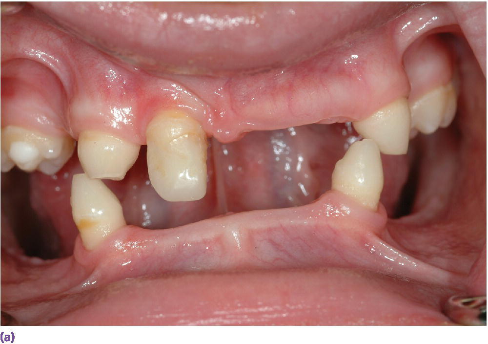
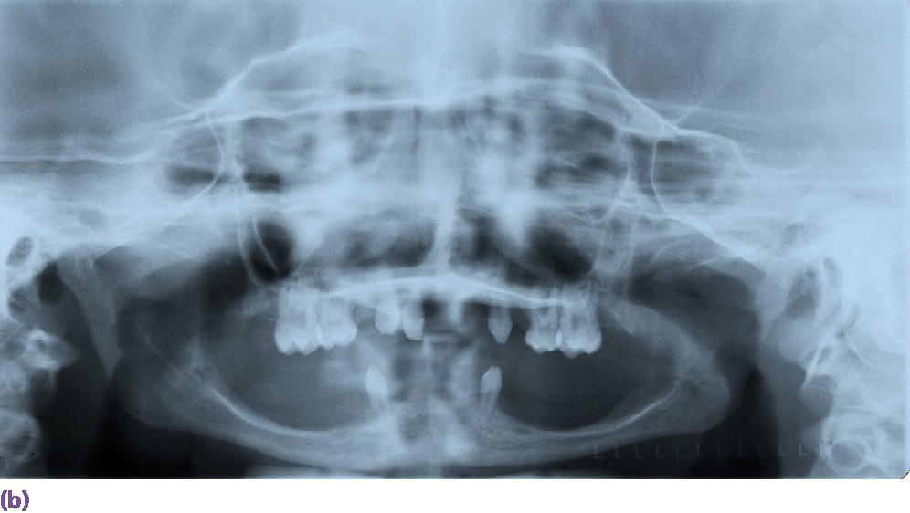
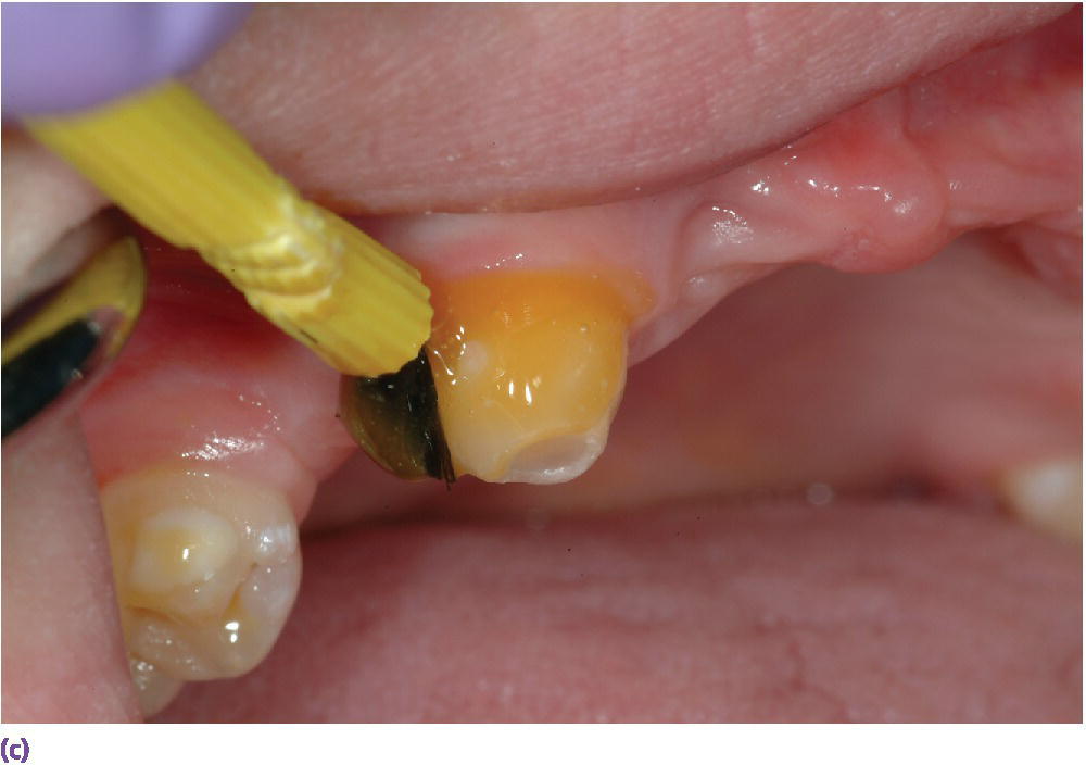
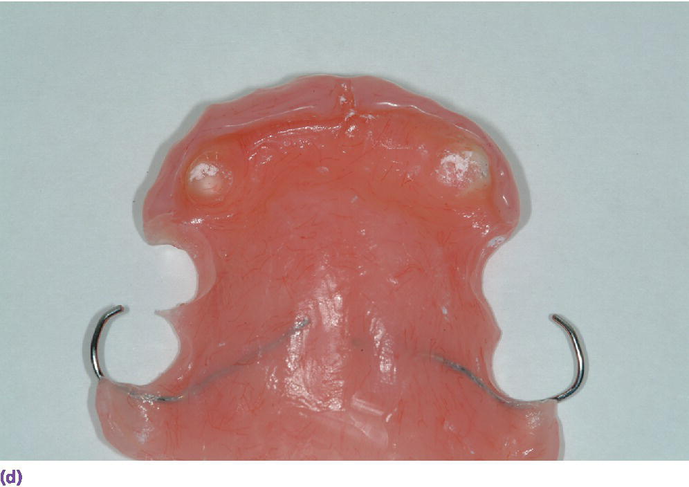
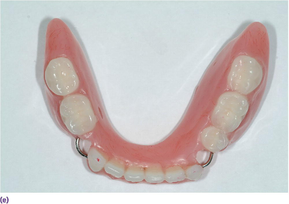
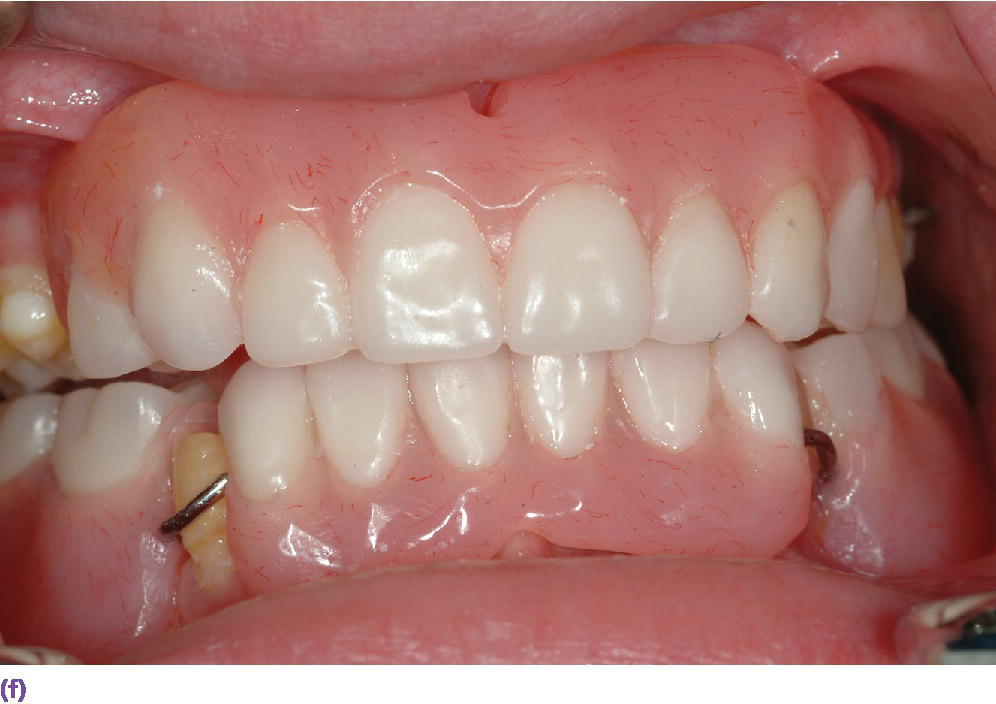
Figure 13.1 X‐linked hypohidriotic ectodermal dysplasia. (a) Intraoral appearance shows the typical oligodontia, cone‐shaped teeth, and retained primary teeth. The class III malocclusion, common in this population, is also evident in this image. (b) Radiographic appearance of the oligodontia. (c) Hyposalivation is common in this population since the salivary glands are derived in the same manner that sweat glands are formed. Use of frequent 5% NaF applications is recommended. (d) Provisional removable overdenture partial denture prosthesis for the maxilla. (e) Provisional removable partial denture for the mandible. (f) Prostheses in place at increased vertical dimension to match appropriate profile and need for interocclusal distance.
In regard to the dental malformations, the issues are usually esthetics, self‐perception, and awareness relative to unaffected peers, loss of perceived function, lack of alveolar ridge development, arch length/tooth size discrepancies, and hyposalivation. The latter can place the patient at a higher risk of dental caries (Figure 13.1c). Various groups have convened consensus conferences to develop expert experience‐based guidelines for tooth replacement therapy, with or without oral implant options primarily for the skeletally immature adult.6–11 Again, the data on outcomes are not strong and, as an evidence‐based procedure, the clinician should proceed very carefully. A leading principle is to provide age‐appropriate therapies holistically respecting the integrity of the patient and the caregiver’s autonomy, cultural competence, and providing staged therapies allowing patients to adapt to each approach as they are undergoing what growth potential they have (Figure 13.1d–f).12–14 Note, in this discussion, it is often better to use the medical term, “care plan” rather than “treatment plan”, since the latter suggests something is needed (when it may not at this time) and indicates the long‐term interests of the patient and their holistic outcomes of care are more important (family‐ and patient‐centered care) than any clinical perception of care.13–19
In the young child (age 0–6 years), a care plan involving a removable prosthesis is usually considered, starting around the age of 3 years. A pediatric dentist is often the lead consultant and should see any patient starting around 1 year of age. In situations where there are multiple age‐appropriate teeth, it may be useful to consult with a prosthodontist to assist in the staging of care. In this way, the affected individual builds a bond of trust with the entire care team with enhanced communication between the members of the care team and enhanced transition of care as the patient reaches adulthood. In the young child, a removable or pediatric fixed prosthesis may be considered.9 It is very helpful that the dentures and denture teeth be age appropriate in shape, size, and dimensions (Figure 13.2).20,21
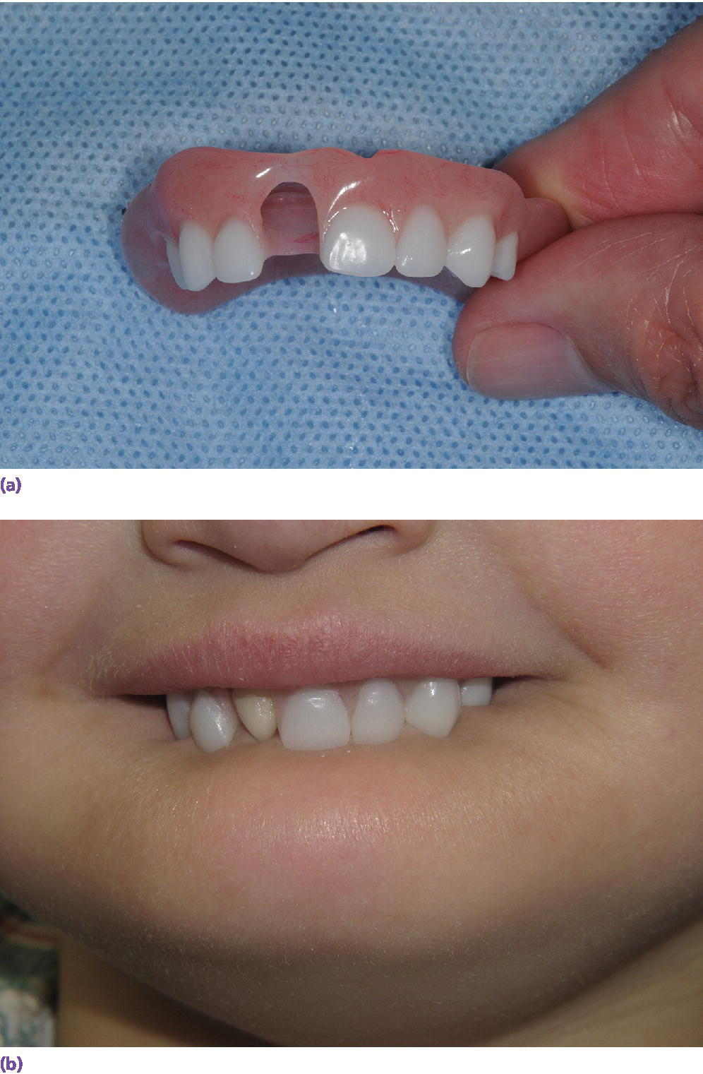
Figure 13.2 (a, b) Maxillary removable partial prosthesis. Age‐appropriate sized replacement teeth used on the maxillary partial denture in a 5‐year old.
In the young child, the prosthetic steps are often simplified relative to the adult complete or partial denture approaches, due to timing, need for frequent replacement, limited family resources, and patient and family tolerance. In a young patient presenting with multiple missing primary and permanent teeth, making a final impression, often with a well‐fitting stock tray and a fast‐setting impression material (e.g., occlusal registration material) may be sufficient. At the impression appointment, jaw relationships are made using a measure of the vertical dimension of rest in order to determine the vertical dimension of occlusion. A construct using about 2 mm of interocclusal distance is used with a measurement from the height of the posterior maxillary vestibule to the commissures of the lips (nodes of the orbicular oris) at approximately the vertical dimension of the anterior/posterior occlusion (canine and first and second primary molar). In a young child, this often translates to about 7 mm from the zygoma in the vestibule to the proposed posterior occlusal plane, coincident if possible with the corners of the mouth. This will develop in the laboratory phase of setting the prosthetic pediatric teeth (Milk Teeth, Nissin Dental Products, Japan, www.kilgoreinternational.com). If this approach is used, it is important to closely evaluate the patient at recall appointments and adjust the denture to allow eruption of any remaining teeth. This especially important since the growth of dento‐alveolar bone creates about two‐thirds of the vertical height of the mid‐ and lower face.22 In the case of total anodontia, the mandibular region, from approximately the second primary molar to second primary molar, completes basilar growth around 5 years of age with ongoing growth at the condylar growth plates and on the distal aspect of the ramus (allowing the mandible to become more upright with growth and carrying the symphysis area passively forward). As such, in the congenitally edentulous mandible, after about the age of 5, the use of oral implant(s) may be considered. Do note if there are teeth in the arch and/or erupting permanent teeth, implant placement should be delayed until at least skeletal maturity. In females this is typically at least 18 years or older and, in males, 20 years or older. As discussed later, the most important aspect of premature implant placement is disruption of normal tooth eruption and changes in tooth position due to the ankylosed oral implant. For this application, it must be clearly determined from the patient, not the parents, the perceived functional, esthetic, phonetic, and emotional/social advantages of this invasive therapy. It is important to hear from the patient the request and desire for the perceived improved function, since the procedures will entail a general anesthesia protocol with the associated elevated risks in this population. With an understanding that one or two two‐part or modular‐style narrow‐diameter implants can be used (e.g., 3 mm diameter), an overdenture‐style approach can be considered in this age group. Obviously, implants should not be used in the partially edentulous jaw in the growing child since the implant will become more submerged as the teeth erupt around it and/or the implant can affect the eruption path (the so‐called halo affect) of the adjacent teeth. Depending on the location and impact on eruption of adjacent teeth, ankylosed primary teeth may act as useful overdenture abutments and should be considered as a useful solution to prosthesis retention.
When the child reaches school age (7–12 years), the transition from primary to permanent dentition normally occurs. It is important to provide replacement teeth to allow for phonetics, mastication, and social/peer awareness.20,23,24 Typically a more removable approach is used in this population allowing ongoing growth (Figure 13.1). This is often the time to consider an orthodontic evaluation, especially as permanent teeth erupt. Retained primary teeth that do not have a succedaneous replacement should be retained to preserve the alveolar bone. Evaluation for ankylosed primary teeth needs to be carried out, especially in bounded spaces with permanent teeth on both sides, commonly the canines and first molars. A key aspect is to evaluate if the ankylosed primary teeth are interfering with the eruption of the adjacent permanent teeth; if so, they should be surgically removed. If not, then they should be maintained until at least skeletal maturity.
In adolescents to young adults (13 years to skeletal maturity), orthodontic therapy should be considered if there are teeth in the arch and space distribution is needed. Comprehensive care in this population should preferably be a team approach between pediatric dentists, prosthodontists, and orthodontists. Orthodontic care may be conventional full‐banded appliances or assisted with temporary anchoring devices (TADs or non‐integrated “temporary implants”) to support anchorage for orthodontic care (Figure 13.3). In cases of minor hypodontia (e.g., missing maxillary lateral incisors), esthetic tooth replacement can be performed either with a removable partial denture, an orthodontic retainer, or with a unilateral bonded pontic. In the bonded pontic approach, this is typically made with a zirconia core, pressed with hydrofluoric acid etchable glass ceramic (e.g., EMaxPress, Ivoclar Vivadent, Amherst NY) and bonded to the mesial aspect of the canine with a veneer‐based cement system (e.g., RelyX Veneer, 3M ESPE, St. Paul, MN).25 An important goal for the transitional period is a stable, esthetic, and functional dentition without overtreatment or loss of retained primary teeth that are developing or maintaining alveolar bone. It is most important that the maximal potential of craniofacial growth be accomplished during this period.
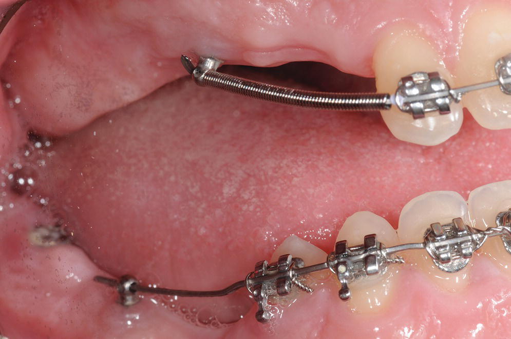
Figure 13.3 Use of TADs to assist in anchorage for orthodontic movement of the four anterior teeth that the patient formed.
If denture retention is insufficient, from the patient’s perceptive, oral implant therapy can be considered in the edentulous anterior mandible during this period. It is best to plan for a definitive final prosthetic design in selecting the implant sites.21 It is also preferable to avoid extensive bone grafting for site development in this age period.
Oral rehabilitation in the skeletally mature adult needs to be based on a comprehensive evaluation of systemic health and oral health risk factors, with a strong weighting on patient‐oriented outcomes, desires, and values. In this population, a multidisciplinary team is critical with additional orthodontic and oral surgical phases of care (e.g., LeFort midface orthognathic procedures).26 The prosthetic outcomes need to be carefully planned by the prosthodontist and strategic decisions made for retention of primary (or sometimes permanent) teeth, use of root canal therapy and removable partial overdentures for multiple missing contiguous teeth, and/or the need for extensive bone grafting for a multiple oral implant rehabilitation (Figure 13.4). The patient needs to understand that any prosthodontic intervention will have significant maintenance issues and will probably need to be replaced multiple times in the patient’s lifetime.15
Stay updated, free dental videos. Join our Telegram channel
VIDEdental - Online dental courses


