Use of the microbiology laboratory

Chapter 13

Use of the microbiology laboratory

Clinical oral microbiology is a relatively new discipline and involves the study of specimens taken from patients suspected of having oral infections. The end result is a report which should assist the clinician in reaching a definitive diagnosis and help in deciding on therapy. It is clear from this that clinicians should be conversant with the appropriate techniques of specimen taking and comprehend at least the principles of microbiological analysis. The purpose of this chapter, therefore, is to outline the ways and means by which clinicians could make the optimal use of the laboratory. In addition, specific laboratory procedures such as sensitivity testing are described to assist clinicians to understand the laboratory reports which they receive.

Requesting a microbiology report

A spectrum of decisions and actions by a number of individuals are involved from the time the clinician decides to take a microbiological sample until he receives the laboratory report. This spectrum of events can be conveniently divided into three categories: (1) ordering, collecting and transportation of specimens; (2) microbiological laboratory processing and reporting; and (3) interpretation and use of information (Figure 13.1).

Clinical request

Factors directly influencing the quality of the sample include the clinical condition of the patient and current antibiotic therapy. If the patient is not suffering from a microbial infection then sampling for pathogens would be futile, for example, in patients with well-defined allergic reactions or traumatic lesions which are not secondarily infected. Successful isolation of pathogens from patients currently on antibiotic therapy would be difficult due to suppression of the pathogen(s) or due to qualitative and quantitative alterations in the commensal flora. Hence the importance of collecting microbial specimens prior to antibiotic therapy whenever possible. However, exceptions to this are where the patient is seriously ill, immunologically compromised or not responding to a specific antibiotic, in which case the necessity of obtaining an interim report as a guide to further management justifies such action.

Collection and transport of specimens

The resources of a microbiology laboratory can be efficiently utilized by sending appropriate specimens which are properly collected. Specimens should be as fresh as possible for optimal isolation of the pathogens. Many organisms, including some anaerobes and most viruses, do not survive for long in clinical specimens kept at room temperature. In addition, some organisms such as coliforms and staphylococci may multiply at room temperature and subsequent analysis of such specimens may lead to inaccurate and misleading results. Therefore, specimens should be transported as soon as possible to the laboratory in an appropriate transport medium. In the absence of a transport medium, dehydration of the specimen and exposure of the organisms to aerobic conditions occur, with the resultant death or reduction in their numbers. Various transport media are available and it is important to establish that the chosen medium is compatible with the microorganisms which are believed to be present in the clinical sample, e.g. a viral transport medium should not be used for transporting samples which are destined for bacterial culture, and prereduced transport media are required for strictly anaerobic bacteria. In circumstances when delay in transportation is inevitable, storage of the specimen at 4 °C for a short period may help to maintain the relative numbers and viability of organisms in the specimen.

When samples are transported by post they should be placed in robust, leak-proof containers and sent by first class letter post (not parcel post) with the legend ’Pathological specimens—handle with care’ prominently displayed. Improperly packed specimens are an infective hazard for those who handle them. Information and containers for the postal transport of microbial samples can be obtained from the area medical/oral microbiology laboratory.

Provision of clinical information

The appropriate tests for each specimen received in the laboratory have to be selected by the microbiologist according to clinical information given on the accompanying request form. In brief, information such as age, main clinical condition, date of onset of illness, information about recent/current antibiotic therapy, antibiotic allergies, and history of previous specimens are all important to rationalize laboratory investigations and should be supplied with the specimen.

Investigations for some diseases require to be adequately discussed with the microbiologist beforehand in order to make the best use of laboratory resources. These include chronic infections where an actinomycotic or mycobacterial aetiology is suspected, tests for viral infections, and special investigations for specific antibiotics (e.g. assessment of minimum inhibitory concentration of antibiotics when treating infective endocarditis patients).

Laboratory analysis

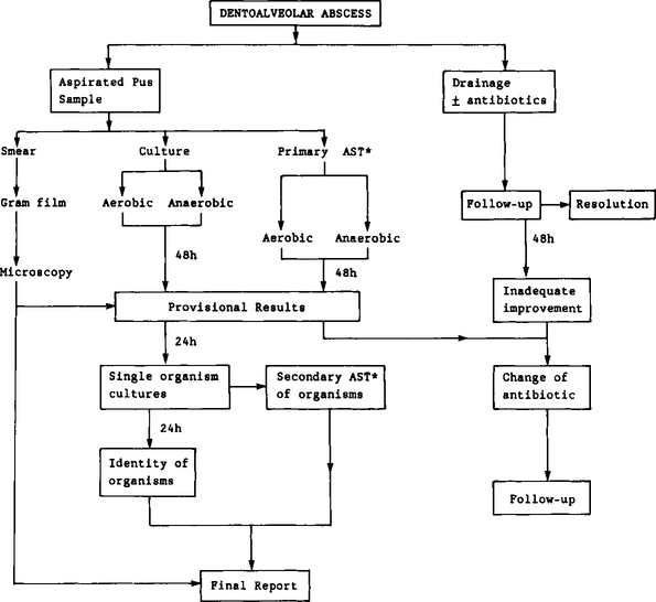
In order to give the reader an insight into the laboratory analysis of specimens, a brief account of the analytical pathway of a pus specimen is given below and is shown in Figure 13.2.

As soon as a pus specimen is received in the laboratory it is given a laboratory identification number and inoculated on to blood agar for culture under aerobic and anaerobic conditions. Two further plates (one aerobic and the other anaerobic) are set up to assess the primary antibiotic sensitivities of the putative pathogens. Finally a smear of pus is made on a glass slide, heat dried and Gram stained for microscopic examination.

As most of the oral pathogens are slow-growing anaerobes it is generally accepted that an incubation period of 2 to 3 days at 37° is required to produce visible colonies of these organisms. After 48 hours the plates are inspected for growth and if a single colony type is present its morphology is determined and identified. However, as a majority of dentoalveolar infections are polymicrobial in nature, samples usually yield a mixture of organisms which require to be isolated in pure culture to enable identification. The primary sensitivity plates are examined at 48 h or earlier (24 h) if the clinician requests an urgent sensitivity report.

At this stage, when about 2–3 days have elapsed, the microbiologist will be in a position to give the clinician an overall primary sensitivity result and presumptive identification of the organisms present. It may take a further 2–3 days to issue the final report as this is the period required for identifying bacteria and for computing reports.

The final report and its interpretation

Generally speaking, the clinician should receive the final report within a few days after the delivery of the specimen to the laboratory. However, in certain situations, e.g., when virus culture is requested, this may take a longer period. In this situation the clinician should make a telephone enquiry for a provisional verbal report. This is of particular help when the patient’s condition is not improving and the clinician can discuss the problem with the clinical microbiologist with regard to a possible change in the antimicrobial therapy and management, or the need for additional specimens. Sometimes the final report may never reach the clinician due to an inefficient data flow system or when more than one clinician is involved in the management of the patient.

The interpretation of the final report by the clinician may sometimes be difficult, especially in answering the question: ’What does it mean?’. There is much confusion when, as is often encountered in oral specimens, the organism reported as the putative pathogen is a member of the commensal oral flora. Such problems of interpretation mainly arise in circumstances where the quality of the specimen, the history of its transport, and clinical data are unsatisfactory. Whenever there is a particular difficulty with such interpretation or, indeed, when choosing an appropriate antibiotic, good collaboration between the clinician and the clinical microbiologist is of the greatest importance.

Specimen collection from oral infections

Bacteriological specimens

Dentoalveolar abscess

It has been the practice for many years to sample the pus from a dentoalveolar abscess by incising the abscess (usually intraorally) and collecting the pus on a swab. Such a specimen is usually grossly contaminated with resident oral commensal microorganisms. Although the manner in which a specimen is taken is dependent to some extent on the nature of the abscess, the optimal way to collect pus is by needle aspiration. The mucosal surface is first isolated and decontaminated with 0.2% chlorhexidine gluconate (Hibitane); patient discomfort at this stage or during subsequent needle aspiration can be relieved by surface anaesthesia using an ethyl chloride spray. Ideally, an aspirated pus specimen should be transported rapidly to the laboratory in the recapped syringe itself. Contamination of sample and exposure of organisms to aerobic and dehydrating conditions is minimized by this procedure although care should be taken when recapping to avoid a needle-stick injury.

It is important to note that aspiration of pus per se is not a satisfactory method of achieving drainage: subsequent incision of the abscess according to surgical principles may be required in some situations in order to drain the residual pus.

When aspiration fails to yield any material, incision and collection of pus is the only available option. In order to minimize contamination the area should be isolated and disinfected with 0.2% chlorhexidine before collection. In spite of these preventive measures, such samples almost invariably become contaminated.

Collection of parotid pus

In general, collection of uncontaminated parotid pus for microbiological examination is difficult due to: (1) relatively poor accessibility of the parotid duct; and (2) the absence of copious amounts of pus in the lesion. Therefore, two main techniques are used in collection of parotid pus.

In the presence of adequate quantities of pus it can be collected by aspiration through a small bore plastic catheter (attached, aseptically, to a sterile 2 ml syringe) inserted into the duct. The pus should then be transferred to the clinic in the syringe itself. The second method entails injecting about 0.5 ml sterile saline via the catheter and the ’saline wash’ of the parotid duct system is then aspirated and sent to the laboratory as described above. This method is particularly useful when there is no frank discharge of pus, although care should be taken not to instil a large volume of saline and to carry out this procedure with minimal pressure to prevent further spread of infection within the gland.

Only gold members can continue reading. Log In or Register to continue

Stay updated, free dental videos. Join our Telegram channel

Mar 12, 2016 | Posted by in General Dentistry | Comments Off on Use of the microbiology laboratory

VIDEdental - Online dental courses

Get VIDEdental app for watching clinical videos