This case report describes the treatment of a 16-year-old girl with ankylosed maxillary central incisors that were noticeably infraoccluded and labially displaced. We performed a segmental osteotomy with an autogenous bone graft in a single-stage surgery to align and level the ankylosed teeth. The dento-osseous segment was successfully repositioned with satisfactory periodontal results.
Ankylosis is the abnormal adhesion of alveolar bone to dentin or cementum. It is a common phenomenon after trauma. When avulsed teeth are replanted, the repair process sometimes results in ankylosis. In some cases, orthodontic movement combined with surgical luxation might be possible. However, if the ankylosed teeth do not respond to orthodontic force, additional surgical procedures are needed to correct their positions. When sufficient periodontal tissue surrounds the ankylosed teeth, a segmental osteotomy—sectioning and repositioning the alveolar bone including the ankylosed teeth—can be used. However, the treatment of ankylosed maxillary central incisors by segmental osteotomy has not been reported. In this case report, we present this treatment.
Diagnosis and etiology
A 16-year-old girl visited the orthodontic department at Yonsei University Dental Hospital in Seoul, Korea. Her chief concern was crowded teeth. She had trauma to the maxillary anterior region from a fall at age 14. The trauma resulted in avulsion of the maxillary central incisors, and they had been left dry for 35 minutes before being replanted. Her medical history was noncontributory, and no habits were reported.
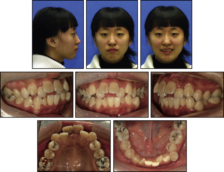
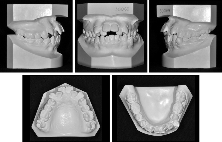
The facial photograph showed a convex profile with a slight mandibular retrusion and protrusion of the upper lip in the lateral view. From the frontal view, facial asymmetry was noticeable. She also had hyperactivity of the mentalis muscle ( Fig 1 ). Intraorally, the maxillary central incisors were in infraocclusion. She had an Angle Class I molar relationship on the left side and a Class II molar relationship on the right side, with Class II canine relationships on both sides. Overjet was 6.0 mm, and overbite was –3.5 mm. There was an 8.0-mm arch-length discrepancy in each arch. The dental midlines were not coincident ( Fig 2 ).


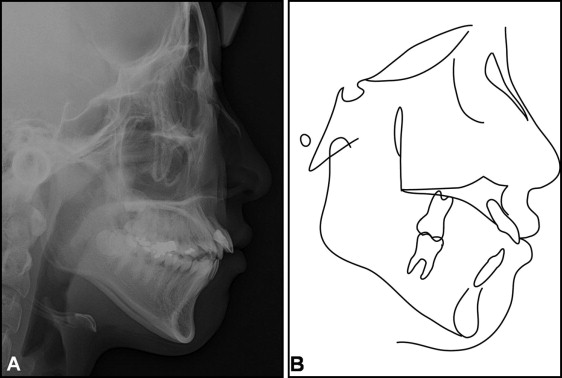
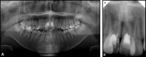
The cephalometric analysis showed a skeletal Class II anteroposterior discrepancy with an ANB angle of 6.0° and a hyperdivergent facial pattern. The maxillary central incisors were proclined ( Fig 3 ). The panoramic radiograph showed that all teeth were present ( Fig 4 , A ). The periapical radiograph showed external root resorption and incomplete endodontic treatment of both maxillary central incisors ( Fig 4 , B ).


Treatment objectives
The patient did not want to correct her facial asymmetry and slight mandibular retrusion. Thus, the facial treatment objectives were to increase her nasolabial angle and to improve her smile esthetics. The dental treatment objectives were to improve the maxillary anterior gingival margins, level the maxillary dentition, resolve the crowding in both dental arches, establish normal overjet and overbite, coordinate the dental midlines, and obtain Class I molar and canine relationships.
Treatment alternatives
Four treatment alternatives were considered.
- 1.
Extract the 4 first premolars and the ankylosed maxillary central incisors, and replace the ankylosed teeth with implants or a bridge. Because of the vertical alveolar bone defects, bone and gingival grafts would be needed. Esthetic prosthetic replacements could have limitations.
- 2.
Extract the ankylosed maxillary central incisors and the mandibular first premolars. The maxillary lateral incisors would be moved into the extraction sites of the maxillary central incisors. No additional surgical procedure would be necessary. However, this treatment would compromise the anterior esthetics and the occlusal guidance. Reshaping, prosthetic treatment, and soft-tissue surgery would be needed for the maxillary lateral incisors, canines, and premolars.
- 3.
Extract the 4 first premolars and perform segmental distraction osteogenesis. Gradual distraction would allow regeneration of the hard and soft tissues. However, the distraction device is normally bulky and inconvenient, and accurate 3-dimensional adjustment of the dento-osseous segment could be difficult, even though the segment is adjusted before the final consolidation phase.
- 4.
Extract the 4 first premolars and perform segmental osteotomy. In a single surgical stage, the dento-osseous segment could be moved to the predetermined position. However, a bone graft would be required in the bony gap. If metal plates and screws were necessary to fixate the segment, an additional surgery would be needed to remove them.
The fourth option was chosen.
Treatment progress
After the patient received oral-hygiene instructions, her maxillary and mandibular first premolars were extracted. Edgewise brackets (0.018 × 0.022 in) were bonded on both dental arches except for the ankylosed maxillary central incisors. An orthodontic miniscrew implant (Orlus; Ortholution, Seoul, Korea), 1.8 mm in diameter and 7.0 mm long, was placed in the interdental space between the maxillary left second premolar and first molar. The miniscrew implant was used to reinforce the posterior anchorage. After aligning and leveling the teeth, we placed 0.016 × 0.022-in stainless steel wires in both dental arches, and the remaining premolar extraction spaces were closed. Before surgery, 2 stainless steel sectional archwires (0.017 × 0.025 in) were placed in the maxillary arch. Model surgery was performed, and an interocclusal splint was made for the anterior region.
After a vestibular incision, a vertical and occlusally diverging osteotomy was performed with a number 701 fissure bur and a slim osteotome ( Figs 5 , A and B , and 6 , A ). The dento-osseous segment was repositioned by using the preformed splint. After extraction of the mandibular right third molar, autogenous bone taken from the external oblique ridge of the mandible was inserted into the bony gap ( Figs 5 , C and D , and 6 , B ). Fixation with plates and screws was not necessary because there was no mobility of the segment. The splint was wired to the brackets for stability. Then, 28 days postoperatively, the splint was removed, and orthodontic finishing was performed. Lingual fixed retainers were placed on the lingual surfaces of the incisors, canines, and second premolars in both arches. Maxillary and mandibular circumferential retainers were placed.


