Introduction
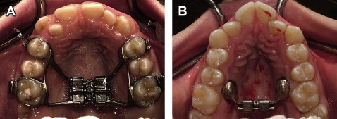
The purpose of this study was to compare the transverse, vertical, and anteroposterior skeletal and dental changes in adolescents receiving expansion treatment with tooth-borne and bone-anchored expanders. Immediate and long-term changes were measured on cone-beam computed tomography (CBCT) images.
Methods
Sixty-two patients needing maxillary expansion were randomly allocated to 1 of 3 groups: traditional hyrax tooth-borne expander, bone-anchored expander, and control. CBCT images were taken at baseline, immediately after expansion, after removal of the appliance (6 months), and just before fixed bonding (12 months). Repeated measures multivariate analysis of variance (MANOVA) was applied to the distances and angles measured to determine the statistical significance in the immediate and long time periods. Bonferroni post-hoc tests were used to identify significant differences between the treatment groups.
Results
Immediately after expansion, the subjects in the tooth-borne expander group had significantly more expansion at the crown level of the maxillary first premolars ( P = 0.003). Dental crown expansion was greater than apical expansion and skeletal expansion with both appliances. The control group showed little change (growth) over the 6-month interval. At 12 months, no group had a statistically significant difference in angle changes, suggesting symmetric expansion. Both treatment groups had significant long-term expansion at the level of the maxillary first molar crown and root apex, first premolar crown and root, alveolus in the first molar and premolar regions, and central incisor root. Tooth-borne expansion resulted in significantly more long-term expansion at the maxillary premolar crown and root than did bone-borne expansion.
Conclusions
Both expanders showed similar results. The greatest changes were seen in the transverse dimension; changes in the vertical and anteroposterior dimensions were negligible. Dental expansion was also greater than skeletal expansion.
Editor’s summary
With the growing popularity of CBCT imaging, it is tempting to try to determine once and for all whether we can increase the amount of skeletal expansion achieved with rapid palatal expansion procedures, with stable results. The purpose of this study was to use CBCT to determine the transverse, vertical, and anteroposterior skeletal and dental changes in adolescents receiving expansion with either tooth-anchored maxillary expansion (TAME) or bone-anchored maxillary expansion (BAME). Over an 18-month period, 62 patients who needed maxillary expansion were randomly allocated into 3 groups. One group received tooth-anchored expanders with bands on the first permanent molars and first premolars. The second group received bone-anchored devices, composed of 2 custom-milled stainless steel onplants, 2 miniscrews, and an expansion screw. The third group delayed treatment for 12 months to serve as the control group. Changes were measured on CBCT images taken at several stages: baseline, immediately after expansion, 6 months after placement of the appliance, and 12 months after placement of the appliance.
Midpalatal suture separation was seen on the CBCT images for both TAME and BAME groups. Approximately 4 mm (70%) of the expansion at the maxillary molars was maintained at 12 months with either appliance. Bone-borne expansion was developed to eliminate some of the negative effects (dental expansion, periodontial recession, and possible root resorption) of tooth-borne systems. However, based on the results of this study, both types of expansion led to similar outcomes. Force application at the teeth and on the roots is greater in the tooth-anchored method; in the bone-anchored method, pressure on the bone is greater. For this reason, the results are surprising. The authors discovered similar tipping of the molars with both appliances. Dental tipping was greater for the first premolars in the TAME group because the force application is on the molars and premolars; with the BAME method, it is at the level between the molars and second premolars. No difference between apical expansion of the molar apices was found between appliances; according to the authors, the rigidity of the tooth-anchored expander would result in buccal root movement, whereas, for bone-anchored expansion, the application of force at the bone surface near the molar root caused bone bending, with movement of the root apex toward the outer surface of the alveolus.
These findings indicate that bone-anchored maxillary expansion can be an alternate choice for achieving expansion when several teeth are missing or other complications limit the use of tooth-anchored devices.


