Three-dimensional (3D) models of teeth and soft and hard tissues are tessellated surfaces used for diagnosis, treatment planning, appliance fabrication, outcome evaluation, and research. In scientific publications or communications with colleagues, these 3D data are often reduced to 2-dimensional pictures or need special software for visualization. The portable document format (PDF) offers a simple way to interactively display 3D surface data without additional software other than a recent version of Adobe Reader (Adobe, San Jose, Calif). The purposes of this article were to give an example of how 3D data and their analyses can be interactively displayed in 3 dimensions in electronic publications, and to show how they can be exported from any software for diagnostic reports and communications among colleagues.
Tessellated surface models are used not only in orthodontics or maxillofacial surgery, but also in many other scientific fields such as astronomy, biology, engineering, architecture, and cartography. In medicine, tessellated surfaces are most often composed of triangular faces. With this method, complex 3-dimensional (3D) scenes are fluently visualized by 3D computer graphics. The possibilities are nearly unlimited, and therefore the challenge of increasing digitalization is to keep things simple and friendly to use. Wouldn’t it be nice to see the patient records of an orthodontic case report in an article in an interactive 3D visualization? Or to send the 3D digital models with their corresponding analysis to a colleague by e-mail without the need for new software or online access?
In this article, we describe a simple way to present 3D surface data with their analyses in articles or diagnostic reports. Electronic articles are generally published in the portable document format (PDF) and mostly consist of text and 2-dimensional pictures. However, an interactive multimedia object with 3D content can be displayed in a PDF document instead of a 2-dimensional picture. Three-dimensional software does not yet allow direct export of tessellated surfaces with analyses and annotations to a PDF document, but often to other open file formats such as STL, PLY, VRML, DAE, and OBJ. Up to now, the image-processing steps for embedding 3D surface data of a biologic specimen in a 3D interactive PDF document, as well as the incorporation of 3D surface data in astronomy research articles with a free open-source software such as basic MiKTeX (version 2.8.3761; MiKTeX, Berlin, Germany; www.miktex.org ), have been already described. In this method, knowledge of LaTeX document markup language is necessary (LaTeX2e, 10/15/2009, LaTeX project team, www.latex-project.org ). In chemistry, 3D molecular structures have been documented by importing VRML files to a 3D interactive document. In contrast to surface data, embedding volume data is not yet as simple. Image stacks from histologic sections, confocal laser scanning microscopy, and microcomputed tomography have been volume rendered and then processed to each 3 stacks of PNG images. These images are displayed in a 3D interactive PDF document and represent closely the original volume rendering. However, this method seems to be complicated and additionally needs a Java script code (ECMA 262 5th Edition, December 2009, Ecma International, Geneva, Switzerland, www.ecma-international.org ) to visualize the data in the PDF document. On the other hand, if the surface data are not present a priori, transformation of volume data to surface data by segmentation based mostly on thresholding algorithms, computer-assisted hand work, or other methods is possible, but quality and artefacts must be considered.
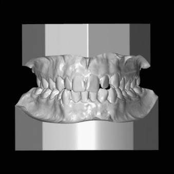
An alternative way that is suitable for embedding 3D surface data of dental and medical records in 3D interactive PDF files is demonstrated in this article with the example of orthodontic digital models. The 3D surfaces ( Figs 1 and 2 ) were exported from the software Digimodel (version 2.37; OrthoProof BV, Nieuwegein, The Netherlands, www.orthoproof.com ), converted to the open source universal 3D file format (U3D), by using the free open-source software meshlab (version 1.2.3; Visual Computing Lab, ISTI-CNR, Pisa, Italy; meshlab.sourceforge.net ) and the free software DAZ Studio (version 3.1.2.19; DAZ Productions, Draper, Utah) and then assembled to a PDF multimedia model by using the commercial software Adobe Acrobat X Pro (version 10.0; Adobe, San Jose, Calif; www.adobe.com ). Direct Import of 3D data to Adobe Acrobat X Pro requires data of the formats U3D or PRC. The plug-in 3D PDF converter (Tetra 4D, Cheyenne, Wyo; www.tetra4d.com ) enhances the import functions of Adobe Acrobat X Pro. More supported file formats for import as well as import and compression settings have become available. Compression by remeshing or simplification of surface data should only be applied with caution. The boundaries of the mesh should be preserved within clearly specified limits. For example, geometric constructions from computer-aided design software can be imported by conversion to a triangulated mesh with a boundary representation at a tolerance of 0.01 mm.