Introduction
The palatine rugae have been suggested as stable reference points for superimposing 3-dimensional virtual models before and after orthodontic treatment. We investigated 3-dimensional changes in the palatine rugae of children over 9 years.
Methods
Complete dental stone casts were biennially prepared for 56 subjects (42 girls, 14 boys) aged from 6 to 14 years. Using 3-dimensional laser scanning and reconstruction software, virtual casts were constructed. Medial and lateral points of the first anterior 3 rugae were defined as the 3-dimensional landmarks. The length of each ruga and the distance between the end points of the rugae were measured in virtual 3-dimensional space. The measurement changes over time were analyzed by using the mixed-effect method for longitudinal data.
Results
There were slight increases in the linear measurements in the rugae areas: the lengths of the rugae and the distances between them during the observation period. However, the amounts of the increments were relatively small when compared with the initial values and individual random variability. Although age affected the linear dimensions significantly, it was not clinically significant; the rugae were relatively stable.
Conclusions
The use of the palatine rugae as reference points for superimposing and evaluating changes during orthodontic treatment was thought to be possible with special cautions.
The palatine rugae are ridges situated in the anterior palatal mucosa on each side of the medial palatal raphe and behind the incisive papilla. At the time of development in the embryonic stage, these structures are relatively prominent and somewhat symmetrically occupy most of the palatal shelves. Around the end of intrauterine life, their patterns become less regular, and the posterior structures begin to disappear. At birth, the well-formed anterior rugae remain in a compressed form. The shape, length, width, prominence, number, and orientation of the palatal rugae vary considerably among people. In general, there is no bilateral symmetry of the rugae pattern in a person.
The palatine rugae are known to be both stable and unique. The clinical significance of the palatine rugae is that they have been widely considered to be stable landmarks during orthodontic treatment. In addition, the use of palatine rugae for forensic identification has been well established. The unique pattern of palatine rugae in each person, similar to fingerprints, makes them useful for postmortem identification. These structures are considered to be stable throughout life, once all growth has ceased. The anatomic position of the rugae surrounded by other orofacial structures is another advantage for their use because they are relatively well protected from trauma and high temperatures.
Several authors have reported on their dimensional changes and relative positions in the evaluation of rugae as suitable reference points for longitudinal cast analysis. Other researchers have investigated the use of palatine rugae as reference points for measuring tooth movement even as a substitute for cephalometric superimposition.
However, there is considerable debate on the stability of rugae positions. The dentoalveolar processes undergo visible alterations as they grow, are a part of the craniofacial complex, and are influenced by changes in various parts of the skull. The palatine rugae are not an exception. It was reported that the vertical position is affected by the continuous eruption of neighboring teeth, and the stability of rugae is different according to their position. Furthermore, information about the development of palatal height in normal subjects is scarce.
Technology with 3-dimensional (3D) scanners and reconstructed virtual models has been widely used in dentistry for various applications. Studies of 3D reconstructions have resulted in accurate and reliable techniques for restorative procedures and facial analyses to aid clinicians in planning more effective treatments. In addition, the use and study of these 3D reconstructions allows for the measurement of specific distances that would be impossible or difficult to analyze by using conventional means. For example, the distance along the model surface and accurate orthographic measurements can be easily obtained with this new method.
We investigated the longitudinal changes occurring in the palatine rugae of children followed from 6 to 14 years of age using 3D reconstructed virtual models. The aim of this study was to evaluate the 3D longitudinal stability of the palatine rugae as reference landmarks. The pattern of individual variations in the longitudinal changes was also discussed by using the mixed-effect analysis method.
Material and methods
The material for this study consisted of dental casts from 56 children, including 14 boys and 42 girls of Korean origin followed from 6 to 14 years of age. From 1995 to 2003, complete dental cast sets were prepared biennially. These subjects were judged to have clinically good rather than ideal occlusions. Casts with missing teeth other than third molars and more than 3 mm of crowding or an arch length discrepancy, and subjects who did not have full sets of casts over the span of 9 years were excluded. All subjects and the parent or guardian provided written informed consent, and the institutional review board for the protection of human subjects of Seoul National University reviewed and approved the research protocol (S-D2010014).
All casts selected were scanned by using a 3D scanner (optoTOP-HE; Breckmann, Meersburg, Germany) with a point accuracy of ±0.001 mm, and resolutions of 0.040 mm in the x and y directions and 0.002 mm in the z direction. Each cast was scanned from 10 or more different views that were then combined and rendered into a 3D image by using specialized software (Rapidform XO; INUS Technology, Seoul, Korea). The virtual 3D models were measured and analyzed by using specialized software (Rapidform 2004; INUS Technology). The virtual casts of the same subject at different ages were simultaneously displayed on the dual monitor system to maintain the consistency of the reference points. For reproducibility, the reference points were created on the virtual casts via consensus between 2 observers. Reference points were created at 2 separate times by the same 2 observers (K.H.K. and P.Y.S.) over a 3-week period.
Landmarks on the palatine rugae and the incisive papilla were created as reference points based on previous studies. The point related to the median palatine raphe was not used because it is indistinguishable and ambiguous. All measurements were restricted to the 3 most anterior rugae. Additional or rudimentary rugae were excluded from this investigation.
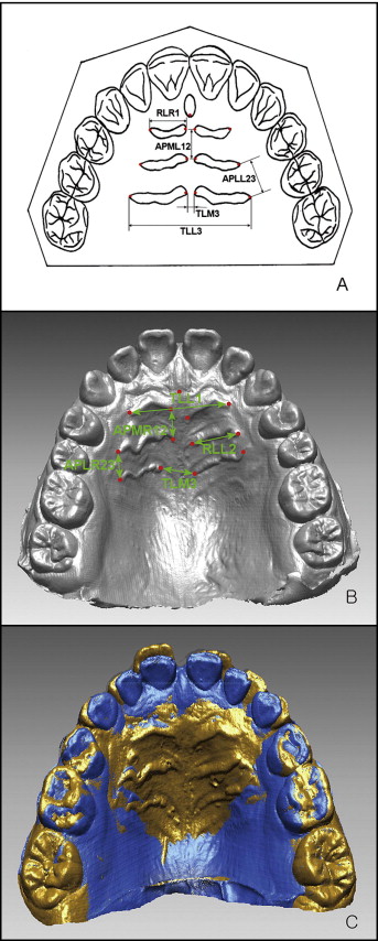
The following parameters were measured on the virtual casts to determine the linear and angular changes in the palatine rugae. First, the distance between the medial and lateral points of the individual rugae or the lengths of individual rugae (RLR and RLL) were measured. The distances between the medial points (TLM) and between the lateral points of the right and left rugae (TLL), as well as right and left anteroposterior linear distances between the medial (APML and APMR) and between the lateral points (APLR and APLL) of the first, second, and third rugae, were measured. Finally, the rugae angle formed by the most distal point of the incisive papilla and the lateral points of the right and left third rugae was measured. These abbreviations are briefly described in Table I , and detailed descriptions of the measurements and an example of virtual model superimposition with rugae anatomies are depicted in Figure 1 . All distances measured were 3D Euclidean distances. To test the reliability, 10 of these 3D scams were randomly selected and measured again on separate days 6 months after the initial measurements.
| Variable | Definition |
|---|---|
| TLM1 (mm) | Transverse linear distance between the medial points of the right and left first rugae |
| TLM2 (mm) | Transverse linear distance between the medial points of the right and left second rugae |
| TLM3 (mm) | Transverse linear distance between the medial points of the right and left third rugae |
| RLR1 (mm) | Length of the right first rugae |
| RLR2 (mm) | Length of the right second rugae |
| RLR3 (mm) | Length of the right third rugae |
| RLL1 (mm) | Length of the right first rugae |
| RLL2 (mm) | Length of the right second rugae |
| RLL3 (mm) | Length of the right third rugae |
| TLL1 (mm) | Transverse linear distance between the lateral points of the right and left first rugae |
| TLL2 (mm) | Transverse linear distance between the lateral points of the right and left second rugae |
| TLL3 (mm) | Transverse linear distance between the lateral points of the right and left third rugae |
| APLR12 (mm) | Anteroposterior distance between the lateral points of the right first and second rugae |
| APLR23 (mm) | Anteroposterior distance between the lateral points of the right second and third rugae |
| APLR13 (mm) | Anteroposterior distance between the lateral points of the right first and third rugae |
| APMR12 (mm) | Anteroposterior distance between the medial points of the right first and second rugae |
| APMR23 (mm) | Anteroposterior distance between the medial points of the right second and third rugae |
| APMR13 (mm) | Anteroposterior distance between the medial points of the right first and third rugae |
| APML12 (mm) | Anteroposterior distance between the medial points of the left first and second rugae |
| APML23 (mm) | Anteroposterior distance between the medial points of the left second and third rugae |
| APML13 (mm) | Anteroposterior distance between the medial points of the left first and third rugae |
| APLL12 (mm) | Anteroposterior distance between the lateral points of the left first and second rugae |
| APLL23 (mm) | Anteroposterior distance between the lateral points of the left first and second rugae |
| APLL13 (mm) | Anteroposterior distance between the lateral points of the left first and third rugae |
| Rugae angle (°) | The angle formed by the most distal point of the incisive papilla and the lateral points of the right and left third rugae |

Descriptive statistics on the changes in the palatal rugae parameters were calculated biennially. Each subject provided multiple repeated observations for the linear and angular measurements in the palatal rugae region. Since the serial measurements were correlated according to each subject, a mixed model was applied. The analysis model for the data was written as y ij = μ + β 1 sex i + β 2 age ij + β 3 sex i ∗age ij + b i1 + b i2 ∗age ij + e ij , where μ is the total mean, β 1 is the sex effect, β 2 is the age effect, β 3 is the interaction effect between sex and age, and b i (i = 1, 2, . . . , 56) is the random effect by individual subject. Statistical analyses were performed by using the language R, and a P value less than 0.05 was predetermined to be statistically significant.
Results
The intraexaminer reliability coefficients ranged from 0.999 to 1.000. In terms of root mean squares, the random errors of estimation were lower than 0.05 mm for linear measurements and 0.45° for angular measurements. No variable showed a statistically significant difference between the test and retest measurements.
Means, standard deviations, and average differences in the means of the 4 intervals for all measurements are summarized in Table II . The changes in the measurements from ages 6 to 14 years were quite small, with slightly increasing tendencies over time. The average 2-year change ranged from a minimum of 0.12 mm (TLM1) to a maximum of 0.67 mm (TLL3). The average 2-year change from the initial value at age 6 showed an average ratio of 3.8% (1.1%-8.4%). Only 3 variables among the 24 linear measurements—APLR23, APLR13, and APLL23—showed more than a 5% increase during that 6-year time period. In other words, the lengths of the rugae and the distances between the adjacent rugae increased during the observation period, even though the changes were minimal. The ratio of the average 2-year change to the initial value at age 6 was the maximum of 8.4% for APLR23. The sole angular measurement, the rugae angle, however, did not show a significant change over time ( P = 0.237) ( Table II ).
| Variable | Age (y) | Average 2-year change ∗ | Ratio † | ||||
|---|---|---|---|---|---|---|---|
| 6 | 8 | 10 | 12 | 14 | |||
| TLM1 (mm) ‡ | 3.22 (1.25) | 3.38 (1.27) | 3.43 (1.32) | 3.60 (1.38) | 3.71 (1.41) | 0.12 (0.06) | 0.038 |
| TLM2 (mm) | 6.66 (3.84) | 6.40 (3.84) | 7.01 (3.90) | 7.28 (3.86) | 7.44 (3.83) | 0.20 (0.13) | 0.029 |
| TLM3 (mm) | 6.86 (2.89) | 7.30 (2.89) | 7.31 (2.88) | 7.61 (3.04) | 7.75 (3.00) | 0.22 (0.19) | 0.033 |
| RLR1 (mm) | 10.35 (1.23) | 10.68 (1.19) | 10.81 (1.24) | 10.92 (1.23) | 10.95 (1.35) | 0.15 (1.28) | 0.014 |
| RLR2 (mm) | 9.54 (2.38) | 9.80 (2.59) | 10.75 (2.71) | 10.31 (2.79) | 10.50 (2.87) | 0.24 (0.03) | 0.025 |
| RLR3 (mm) | 10.44 (2.63) | 11.00 (2.57) | 11.43 (2.70) | 11.95 (2.77) | 12.30 (2.86) | 0.47 (0.10) | 0.045 |
| RLL1 (mm) | 10.21 (1.62) | 10.54 (1.65) | 10.54 (1.66) | 10.71 (1.64) | 10.97 (1.68) | 0.19 (0.14) | 0.019 |
| RLL2 (mm) | 9.26 (2.94) | 9.61 (3.06) | 9.65 (2.95) | 10.04 (3.17) | 10.29 (3.24) | 0.26 (0.16) | 0.028 |
| RLL3 (mm) | 10.94 (2.59) | 11.09 (2.39) | 11.54 (2.67) | 12.09 (2.87) | 12.30 (2.93) | 0.34 (0.19) | 0.031 |
| TLL1 (mm) | 21.12 (2.13) | 21.80 (2.18) | 21.85 (2.27) | 21.93 (2.32) | 22.05 (2.33) | 0.23 (0.30) | 0.011 |
| TLL2 (mm) | 22.17 (1.93) | 22.74 (2.11) | 22.89 (2.05) | 23.30 (2.36) | 23.45 (2.37) | 0.32 (0.21) | 0.014 |
| TLL3 (mm) | 23.28 (2.77) | 24.20 (2.94) | 24.78 (2.91) | 25.60 (3.00) | 25.97 (2.99) | 0.67 (0.24) | 0.029 |
| APLR12 (mm) | 3.98 (1.63) | 3.88 (1.59) | 4.29 (1.83) | 4.53 (1.96) | 4.71 (1.96) | 0.18 (0.21) | 0.045 |
| APLR23 (mm) | 4.11 (1.99) | 4.53 (2.17) | 4.88 (2.38) | 5.21 (2.62) | 5.49 (2.68) | 0.34 (0.06) | 0.084 |
| APLR13 (mm) ‡ | 7.47 (2.29) | 7.79 (2.35) | 8.55 (2.63) | 9.11 (2.93) | 9.47 (3.06) | 0.50 (0.20) | 0.067 |
| APMR12 (mm) | 4.33 (1.58) | 4.41 (1.61) | 4.57 (1.69) | 4.75 (1.72) | 4.92 (1.80) | 0.15 (0.04) | 0.035 |
| APMR23 (mm) | 4.30 (1.55) | 4.31 (1.65) | 4.62 (1.63) | 4.79 (1.77) | 4.96 (1.78) | 0.17 (0.12) | 0.038 |
| APMR13 (mm) | 6.11 (2.41) | 6.03 (2.62) | 6.72 (2.67) | 7.00 (2.82) | 7.06 (2.80) | 0.24 (0.33) | 0.039 |
| APML12 (mm) | 4.25 (1.89) | 4.36 (1.88) | 4.41 (1.87) | 4.72 (1.92) | 4.93 (2.03) | 0.17 (0.11) | 0.041 |
| APML23 (mm) | 4.30 (1.69) | 4.47 (1.83) | 4.80 (1.87) | 4.92 (2.00) | 4.98 (1.97) | 0.17 (0.12) | 0.040 |
| APML13 (mm) | 6.04 (2.38) | 6.31 (2.39) | 6.70 (2.57) | 6.99 (2.71) | 7.16 (2.81) | 0.28 (0.09) | 0.047 |
| APLL12 (mm) | 3.51 (1.31) | 3.56 (1.42) | 3.89 (1.56) | 4.02 (1.59) | 4.11 (1.70) | 0.15 (0.12) | 0.043 |
| APLL23 (mm) | 4.01 (1.82) | 4.12 (2.01) | 4.51 (2.12) | 4.76 (2.11) | 4.99 (2.10) | 0.24 (0.12) | 0.061 |
| APLL13 | 6.84 (2.03) | 7.02 (2.21) | 7.62 (2.48) | 7.91 (2.38) | 8.15 (2.46) | 0.33 (0.19) | 0.048 |
| Rugae angle (°) | 98.60 (12.16) | 98.53 (12.85) | 95.91 (11.82) | 97.09 (12.13) | 96.25 (12.20) | −0.59 (11.60) | −0.006 |
Stay updated, free dental videos. Join our Telegram channel

VIDEdental - Online dental courses


