Atlases of human anatomy are fascinating volumes. They are particularly appealing and useful to those with special professional interests: figurative artists, biologists, anatomists, and clinicians. In this new anatomic atlas of the face crafted largely by Ralf J. Radlanski, an orthodontist and professor at Freie Universität Berlin, and Karl H. Wesker, a medical illustrator, we may begin to appreciate the many striking advances in atlas production that have occurred in the 470 years since the publication of De humani corporis fabrica , the landmark textbook of human anatomy by Andreas Vesalius.
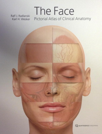
Radlanski and Wesker, with help from 11 listed contributors, have fashioned their facial atlas with significant boosts from modern imaging technology. There is a pleasing consistency throughout the book: The anatomic drawings are built on templates made from magnetic resonance images of 1 attractive, angular face, that of Dr Radlanski’s daughter Jana. Mr Wesker’s softly shaded drawings, many of which are life-size or larger, are color coded in a natural and uniform way and are clear to comprehend, supported by descriptive labels and detailed supplemental text. The artist’s skill in placing subtle shadows between contiguous, layered structures and in external and internal anatomic sections gives many of the more complicated figures a marvelous 3-dimensional quality that aids the viewer’s visual understanding.
This is the first English edition, after publication of the atlas in German in 2011. The book is quarto size, 12 × 9.5 in (305 × 242 mm), and weighs 4.5 lbs (2 kg). Unfortunately, the hardcover boards appear to be too thin and bendable for such a large, heavy volume.
Several parts of the atlas will engage even the casual nonprofessional reader. A section on facial expression carefully illustrates the muscle groups acting in concert to produce emotional expressions, such as disgust, happiness, pouting, and other extreme states. Another section shows how the human facial integument changes with age. It includes informative histologic and descriptive facial drawings and photographs of the features of facial aging. A reader may wish these valuable sections provided more citations to original research sources on which the figures were based.
The face: Pictorial atlas of clinical anatomy would be a handsome addition for an orthodontist or other facial clinician with an expansive professional library. However, in this era of immediate electronic access to reliable medical resources and images, purchase of this hard-copy reference book might be considered elective.
Stay updated, free dental videos. Join our Telegram channel
VIDEdental - Online dental courses


