Introduction
The purpose of this study was to evaluate the long-term stability of quad-helix/crib treatment in subjects with dentoskeletal open bite.
Methods
Twenty-eight subjects (11 boys, 17 girls; mean age, 8.2 ± 1.3 years) were treated consecutively with quad-helix/crib appliances. The patients were reevaluated at the end of active treatment with the quad-helix/crib (mean age, 9.7 ± 1.6 years) and at least 5 years after the completion of treatment (mean age, 14.6 ± 1.9 years). A control group of 20 untreated subjects with the same dentoskeletal disharmony was used for the statistical comparison (Mann-Whitney U test).
Results
In the long term, the quad-helix/crib group showed a significant reduction in the ANB angle (−1.3°), a downward rotation of the palatal plane (1.8°), a greater increase in overbite (2.1 mm), and a decrease in overjet (−1.5 mm) when compared with the controls.
Conclusions
In the long term, the use of the quad-helix/crib appliance led to successful outcomes in about 93% of the patients considered. Correction of dentoskeletal open bite was associated with a clinically significant downward rotation of the palatal plane.
Anterior open bite is characterized by a localized absence of occlusion between the incisal edges of the maxillary and mandibular teeth when the remaining teeth are in occlusion. This malocclusion occurs because of interferences during normal dental eruption and alveolar development. Several factors are involved in the etiology of anterior open bite. Thumb sucking and increased vertical skeletal relationships are significant risk factors for the establishment of an anterior open bite. Subjects with dentoskeletal open bite and sucking habits often have concomitant transverse discrepancies. Many authors have emphasized that a skeletal open bite should be treated early in the mixed dentition to allow for normal development of the anterior dentoalveolar region.
Various treatment approaches can be found in the literature with regard to early treatment of anterior open bite. The elimination of persisting sucking habits and the control of the vertical dimension must be therapeutic objectives. The correction of maxillary constriction is an additional target for treatment in patients with open bite.
The use of a palatal crib has been proposed as an excellent treatment option, because it prevents thumb or pacifier sucking, as well as tongue thrust. According to Haryett et al, the palatal crib is effective for the elimination of a thumb-sucking habit in 85% to 90% of subjects. Studies reporting the success of early treatment in subjects with anterior open bite when compared with a well-matched control group, however, are scarce in the literature. Only 2 studies have incorporated untreated controls with the same type of dentoskeletal disharmony, but they were confined to short-term observations. The authors of both studies analyzed the effects of a removable palatal crib associated with a vertical-pull chincup; this treatment protocol did not produce significant changes in the skeletal maxillary and mandibular components. The results of both studies showed that the effects of therapy primarily were dentoalveolar.
A proposed treatment protocol aimed to eliminate the thumb-sucking habit and to correct both the anterior open bite and the maxillary transverse deficiency in growing high-angle subjects is a quad-helix (Q-H) appliance with the addition of a palatal crib (Q-H/C). When compared with the effects of a removable appliance (open-bite bionator or removable palatal crib), the Q-H/C appliance was shown to be significantly more effective in the improvement of overbite in the short term. The treatment and posttreatment effects of a Q-H/C appliance showed a clinical effectiveness in correcting the dental open bite of 85% of the patients after a follow-up of 2 years. This favorable result was associated with clinically significant improvement in the maxillomandibular vertical skeletal relationships. No data, however, are available in the literature about the outcomes of the Q-H/C appliance reevaluated at a follow-up of at least 5 years.
The purpose of this study, therefore, was to evaluate the long-term stability of Q-H/C treatment in subjects with thumb-sucking habits and anterior dentoskeletal open bite. Both active treatment and posttreatment effects were analyzed in consecutively treated patients, and these results were compared with the growth changes in an untreated control group with the same dentoskeletal disharmony during a follow-up period of at least 5 years.
Material and methods
The Q-H/C sample comprised 28 subjects (17 girls, 11 boys) who were treated consecutively at the Department of Orthodontics at the University of Rome “Tor Vergata” or in a private orthodontic practice in Rome. Lateral cephalograms of treated patients were analyzed regardless of treatment results. Each patient had the following features: thumb-sucking habit before treatment; negative overbite; constricted maxillary arch as consequence of thumb sucking; full eruption of first permanent molars and permanent incisors (to prevent the “pseudo-open bite” due to undererupted permanent incisors) ; no permanent teeth extracted before or during treatment; 3 consecutive lateral cephalograms of good quality with adequate landmark visualization and minimal or no rotation of the head, taken before treatment (T1), at the end of the active treatment with the Q-H/C (T2), and at a follow-up observation at least 5 years after the completion of treatment (T3); and treatment with the Q-H/C appliance for at least 12 months.
All subjects were at a prepubertal stage of skeletal maturity according to the cervical vertebral maturation method (CS 1 or CS 2) at T1. The overall observation period was 6.4 ± 1.4 years, which included a follow-up period of at least 5 years during which the Q-H/C patients were treated with fixed appliances. No active biomechanics or vertical elastics to extrude the incisors were applied during fixed appliance therapy. No intraoral Class II elastics were used.
All subjects had reached postpubertal skeletal maturity at T3 (CS 4-6). The stages of cervical vertebral maturation were determined by a calibrated examiner (L.F.) trained in this method. All patients were in the permanent dentition at T3.
A control group of 20 subjects (10 girls, 10 boys) with anterior open bite was retrieved from the archives of the University of Michigan Growth Study and the Denver Child Growth Study. The control group matched the Q-H/C group for negative overbite at T1, chronologic age, and skeletal maturation at the various time periods and for the duration of intervals.
The mean ages at the 3 time periods in both the Q-H/C and control groups and the duration of either treatment or observation intervals are given in Table I .
| Chronologic age (y) | Q-H/C group (n = 28) | Control group (n = 20) | ||
|---|---|---|---|---|
| Mean | SD | Mean | SD | |
| T1 | 8.2 | 1.3 | 8.1 | 0.4 |
| T2 | 9.7 | 1.6 | 9.8 | 0.4 |
| T3 | 14.6 | 1.9 | 14.5 | 0.7 |
| T1-T2 | 1.5 | 0.4 | 1.7 | 0.4 |
| T2-T3 | 4.9 | 1.3 | 4.7 | 0.6 |
| T3-T1 | 6.4 | 1.4 | 6.4 | 0.7 |
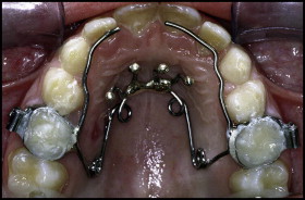
The Q-H appliance used in this study was made of 0.036-in stainless steel wire soldered to bands on the second deciduous molars or the first permanent molars ( Fig ). The lingual arms of the appliance extended mesially to the deciduous canines or to the permanent incisors. The anterior helices were brought as far forward on the palate as possible. Spurs to prevent thumb sucking were formed from 3 segments of 0.036-in stainless steel wire soldered to the anterior bridge of the Q-H. The wire segments were inclined lingually to prevent impingement on the sublingual mucosa. Activation of the Q-H was equivalent to the buccolingual width of 1 molar. The appliance was reactivated once or twice during treatment to achieve overcorrection of the transverse relationships.
The T1, T2, and T3 cephalograms were hand traced by 1 investigator (V.G.) and then verified for landmark location by a second investigator (L.F.). Any disagreements were resolved by retracing the landmark or structure to the satisfaction of both observers. Cephalometric software (Viewbox, version 3.0; dHAL Software, Kifissia, Greece) was used for a customized digitization regimen that contained 21 variables (11 linear, 10 angular). The magnification factor of the cephalograms was standardized at 8%.
Statistical analysis
Descriptive statistics (mean differences and standard deviations) were calculated for all cephalometric measurements at T1, and for the changes from T1 to T2, T2 to T3, and T1 to T3 in both groups.
The homogeneity between the Q-H/C and control groups for skeletal maturity at each observation time and mean duration of observation intervals allowed for comparisons without annualizing the data. Matching between treated and control subjects was tested also by means of propensity score analysis. This matching protocol in observational studies allows researchers to mimic randomization by creating a sample of subjects who did not receive treatment comparable on all observed covariates with the sample of subjects who received treatment. The program “psmatching” (available at http://sourceforge.net/projects/psmspss/files ) was used to calculate propensity scores and to test matching between the treated and control samples (SPSS version 21.0; IBM, Armonk, NY). Propensity scores were calculated for some clinically relevant covariates (Wits, Frankfort horizontal to palatal plane, palatal plane to mandibular plane, and overbite) whereas all other variables were entered as additional covariates. An overall balance chi-square test developed by Hansen and Bowers was applied to test group matching. This test examines all covariates that were used to estimate the propensity scores and all variables that were defined as additional covariates.
Changes in the 2 groups were compared by nonparametric tests, since normal distribution (Kolmogorov-Smirnov test) or equality of variances (Levene test) could not be assessed for all variables. In general, parametric tests are more powerful than nonparametric statistics. However, the assumptions required for parametric tests are particularly important when sample sizes are small, with small usually thought to be fewer than 30 in each group; if the assumptions cannot be verified, then nonparametric methods should be used.
Before making the comparisons of the longitudinal changes, significant differences between the craniofacial starting forms at T1 were assessed with the Mann-Whitney U test between the Q-H/C and control groups. To assess the differences between the Q-H/C and control groups with regard to T1 to T2, T2 to T3, and overall T1 to T3 changes, Mann-Whitney U tests ( P <0.05; P <0.01; and P <0.001) were used. Chi-square tests with the Yates correction were performed to compare the prevalence rates of correction of anterior open bite in the 2 groups at T2 and T3. The correction of anterior open bite at the dentoalveolar level was considered to be obtained when the overbite measurement was equal to or greater than 0 mm.
The data were analyzed with statistical software (SPSS 21.0 and SigmaStat version 3.5; Systat Software, Point Richmond, Calif). Statistical significance was tested at P <0.05. The power of the study was 0.91 for an alpha level of 0.05 and an effect size equal to 1 for the clinically relevant variable palatal plane to mandibular plane angle, as derived from a previous study.
To test the reliability of the measurements, 20 lateral cephalograms randomly selected from various subjects in the study were retraced and remeasured by the same examiner (V.G.) after a 1-month interval. No systematic error was found with the Wilcoxon signed rank test.
Random errors were estimated with Dahlberg’s formula. The errors for linear measurements ranged from 0.1 mm for pogonion to nasion perpendicular, to 1.2 mm for condylion-gonion. The errors for angular measurements ranged from 0.4° for ANB angle, to 1.4° for interincisal angle.
Results
Analysis of the starting forms ( Table II ) showed that the Q-H/C and the control groups had no statistically significant differences in craniofacial characteristics at T1. The only exception was a significantly longer ramus length (Co-Go) at T1 in the Q-H/C group. For the dentoskeletal features at T1, the vertical skeletal relationship was increased, and the sagittal intermaxillary relationship was skeletal Class II in both groups. The overall matching between the treated and control groups was assessed further with propensity scores. The chi-square balance test was not statistically significant (chi-square = 28.154; P = 0.081), thus indicating good overall balance between the 2 groups.
| Cephalometric measurement | Q-H/C group (n = 28) | Control group (n = 20) | Difference | Significance | ||
|---|---|---|---|---|---|---|
| Mean | SD | Mean | SD | |||
| Maxillary skeletal | ||||||
| SNA (°) | 82.1 | 2.9 | 80.7 | 3.2 | 1.4 | NS |
| Point A-nasion perp (mm) | 2.4 | 2.7 | 1.1 | 3.0 | 1.3 | NS |
| Mandibular skeletal | ||||||
| SNB (°) | 76.8 | 3.2 | 75.4 | 2.0 | 1.4 | NS |
| Pg-nasion perp (mm) | −5.5 | 6.6 | −8.5 | 4.0 | 3.0 | NS |
| Co-Gn (mm) | 106.3 | 6.5 | 104.3 | 3.9 | 2.0 | NS |
| Maxillary/mandibular | ||||||
| ANB (°) | 5.3 | 2.4 | 5.4 | 2.0 | −0.1 | NS |
| Wits (mm) | −1.6 | 2.7 | −1.5 | 2.6 | −0.1 | NS |
| Vertical skeletal | ||||||
| FH-PP (°) | −3.1 | 3.4 | −3.4 | 2.8 | 0.3 | NS |
| MPA (°) | 28.5 | 4.2 | 27.9 | 4.4 | 0.6 | NS |
| PP-mandibular plane (°) | 31.5 | 4.9 | 31.3 | 4.0 | 0.2 | NS |
| ANS-Me (mm) | 65.1 | 5.7 | 64.9 | 4.1 | 0.2 | NS |
| Co-Go (mm) | 49.3 | 3.3 | 47.2 | 3.5 | 2.1 | ∗ |
| Gonial angle (°) | 131.7 | 5.1 | 130.2 | 4.2 | 1.5 | NS |
| Interdental | ||||||
| Overjet (mm) | 2.8 | 2.9 | 3.6 | 1.8 | −0.8 | NS |
| Overbite (mm) | −3.3 | 1.6 | −2.2 | 2.3 | −1.1 | NS |
| Interincisal angle (°) | 122.0 | 9.7 | 125.7 | 11.0 | −3.7 | NS |
| Molar relationship (mm) | 0.4 | 1.9 | 0.8 | 1.3 | −0.4 | NS |
| Maxillary dentoalveolar | ||||||
| U1-Point A vert (mm) | 4.4 | 2.3 | 3.8 | 1.8 | 0.6 | NS |
| U1-FH (°) | 117.0 | 7.8 | 114.3 | 6.8 | 2.7 | NS |
| Mandibular dentoalveolar | ||||||
| L1-Point A Pg (mm) | 2.1 | 2.1 | 1.9 | 2.2 | 0.2 | NS |
| L1-MPA (°) | 93.1 | 6.0 | 92.6 | 7.1 | 0.5 | NS |
The statistical comparisons of the T1 to T2 changes ( Table III ) showed no statistically significant differences between the Q-H/C and control samples for any maxillary or mandibular skeletal measurements in the sagittal plane. For the vertical skeletal measurements, the Q-H/C group exhibited greater downward rotation of the palatal plane than did the control group (1.9 mm). A significant effect of therapy was found for the dentoalveolar variables. The Q-H/C group showed a significantly greater increase in overbite (2.2 mm more than the control group) associated with 5.7° of lingual tipping of the mandibular incisors relative to the mandibular plane with respect to the controls.
| Cephalometric measurement | Q-H/C group (n = 28) | Control group (n = 20) | Difference | Significance | ||
|---|---|---|---|---|---|---|
| Mean | SD | Mean | SD | |||
| Maxillary skeletal | ||||||
| SNA (°) | 0.2 | 1.8 | 1.0 | 4.3 | −0.8 | NS |
| Point A-nasion perp (mm) | 0.2 | 1.7 | 1.0 | 4.0 | −0.8 | NS |
| Mandibular skeletal | ||||||
| SNB (°) | 1.1 | 1.3 | 1.2 | 3.2 | −0.1 | NS |
| Pg-nasion perp (mm) | 2.6 | 2.2 | 2.3 | 6.2 | 0.3 | NS |
| Co-Gn (mm) | 4.1 | 3.3 | 4.3 | 1.9 | −0.2 | NS |
| Maxillary/mandibular | ||||||
| ANB (°) | −0.9 | 1.3 | −0.1 | 1.8 | −0.8 | NS |
| Wits (mm) | −0.2 | 2.4 | 1.9 | 3.7 | −2.1 | NS |
| Vertical skeletal | ||||||
| FH-PP (°) | 1.7 | 3.6 | −0.2 | 2.5 | 1.9 | ∗ |
| MPA (°) | −0.6 | 3.3 | −0.5 | 3.6 | 0.1 | NS |
| PP-mandibular plane (°) | −2.2 | 2.2 | −0.3 | 2.1 | −1.9 | ∗ |
| ANS-Me (mm) | 0.9 | 1.6 | 1.9 | 1.5 | −1.0 | NS |
| Co-Go (mm) | 1.8 | 2.8 | 2.1 | 2.2 | −0.3 | NS |
| Gonial angle (°) | −1.7 | 2.7 | −1.2 | 3.1 | −0.5 | NS |
| Interdental | ||||||
| Overjet (mm) | 0.3 | 2.2 | 1.0 | 1.3 | −0.1 | NS |
| Overbite (mm) | 4.2 | 1.8 | 2.0 | 1.6 | 2.2 | † |
| Interincisal angle (°) | 6.2 | 9.6 | −1.7 | 6.3 | 7.9 | NS |
| Molar relationship (mm) | 0.5 | 1.8 | 0.1 | 1.8 | 0.4 | NS |
| Maxillary dentoalveolar | ||||||
| U1-Point A vert (mm) | 0.5 | 1.8 | 1.1 | 1.4 | −0.6 | NS |
| U1-FH (°) | 0.3 | 6.1 | 1.2 | 5.6 | −0.9 | NS |
| Mandibular dentoalveolar | ||||||
| L1-Point A Pg (mm) | −0.8 | 1.9 | 0.5 | 1.2 | −1.3 | NS |
| L1-MPA (°) | −3.7 | 5.8 | 2.0 | 2.8 | −5.7 | † |
After active treatment (T2), the prevalence rates for correction of overbite were 86% (24 subjects) in the Q-H/C group and 50% (10 subjects) in the control group. The comparison was statistically significant (chi-square = 5.58; P = 0.018).
No significant differences in posttreatment changes (T2-T3) were found between the Q-H/C and control groups ( Table IV ).
| Cephalometric measurement | Q-H/C group (n = 28) | Control group (n = 20) | Difference | Significance | ||
|---|---|---|---|---|---|---|
| Mean | SD | Mean | SD | |||
| Maxillary skeletal | ||||||
| SNA (°) | −0.8 | 2.6 | 0.5 | 4.4 | −1.3 | NS |
| Point A-nasion perp (mm) | −0.5 | 2.7 | 0.7 | 4.7 | −1.2 | NS |
| Mandibular skeletal | ||||||
| SNB (°) | 0.2 | 2.4 | 0.9 | 3.7 | −0.7 | NS |
| Pg-nasion perp (mm) | 1.2 | 4.9 | 2.3 | 7.9 | −1.1 | NS |
| Co-Gn (mm) | 10.1 | 6.4 | 11.5 | 3.2 | −1.4 | NS |
| Maxillary/mandibular | ||||||
| ANB (°) | −1.0 | 2.0 | −0.5 | 1.6 | −0.5 | NS |
| Wits (mm) | 0.8 | 4.2 | 0.4 | 4.9 | 0.4 | NS |
| Vertical skeletal | ||||||
| FH-PP (°) | −0.6 | 3.5 | −0.5 | 1.9 | −0.1 | NS |
| MPA (°) | −2.7 | 3.0 | −2.3 | 2.5 | −0.4 | NS |
| PP-mandibular plane (°) | −2.1 | 2.7 | −1.8 | 2.9 | −0.3 | NS |
| ANS-Me (mm) | 5.4 | 3.8 | 5.7 | 2.5 | −0.3 | NS |
| Co-Go (mm) | 5.4 | 5.2 | 6.5 | 3.3 | −1.1 | NS |
| Gonial angle (°) | −4.3 | 3.1 | −3.2 | 3.3 | −1.1 | NS |
| Interdental | ||||||
| Overjet (mm) | −0.9 | 2.0 | 0.0 | 2.0 | −0.9 | NS |
| Overbite (mm) | 0.7 | 1.6 | 0.6 | 1.8 | 0.1 | NS |
| Interincisal angle (°) | −2.1 | 12.0 | 1.5 | 7.0 | −3.6 | NS |
| Molar relationship (mm) | 0.8 | 2.7 | 0.8 | 2.0 | 0.0 | NS |
| Maxillary dentoalveolar | ||||||
| U1-Point A vert (mm) | 1.1 | 2.2 | 1.2 | 1.6 | −0.1 | NS |
| U1 to FH (°) | −1.2 | 7.1 | −0.5 | 4.6 | −0.7 | NS |
| Mandibular dentoalveolar | ||||||
| L1-Point A Pg (mm) | 1.9 | 2.6 | 0.4 | 1.4 | 1.5 | NS |
| L1-MPA (°) | 4.5 | 6.8 | 0.4 | 3.8 | 4.1 | NS |
Stay updated, free dental videos. Join our Telegram channel
VIDEdental - Online dental courses


