At time of presentation
1. If patient has signs and symptoms of TMD, then the patient should be informed that orthodontic treatment will not resolve those problems.
2. Current TMD signs and symptoms should be noted, and a full TMD history and clinical examination should be undertaken and recorded.
3. If the existing TMD is acute and severe, the commencement of orthodontic treatment should be postponed until the condition is either resolved or stabilized.
During treatment
1. Acknowledge and recognize the signs and symptoms of TMD.
2. Reassure and educate the patient that TMD is not necessarily a progressive problem and in most cases symptoms will improve over time with conservative treatment.
3. Active orthodontic treatment should be postponed and TMD signs and symptoms should be managed by either the orthodontist or an expert TMD colleague.
4. Once signs and symptoms have been alleviated or controlled, active orthodontic treatment may be resumed with consideration to modification of treatment (reduction of forces on headgear, remove or lighten elastics, use of oral TMD treatment appliance).
After treatment
The patient should be monitored for signs and symptoms throughout the retention period. If symptoms arise, appropriate management should be provided.
3.3 Previous and Current TMD Screening Forms or Recommended Protocols
The first formal attempt to present a structured questionnaire for screening all dental patients for the presence of a TMD appeared at the end of the book summarizing the proceedings of the ADA President’s Conference that was held in 1982, and it also was cited in the ADA journal [10]. The recommended history questions and examination procedures from that conference are presented in Table 3.2. While some of the questions might have been helpful for screening purposes, others were so broad (ever had injury? ever had arthritis?) as to be practically meaningless. This form was not widely distributed or accepted by the profession, mainly due to its limited publication, and therefore it was not widely used by clinical dentists. Subsequent attempts to present recommended approaches to TMD screening have met with mixed success. In 1986 the TMJ Scale, a commercially developed questionnaire for screening TMD in private dental offices, was described in a popular journal; however, this 97-question form was far too cumbersome for routine use in dental practices [11]. In that same year, Kleinknacht et al. [12] presented a 14-question approach to screening, but their questionnaire had many shortcomings: the interexaminer reliability (percentage agreement) ranged from 50 to 92 %, there was a lack of reference to any standard diagnosis, and no psychometric properties were presented.
Table 3.2
Recommended (1982) protocol for screening patients for temporomandibular disorders
|
Screening history for temporomandibular disorders
|
Screening examination for temporomandibular disorders
|
|---|---|
|
Do you have difficulty opening your mouth?
|
Inspection for facial asymmetry
|
|
Do you hear noises from the jaw joints?
|
Evaluation of jaw movements
|
|
Does your jaw get “stuck, locked, or go out?”
|
Palpation for muscle or joint tenderness
|
|
Do you have pain in or about your ears or cheeks?
|
Palpation for clicking, crepitus, abnormal movements (incoordination)
|
|
Do you have pain on chewing? Wide opening?
|
|
|
Does your bite feel uncomfortable/ unusual?
|
|
|
Have you had injury to jaw, head, or neck?
|
|
|
Have you ever had arthritis?
|
|
|
Have you previously been treated for TMD?
|
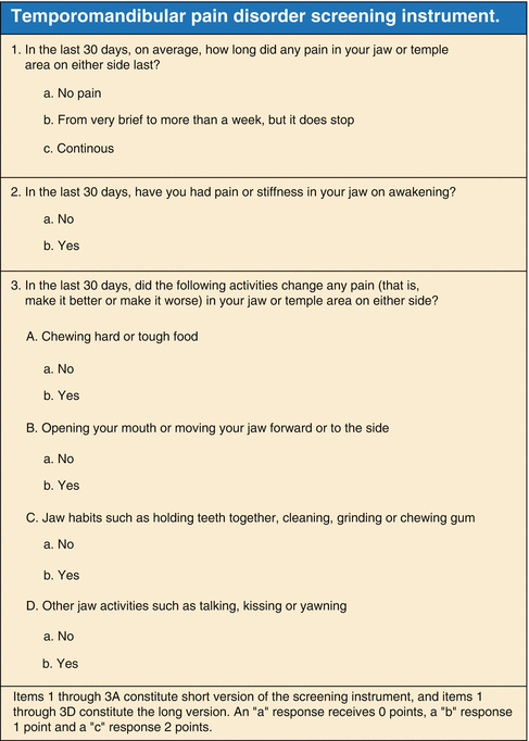
Similar problems afflicted several subsequent attempts to develop a useful screening instrument for detecting TMD problems in ordinary dental patient populations. These past attempts are well summarized in Table 3.1 in an excellent article by Gonzalez et al. [13] so they will not be discussed further here. In that 2011 paper, these authors presented both short (three-item) and long (six-item) versions of a newly developed TMD screening form (Fig. 3.1). By using psychometric methods for item selection, they developed these questionnaires and evaluated them for validity among 504 participants. They concluded that the selected items exhibited excellent content validity. The excellent levels of reliability, sensitivity, and specificity demonstrate the validity and usefulness of this instrument in any clinical office setting.


Fig. 3.1
TMD screening instrument (Gonzalez et al. [13]). Copyright © 2011 American Dental Association. All rights reserved. Reprinted by permission)
In addition to these TMD screening forms presented by various authors in the dental literature, there have been other approaches recommended by several dental organizations (academies, consortiums, institutes, etc.). The American College of Prosthodontists formed a committee that developed a 15-item form, which appears in an article in the inaugural issue of their journal [14]. In 2008, the European Academy of Craniomandibular Disorders (EACD) published a 4-item questionnaire that is quite minimal, with the instruction that any positive answer should lead to more in-depth investigations [15]. Meanwhile, various occlusally oriented institutes and study clubs have developed in-house protocols for detecting TMD problems in newly presenting patients. The Pankey and Dawson groups in Florida emphasize a manipulative methodology in which the mandible is placed in centric relation and occlusal relationships are observed. Also, the mandible is “loaded” by pushing horizontally backward and laterally to see how the TMJs respond to such forces [16]. Other groups (Spear, Kois) use so-called “de-programming splints” to allow the mandible to drift into a “relaxed” muscular position. Based on the outcome of this procedure, judgments are made about the need to change the TMJ relationship via permanent occlusal treatment [17]. The Las Vegas Institute, however, utilizes electronic diagnostic instrumentation to analyze mandibular and occlusal relations; their concept is described as “neuromuscular dentistry.” Based on a combination of electrical stimulators, jaw trackers, electromyographic recorders, and sound recorders, they determine who needs to have occlusal therapy as either a preventive or therapeutic treatment for TMD [18]. However, numerous papers have been published about the flaws and problems associated with the use of these electronic devices [19–23]. It should be obvious that all of these parochial in-house procedures are biased by the underlying philosophy of each organization; therefore, it is hard to accept the outcomes as providing meaningful analyses of good or bad mandibular positions, let alone as a screening method to detect TMDs. In addition, the invasiveness of the irreversible occlusal procedures that follow such analyses demands a much higher level of scientific evidence than is provided by these arbitrary diagnostic practices.
3.4 What to Do If Positive Findings Are Obtained During a Screening Exam?
In the course of screening new orthodontic patients for the presence of TMDs, there will inevitably be some positive answers to clinical questions as well as some positive “findings” from the physical examination of stomatognathic structures. The important question will be: when are those positive findings significant enough to establish a clinical diagnosis of TMD? This dilemma was first confronted by some of the early epidemiologic studies of TMD, in which researchers wanted to establish the prevalence of TMDs in the general population. The numbers reported in early studies by Helkimo and others who used the Index he developed were surprisingly high, often exceeding 50 % of the surveyed population [24]. This was largely due to the inclusion of various minor pain complaints and certain “objective” findings like TMJ clicking or deviated opening. Over time, this approach was criticized by Greene and Marbach [25] as well as several others, and later de Kanter in Holland presented survey numbers for that entire country which were much more reasonable [26]. Terms like “clinically significant” or “requiring treatment” became the threshold for determining whether people had actual TMD problems, or if they merely had variations from the expected ideal or normal findings. As a result, most modern epidemiologic studies of TMD agree that less than 10 % of the general population has a clinical problem that requires professional attention [27–29].
A screening exam for TMD should begin by asking questions about symptoms, both past and present. The first question should be: “Have you ever been diagnosed and/or treated for a TMD problem?” Assuming the answer is NO, the patient should be asked if any type of nondental facial pain has been occurring; however, this question should be elaborated so that the answers to it are meaningful for establishing a TMD diagnosis. What kind of pain has occurred? How often? Where did it seem to be? Did it affect jaw functions like chewing? Was it happening only after extreme functions like long dental appointments, extended gum chewing, etc.? Many people will report having a minimal experience of some type of facial pain history, often related to dento-alveolar problems. Therefore, a positive response to a pain question is far from being enough to classify the person as having a TMD.
The next symptom to ask about is TMJ clicking or popping. If there is a positive response, once again some important qualifying questions should be asked: When did the clicking start? Has it become more frequent or louder? Is it associated with any pain? Does the jaw ever get “stuck” in trying to open or close? Did the patient ever report it to a physician or dentist?
Some questions about functional difficulty should be part of the symptom history. Patients should be asked if they have noticed a limitation in their ability to open widely; however, it is important to ask whether that has always been true, or if it has been developing over time. They should be asked if normal functions like chewing hard food, singing in a choir, yawning widely, chewing gum, or sitting through a long dental appointment produce fatigue and pain; if so, does this symptom linger afterward or go away fairly quickly? Once again, there is a possibility that this is merely a situational problem, but it also is possible that these functional limitations are significant.
After the history portion of the exam is completed, the clinician needs to perform a physical examination to look for signs of TMD (and to correlate those with symptoms if possible). The first phenomenon to look for is clicking, popping, or other TMJ noises. This can be done by manual palpation or by auscultation with a stethoscope; clinicians may be surprised by how often a sound is discovered during the exam, but it was not reported by the patient. If the joint sound is a single click, it often will be louder on opening and softer on closing (reciprocal click) as the condyle goes beyond the posterior band of the articular disk. If the sound is a grating (crepitus) noise, the patient should be asked about a history of arthritis in other joints; if the TMJ is the only joint, questions can be raised about previous painful episodes in that area. Imaging is not required either medico-legally or clinically to document this type of finding in the absence of significant pain and dysfunction.
Continuing with manual palpation, the masticatory muscles and both TMJs can be palpated for tenderness. Once again, however, caution must be exercised so that every positive response does not become an important “finding” of muscle or joint problems. Some sensible questions like “Are you surprised this area is tender? Have you noticed pain or tenderness before in this area?” can be helpful in sorting out the significance of such findings. It is wise to palpate some adjacent structures that should not ever be tender in order to decide whether the patient is simply a strong responder to these kinds of provocations. The next objective assessment to perform is the observation and measurement of mouth opening, lateral excursions, and mandibular protrusion. One might see deviation during opening, but the mandible ends up in the midline at maximum opening, or there may be deflection of the mandible to one side as wide opening is reached. A finding of limited opening requires questioning the patients about their awareness of this fact; many subjects will report that they either were not aware of it or this is how it always has been for them, while others may say that this limitation has become an increasing problem for them. This is a crucial distinction, since the latter finding may be a sign of serious trouble and will need further investigation.
Stay updated, free dental videos. Join our Telegram channel

VIDEdental - Online dental courses


