Fig. 2.1
A simple, logical, evidence-based system for detection and classification of caries (Based on data from ICDAS International Caries Classification and Management SystemTM)
The pit and fissure probing as well as the interdental spaces, with a well sharp-pointed probe, together with a close microscopic inspection allows a good evaluation of the type of fissure or the existence of detectable cavitation (see Chap. 6). To detect early and hidden occlusal and proximal decays in children and pregnant women, a laser fluorescent device (DIAGNOdent, KaVo, Germany) is very useful, because it allows good sensitivity and specificity diagnosis without exposing the patient to X-ray examination; in case of frequent recall sessions for high decay risk, the laser fluorescent device permits a good check and control over time, avoiding excessive X-ray dose exposure (see Chap. 6).
The pulp vitality tests should always be performed, especially in case of trauma and deep caries, and noted in the chart, to compare in subsequent tests.
Intraoral periapical and bitewing X-rays can be an important aid in the diagnosis of occlusal caries lesions underlying previous fillings and hidden proximal caries invisible to direct observation.
The examination of the superficial and deep periodontal tissues and its relationship with the restorative therapy should be performed before starting any conservative therapy (proximal and cervical tooth decay or injury, in juxta- or subgingival position) or esthetic treatment (whitening in case of presence of cervical lesions or gum recession).
The occlusal status and TMJ examination allow to evaluate the presence of functional disorders and/or anatomical factors that may affect the choice of techniques and materials to be used (filling or inlay/onlay, hardness and elasticity of the materials to be used).
An intraoral photographic series is useful for planning the work in case of complex quadrant restorations (direct and indirect multiple restorations) and completes the documentation.
The evaluation of the degree of cooperation of the patient also provides a valuable suggestion to predict its future involvement during and after treatment (oral hygiene and regular visits to the control), to direct the best treatment choice.
2.5.1 Caries Risk Assessment
The caries risk assessment (high, low, average) directs toward an operating procedure rather than another and toward choice for different types of restoration.
The presence of decayed teeth, of missing teeth, or of filled teeth and also the presence of white spot, plaque accumulation, crowding teeth, and bad oral and diet habits (dummy, nighttime use of the bottle, fermentable carbohydrates) and the socioeconomic status [15] are strong caries risk indicators and suggest for higher caries risk assessment and different preventive approaches for children and adolescents (Fig. 2.2).
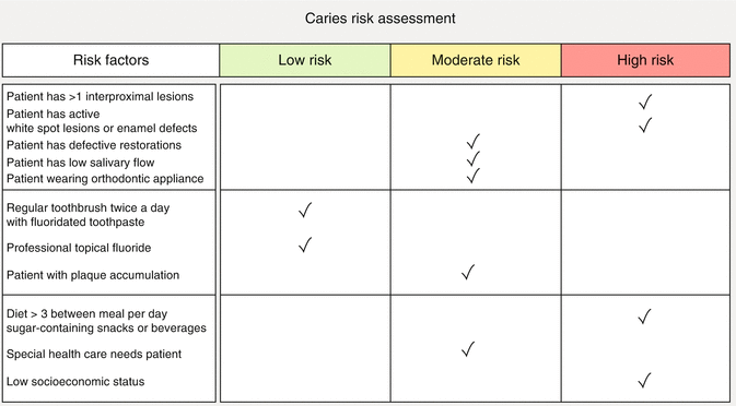

Fig. 2.2
Caries risk assessment guidelines for adolescents (Based on data from AAPD clinical guidelines)
A caries management protocol allows to establish the need to complete the regular (standard) preventive measurements with closest measurements including fluoride supplements and professional topical treatment every 3–6 months during more frequent periodic sessions, monitoring for signs of caries progression and relapse [13] (Fig. 2.3).
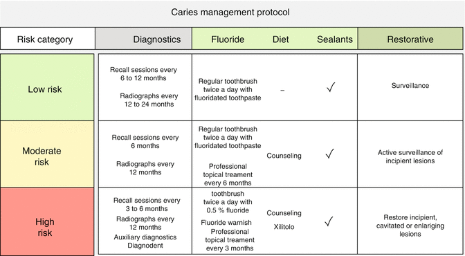

Fig. 2.3
Different caries management protocols according to different caries risk groups (Based on data from AAPD clinical guidelines)
2.6 Treatment Plan
A treatment plan must be explained to the patient who has to provide his informed consent to treatment. The parents or guardians of a minor patient must understand the importance of the prevention phase and provide consent, which must still be motivated and informed on the treatment plan chosen.
Dental caries can be largely prevented; prevention programs should therefore precede, accompany, and follow conservative treatment.
2.7 Methods of Caries Removal
Dentists are familiar with drills because they are the daily more frequently used instruments. These simple informations are necessary only to compare the conventional instrumentation with chemo-mechanical, air abrasion, and laser preparation.
2.7.1 High- and Low-Speed Drills
The high-speed drill or air turbine is the fastest instrument for dental structure removal (more than 200,000 rpm). The high speed and number of revolution per minute allow for minimal vibration and precise ablation of a dental cavity; it is the contact modality and the tactile feedback control that allow a precise ablation [16].
The interaction with the tooth produces abrasion of the dental structure, with formation of debris from dental ablation, and heating of the tooth structures, due to continuous friction.
Several rotating instruments are available for both high- and low-speed handpieces; diamond and multiblade carbide and the newest ceramic burrs also differ in shape, size, length, and grit.
Different shapes permit precise cutting of different forms that reproduce one of the burrs used: ball, pear, cylindric, tapered, pointed, football, or special shape. Different grits (from 100 μm up to 5–10 μm) permit different types of cut, from an unrefined dental tissue removal up to a very fine smoothening of the tooth margins and walls or a finishing of composite restoration.
Conditioning of surface and margin cavity, using 37 % orthophosphoric acid, is required after cavity preparation and margin smoothening to remove the produced debris and smear layer and to leave a surface suitable for bonding procedures; also the rise in pulp temperature may be a concern in case of deep cavities and dental trauma. Adequate water spray irrigation controls the rise in temperature and partially cleans the surface, but on the other hand contributes to the formation of mud and smear layer.
The high-speed drill can be also used to remove previously placed metal restorations (amalgam and gold).
Advantages of high-speed turbine are the fast dental procedure, the different possible shapes of preparation, the possibility to work on both the end and lateral sides of the burr and so in the undercuts of a cavity, and the tactile feedback control.
Disadvantages are related to the production of debris and smear layer during the ablation and the heating due to continuous friction. The heating is responsible for the unpleasant or painful perception. High-speed drill can create microcracks in the enamel and dentin. In deep cavity, high-speed instrumentation does not permit good control and minimal tissue removal, and also the safety is reduced when working on children and disabled patients, because of the potential of the patient’s head to suddenly move during the procedure.
The low-speed drill works slowly from few revolution for minutes with a speed reducer (100–300) up to 20,000–40,000 revolution per minute, allowing better control in deep dentin carious removal, but however the tissue removal is not selective or minimally invasive.
The lower speed increases the perception of vibration but also permits less heating of the tooth and dental pulp; intermittent contact permits to control the pain perception.
However, some patients prefer the minimal vibration of the turbine, while others the lower friction of the low-speed drill; local anesthetic may be needed for comfortable preparation of the tooth or teeth.
Common disadvantages of high- and low-speed drills are the absence of caries selectivity and of antibacterial activity and the possible surrounding soft tissue damage during tooth preparation.
The introduction of adhesive restorative materials and acid-etching techniques changed the concepts of cavity preparation and led to search and development of new cavity preparation systems.
2.7.2 Chemo-Mechanical Preparation
The first chemo-mechanical system was described by Goldman et al. in 1976 [17]. This system, named Caridex, was later followed on the market by Carisolv (Rubicon Life Science, Gothenburg, Sweden) in 1997. It is a hand excavation method that uses a chemical gel (Carisolv) to soften the demineralized affected dentin tissue. The gel agent is a mixture of amino acids (leucine, lysine, glutamic acid), NaOH, NaCl, and NaOCl in purified water that produces an alkaline solution (pH11). The chemical action of these components is aimed to degrade and disrupt the affected collagen fibrils making softened dentin easy to remove; successive hand excavation steps complete the caries removal. The aim of this technique is to produce a selective and minimal tissue removal; this conservative approach may be a help in case of deep decay, reducing the risk of pulp exposure. The chemo-mechanical preparation produces dentin modifications suitable for bonding procedure. The surface does not present smear layer, dentinal tubules are open, and bonding procedure produces deeper resin tags (15 μm versus 10 μm) when compared to burr excavation, confirming the effectiveness of the chemo-mechanical preparation in achieving a selective carious removal and good adhesive surface [18, 19]. The disadvantage of this technique is the longer time needed for the treatment.
2.7.3 Air Abrasion
Air abrasion is a relatively new method for removing tooth structure and dental caries during cavity preparation and has specific characteristics suitable for adhesive restorative materials. Air abrasion utilizes fine particles (10–50 μm) of aluminum oxide for the removal of superficial enamel and dentin caries allowing minimal invasive ablation of the tissue (Fig. 2.4). The dental morphologic pattern produced by air abrasion is very conservative [20, 21], with irregular surfaces due to the impact of abrasion particles [20]; the most relevant morphologic characteristic is a round-shaped and halo contour [20, 22, 23] (Fig. 2.5). This surface aspect seems to improve the adhesion, facilitating the fitting of composite materials, reducing microleakage [24–26]; also the incidence of microcracks within the tooth and fracture of composite material is reduced when comparing a cavity preparation with round contour to another with defined acute angles [25, 26] and so enhancing the longevity of tooth and of adhesive restorations [26, 27].
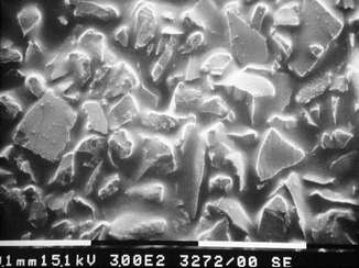
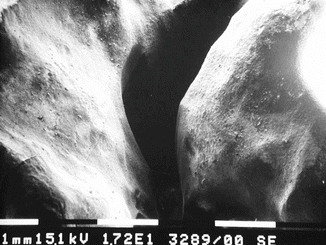

Fig. 2.4
SEM image shows the fine particles (10–50 μm) of aluminum oxide used for air abrasion system (Courtesy of Prof. Vasilios Kaitsas, Italy)

Fig. 2.5
SEM image shows a fissure preparation for sealant application using air abrasion. To note the typical round-shaped and halo contour of the interaction (Courtesy of Prof. Vasilios Kaitsas, Italy)
Air abrasion does not produce smear layer, but the main disadvantage is dust particles remaining from cavity preparation creating a film over the operatory field (teeth and rubber dam); the dust is also a potential health hazard from breathing dust particles, and emphysema of the patient’s orbital area, related to the air pressure applied, has also been described in the literature (Fig. 2.6). Furthermore, as a minor side effect, the dust particles can scratch dental mirrors [28]. As for drills, air abrasion does not provide any bactericidal effect [28].
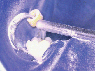

Fig. 2.6
Intraoperative image shows the dust particles remaining from cavity preparation creating a film over all the operatory field (Courtesy of Prof. Vasilios Kaitsas, Italy)
2.7.4 Erbium Family of Lasers
Among the other systems for cavity preparation, the erbium lasers are completely different because of their possibility to work on both hard and soft tissues and the ability to complete the entire conservative procedure. Erbium lasers are suitable for carious removal and are very efficient for deep dentin decontamination. They can expose subgingival cavity margins in case of class 2, class 3, and class 5 caries, easily and effectively performing a laser gingivectomy; laser can also coagulate the gingiva before cavity preparation or coagulate and decontaminate the exposed pulp during a cavity preparation. Erbium lasers can also perform a vital pulpotomy or pulpectomy, if needed, and all these abilities make the laser “unique.”
Also, the morphologic pattern of lased ablated enamel and dentin is suitable for adhesive dentistry because of the absence of debris and smear layer and the increased surface for bonding; surface and margin finishing and conditioning are necessary to improve the bonding characteristics of lased surface (see Chaps. 4 and 5).
Finally, the laser has less impact on the patient because it works in noncontact mode, without vibration, and allows quite lower stimulation of the dental nerve (see Sects. 7.2, 7.3, and 7.4); the noise is also different from the shrill noise of the high-speed handpiece that often created fear in children and phobic patients; it sounds like a “popping sound” and it is better accepted by the patients. Local anesthesia is often unnecessary [29].
Stay updated, free dental videos. Join our Telegram channel

VIDEdental - Online dental courses


